Prediction of Extracellular Proteases of the Human
Pathogen
Helicobacter pylori
Reveals Proteolytic Activity
of the Hp1018/19 Protein HtrA
Martin Lo¨wer1, Christiane Weydig2, Dirk Metzler3, Andreas Reuter4, Anna Starzinski-Powitz1, Silja Wessler2., Gisbert Schneider1.*
1Goethe-University, Institute of Cell Biology and Neuroscience / CMP, Frankfurt am Main, Germany,2Junior Research Group, Paul-Ehrlich Institute, Langen, Germany, 3Goethe-University, Institute of Computer Science, Frankfurt am Main, Germany,4Paul-Ehrlich Institute, Department of Allergology, Langen, Germany
Abstract
Exported proteases ofHelicobacter pylori (H. pylori) are potentially involved in pathogen-associated disorders leading to gastric inflammation and neoplasia. By comprehensive sequence screening of theH. pyloriproteome for predicted secreted proteases, we retrieved several candidate genes. We detected caseinolytic activities of several such proteases, which are released independently from theH. pylori type IV secretion system encoded by the cagpathogenicity island (cagPAI). Among these, we found the predicted serine protease HtrA (Hp1019), which was previously identified in the bacterial secretome ofH. pylori. Importantly, we further found that theH. pylorigeneshp1018andhp1019represent a single gene likely coding for an exported protein. Here, we directly verified proteolytic activity of HtrAin vitroand identified the HtrA protease in zymograms by mass spectrometry. Overexpressed and purified HtrA exhibited pronounced proteolytic activity, which is inactivated after mutation of Ser205 to alanine in the predicted active center of HtrA. These data demonstrate that
H. pylorisecretes HtrA as an active protease, which might represent a novel candidate target for therapeutic intervention strategies.
Citation:Lo¨wer M, Weydig C, Metzler D, Reuter A, Starzinski-Powitz A, et al. (2008) Prediction of Extracellular Proteases of the Human PathogenHelicobacter pyloriReveals Proteolytic Activity of the Hp1018/19 Protein HtrA. PLoS ONE 3(10): e3510. doi:10.1371/journal.pone.0003510
Editor:Raphael H. Valdivia, Duke University Medical Center, United States of America ReceivedJune 25, 2008;AcceptedSeptember 30, 2008;PublishedOctober 23, 2008
Copyright:ß2008 Lo¨wer et al. This is an open-access article distributed under the terms of the Creative Commons Attribution License, which permits unrestricted use, distribution, and reproduction in any medium, provided the original author and source are credited.
Funding:This research was supported by the Beilstein-Institut zur Fo¨rderung der Chemischen Wissenschaften Frankfurt am Main (Germany), the Centre for Membrane Proteomics (CMP) Frankfurt am Main (Germany), the Ju¨rgen-Manchot Stiftung, the Paul-Ehrlich Institut Langen (Germany), and the Deutsche Forschungsgemeinschaft (SFB-579, project A11).
Competing Interests:The authors have declared that no competing interests exist.
* E-mail: [email protected]
.These authors contributed equally to this work.
Introduction
The mucosal epithelium in the human stomach forms the first barrier that prevents infiltration of pathogens into the host organism. The human pathogen H. pylori developed efficient strategies to colonize the gastric epithelium as a unique niche, where it induces the disruption of the epithelial layer contributing to inflammatory diseases (e.g.chronic gastritis, ulceration), mucosa-associated lymphoid tissue (MALT) lymphoma and gastric cancer in humans [1,2]. More virulent H. pylori strains express a combination of key disease-associated virulence factors allowing successful colonization in the stomach [3]. Among those,H. pylori
harborscagpathogenicity island (cagPAI), which encodes a type IV secretion system (T4SS) to inject the bacterial CagA (cytotoxin-associated gene A) oncoprotein into host cells [4]. In vitro, translocated CagA can strongly enhance the disruption of intercellular adhesions [4,5]. This process is believed to contribute to inflammation, carcinogenesis and invasive growth. Although the cellular aspects of CagA have been investigated intensively, the complex mechanisms of the actual interaction ofH. pyloriand the human epithelium are not fully understood yet.
Many pathogens developed elegant mechanisms for tissue destruction by secreting proteins with proteolytic activity.
Exported bacterial enzymes can directly activate hostpro -matrix-metalloproteinases (pro-MMPs) representing a biochemical effi-cient way for matrix degradation. An example is set by the wide range of proteases of the thermolysin family secreted by
Pseudomonas aeruginosaandVibrio cholerathat activateproMMP1, -8, and -9 [6]. It has been further observed that serine proteases associated with lipopolysaccharides can induce MMP-9 activity in macrophages [7]. MMP-9 cleavage was also detected by a secreted zinc metalloproteinase (ZmpC) fromStreptococcus pneumoniae, which indicates that ZmpC may play a role in pneumococcal virulence and pathogenicity in the lung [8].
Proteases might also play a role inH. pylori pathogenesis, and protease secretion has already been described for this organism [9].H. pylorisheds an unknown protease that efficiently degrades PDGF (platelet derived growth factor) and TGF-b(transforming growth factor beta), which can be inhibited with sulglycotide [10]. Some features present in the primary sequence of H. pylori
virulence factor vacuolating cytotoxin A (VacA) are reminiscent of serine proteases [11], although the predicted proteolytic activity of VacA has not been detected yet. In 1997, a H. pylori
metalloproteinase with a native molecular size of approximately 200 kDa was discovered, which was secreted whenH. pyloriwas grown in liquid culture [12]. The authors hypothesized that
surface expression of this metalloprotease activity may be involved in proteolysis of a variety of host proteins in vivo and thereby contribute to gastric pathology [12]. Importantly,H. pylorisecretes a collagenase, encoded by hp0169, which might represent an essential virulence factor forH. pylori stomach colonization [13]. The predicted serine protease and chaperone HtrA (Hp1019) was previously identified as an extracellular protein ofH. pylori [14], but its proteolytic role and substrates are still unknown.
As 658 of the 1,576 identified genes of theH. pylorigenome [15] are annotated as ‘‘hypothetical’’ or as bearing a hypothetical function [16], we aimed at the identification of H. pylori genes possibly coding for secreted proteases by combining genomic data analysis with zymography. Indeed, we found thatH. pylorisecretes unknown proteins exhibiting caseinolytic activity. By calculating similarities to known proteases and using localization prediction methods, we inferred function and localization of these hypothet-icalH. pyloriproteins. We also identified a sequencing error in the
hp1018gene, which after correction encodes for a signal peptide for the putative serine protease HtrA (Hp1019). Eventually, we verified proteolytic activity of HtrA in biochemical approaches. The present study demonstrates the usefulness of sequence-based genome mining for potential drug targets representing one possible route for the prevention of matrix degradation of the mucosal epithelium byH. pyloriand other pathogens.
Results and Discussion
H. pylori secretes caseinolytic proteases
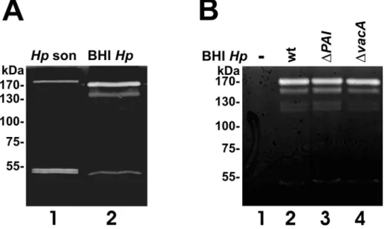
Data are accumulating that bacteria secrete proteases with functional roles in microbial pathogenesis, but knowledge ofH. pylori-secreted proteases and their functions is still limited. To analyze whetherH. pyloriactually secretes proteases, we performed casein zymography to monitor proteolytic activity in the supernatants ofH. pylori lysates (Figure 1A, lane1) and H. pylori
culture medium (Figure 1A, lane 2). At least three casein-cleaving proteases were exported byH. pyloriexhibiting apparent molecular weights of approximately 170 kDa, 140 kDa, and 50 kDa
(Figure 1A, lane 2). Interestingly, the protein band pattern present in the supernatant of theH. pylorimedium obviously differs from the equivalentH. pylori lysate (Figure 1A lanes 1). The detected 170 kDa protease present in the supernatant ofH. pylori(BHIHp) consistently migrated slightly faster than in the H. pylori lysate, while the 140 kDa protein was only present in the supernatant, but absent in the lysate ofH. pylori(Hpson). In contrast to the double band detected in the lysates, we observed only a single proteolytic activity in the supernatant (Figure 1A, lanes 1–2). These data indicate that the export of the proteases might occurvia active signal peptide-dependent translocation, rather than being an artifact of bacterial autolysis in theH. pyloriliquid culture.
Since H. pylori encodes a well-described T4SS and the T4SS-independently secreted pathogenic factor VacA with a hypothe-sized protease function [11], we also included supernatants of isogenicH. pylorimutants which are deficient of T4SS and CagA (DPAI, Figure 1B, lane 3), or VacA (DVacA, Figure 1B, lane 4), and compared them with the H. pylori wildtype strain (wt, Figure 1B, lane 2) and H. pylori-free culture medium (-, Figure 1B, lane 1). Compared to the wildtype strain, theDPAI mutant showed the same secretion pattern of proteins with caseinolytic activity in the extracellular space suggesting that their secretion might occur independently from the T4SS (Figure 1B, lane 3). Although initial publications indicated a predicted serine protease activity of the pathogenic factor VacA [11], we can exclude a caseinolytic effect of VacA since the isogenic vacA -deficientH. pylori mutant showed a similar pattern of proteases (Figure 1B, lane 4). Gelatin zymographies were also performed by us and clearly demonstrated the lack of gelatinolytical H. pylori
proteases (not shown). A positive result here would have demonstrated a closer link to matrix degaradation, as gelatin is a product of collagen, a major extacellular matrix protein.
So far, the identity of the detected H. pylori proteases was unknown. A previously described multi-metalloprotease-like complex secreted byH. pylori with a molecular weight of about 200 kDa [12] might be an explanation for the largest protein seen in the zymogram, since its size is four to six times greater than
Figure 1.H. pylorisecretes bacterial factors with caseinolytic activities. (A)TheH. pyloristrain 26695 was grown in protein-free BHI medium. After 48 hours, the bacteria were harvested and lysed by sonfication (Hpson). 30ml aliquots of the supernatants (BHIHp) and bacterial lysates were
separated by casein zymography and analyzed proteolytic activities.(B)H. pyloristrains wild type (P12, wt),DPAI, andDVacA were grown in protein-free liquid growth medium. 30ml of the aliquots of the medium were analyzed in casein zymograms for proteolytic activities.
comparable proteases of other Gram-negative bacteria [12]. Also, protease DegP ofEscherichia coli, which is a homolog of Hp1019 fromH. pylori, was shown to form hexamers when crystallized [17]. Therefore, as zymography was performed under non-reducing conditions, the upper band(s) might result from smaller proteins forming a macromolecular complex.
In silico genome screening for candidates ofH. pylori secreted hypothetical proteases
Based on the finding thatH. pyloriactively secretes proteases, we then aimed to identify suitable candidates by in silico analysis. Thus, we compared the H. pylori proteome to a set of known proteases from various organisms using sequence alignment techniques. A reference set of known proteases containing 3,566 amino acid sequences was compiled from the UniProtKB/ SwissProt database (version 6.7) [18], which served as queries for exhaustive pairwise alignment to genomic and protein sequence data of H. pylori strain 26695 with 1,576 annotated genes from the NCBI RefSeq database (accession number NC_000915) [16]. For the 1,576 putative H. pylori proteins, 75,524 local alignments were returned by the BLAST algorithm [19]. Alignments yielding an E-value#0.5 were selected and divided into four classes:
Class A: alignments showing complete conservation of the active-site region,
Class B: alignments showing partial conservation of the active-site region,
Class C: alignments with proteases lacking an active-site annotation, and
Class D: all other alignments.
The latter (class D) were not further examined. Information about the localization of the active sites was retrieved from the feature tables of the respective SwissProt entries [18].
Then, we predicted protein localization using prediction systems, which are publicly available on the World Wide Web: SignalP [20], SecretomeP [21], Phobius [22], CELLO [23], PA-SUB [24], and PSORTb [25]. All systems are capable or explicitly designed to analyze amino acid sequences from Gram-negative bacteria. Alignments were selected for further examination when the corresponding predictions for a protein sequence matched one or more of the following criteria:
i) predicted extracellular localization (CELLO, PSORTb, PA-SUB),
ii) predicted signal peptide (SignalP),
iii) predicted signal peptide, but no transmembrane helices (Phobius), and a SecretomePscore$0.5.
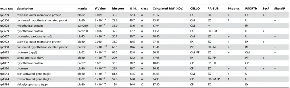
By filtering the alignments with respect to the active residues marked in the sequence of reference protease and the localization prediction, we obtained 47 class A, 39 class B and 32 class C proteins (vide supra) and their corresponding genes. The best-scoring alignments of those proteins to proteases of the reference set were manually inspected. Among those, nine genes have not been described to code for H. pylori proteases yet, but can be aligned with a statistically significant score to proteases of the reference protease sequences (Table 1). Interestingly, the putative translation products of geneshp0289,hp0609, andhp0922form a group of paralogs to VacA cytotoxin [26], which can be seen in a multiple sequence alignment (not shown). Structural similarities of VacA to extracellular IgA proteases ofHaemophilus influenzaehave been described previously [11]. Pairwise sequence identity of these
VacA paralogs to VacA ranges between 25% and 30% which also include the C-terminal autotransporter sequence [27]. As this sequence is sufficient to transclocate the N-terminal part of the protein across the outer membrane2which is often followed by an autoproteolytic event to release the translocated part into the extracellular space [27] 2 it seems likely that some of these proteins possess a proteolytical function.
Finally, although we could not detect caseinolytic activity of VacA in our casein zymography study, we cannot exclude an effect of VacA and its paralogs on other substratesper se. However, the alignments do not reveal conserved active site residues in VacA paralogs. Still they might represent autoproteolytic autotransporter proteins without common protease motifs which have been reported already [27]. Notably, their precursor proteins have a molecular weight of 136 to 311 kDa (according to SwissProt entries O25063, O25330 and O25579) which is in accordance with the molecular weights we observed in the zymography after a possible cleavage of the N-terminal signal peptide and the autotransporter sequence.
H. pyloriharbors five genes that are described in the literature and/or database annotations to code for potential extracellular proteases (Table 1). Processing protease YmxG (Hp0657) and protease pqqE (Hp1012) are predicted to possess a signal peptide (Table 1) and to be extracellular or outer membrane-bound. The protease coded by the gene hp1350 could be extracellular, as SecretomeP and PA-Sub vote for this localization and the existence of a signal peptide is also predicted (Table 1). The product ofhp1019, which is annotated as a serine protease in the respective GenBank file, seems to be a homologue to heat shock protein HtrA fromEscherichia coli. Its active site is fully conserved, and the extracellular localization has been determined previously [14]. The gene product ofhp1584 is annotated as a sialoglyco-protease (gcp). Its amino acid sequence does not contain known export motifs, and the amino acid composition is predicted to be cytoplasmic. However, the PA-SUB and PSORTb predictors categorized the protein as extracellular (Table 1) based on the extracellular localization of the homologous o-sialoglycoprotein endopeptidase of Mannheimia haemolytica (SwissProt identifier GCP_PASHA), which also lacks an N-terminal targeting signal [28]. In fact, very recently Hp0657, Hp1012, Hp1019, and Hp1350 have been identified in the extracellular H. pylori
proteome [29] indicating the high specificity of our bioinformatical prediction of hypothetical extracellularH. pyloriproteases (Table 1). Since we demonstrated that several caseinolytic proteases are secreted byH. pyloriindependently of functional T4SS, it is likely that other secretion systems exist. This is underlined by our observation that nine out of 14 genes either contain a signal peptide, which only explains a transportation to the periplasm, or receive a high SecP prediction score (Table 1). We stress that these predicted features are common for extracellular proteins but do not explain a possible transport pathway. Thus one can speculate that a secretory machinery not yet attributed to H. pylori, or entirely novel ones, might be involved which require export signals of an unknown nature. For example, H. pylori might involve a specific type I (ABC) or a type III transportation system.
H. pyloriHtrA is an active protease
We were then interested in answering the question whether one of the predicted H. pylori proteases accounts for the observed proteolytic activity. In a first step, concentratedH. pylori lysates were separated by zymography under non-reducing conditions followed by protein eluation of proteins from the negatively stained protein bands I and II (Figure 2A). Then, eluated proteins were concentrated and separated by a denaturating SDS PAGE
Secreted Proteases ofH.pylori
Table 1.Results of the Blastp search for proteases genes, of the localization prediction and calculated molecular weights (MW).
locus tag description matrix E-Value bitscore % id. class Calculated MW (kDa) CELLO PA-SUB Phobius PSORTb SecP SignalP
hp0289 toxin-like outer membrane protein blo62 0.009 38.9 22.3 A 31.12 EX EX + EX + +
hp0506 conserved hypothetical secreted protein blo80 8610214 72.8 40.7 A 45.97 OM EX 1 U
+
hp0608 hypothetical protein pam250 761024 36.9 23.6 A 17.94 OM 2 IM
hp0609 hypothetical protein pam250 0.006 37.9 17.7 A 13.51 EX EX, OM U +
hp0657 processing protease (ymxG) blo45 461029 56.7 20.7 A 48.80 OM EX
+ U +
hp0922 toxin-like outer membrane protein blo80 0.080 35.7 30.5 A 27.46 EX EX + EX +
hp0980 conserved hypothetical secreted protein pam30 3610212 65.5 38.6 A 11.41 PP EX, IM + IM +
hp1012 protease (pqqE) blo62 1610220 95.5 23.8 A 50.33 OM, PP EX
+ OM +
hp1019 serine protease (htrA) blo80 4610282 299 42.2 A 47.98 EX EX, PP PP +
hp1037 hypothetical protein pam70 0.061 33.3 30.7 A 40.80 CP CP, EX CP
hp1350 protease blo80 7610281 295 39.7 A 50.55 CP EX
+ U + +
hp1543 toxR-activated gene (tagE) blo80 1610216 81.5 43.5 A 35.63 OM EX 1 U
hp1544 toxR-activated gene (tagE) blo62 561028 52.8 34.0 A 34.93 CP EX,OM,PP 1 U
hp1584 sialoglycoprotease (gcp) blo80 1610239 158 36.4 C 37.80 CP EX EX
Locus tags and descriptions were taken from the corresponding GenBank entries. The columns ‘‘matrix’’, ‘‘E-Value’’, ‘‘bitscore’’ and ‘‘% id.’’ list the alignment data of the according highest scoring alignment. The column ‘‘class’’ refers to our definition of alignment classes. Molecular weight was calculated by a program hosted on the ExPASy server. The columns ‘‘CELLO’’, ‘‘PA-SUB’’, and ‘‘PSORTb’’ give the classifications according to the prediction software. The column ‘‘Phobius’’ gives the number of transmembrane helices and a plus (+) sign if a signal peptide was found. Columns ‘‘SecP’’ and ‘‘SignalP’’ contain a plus sign for a SecretomeP output$0.5 or prediction of a signal peptide, respectively. CP = cytoplasm, IM = inner membrane, PP = periplasm, OM = outer membrane, EX = extracellular, U = unknown.
doi:10.1371/journal.pone.0003510.t001
Secreted
Proteases
of
H.pylori
ONE
|
www.plos
one.org
4
October
2008
|
Volume
3
|
Issue
10
|
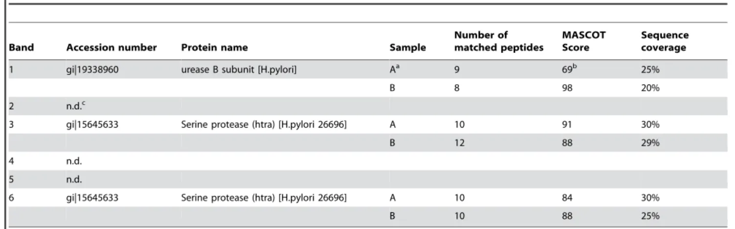
(Figure 2B). We detected four different proteins in the Coomassie-stained SDS PAGE, which were isolated from protein band I in the zymogram (compare Figure 2A, band I and Figure 2B, lane I). Electrophoretic separation of proteins from protein band II (Figure 2A) resulted in two different proteins (Figure 2B, lane II). The identity of these proteins was determined by MALDI-TOF-MS. The accession number, denomination and a summary of the MS data are presented in Table 2. The results of the MS analyses are shown for a single database entry for each band. However, due to the high degree of sequence identity between proteins isolated from differentH. pyloristrains significant hits were obtained also for other urease and serine proteases,e.g.serine protease fromH. pyloristrain J99 or Ure B from database entry gi/51989332.
Hp1018 encodes a signal peptide for an active Hp1019 protease
Hp1019 has been previously predicted as a secretedH. pylori
protease with unknown function [14,29]. However, its proteolytic activity had not been demonstrated. Considering the protein sequence ofH. pyloriHtrA, it lacks an annotated N-terminal signal
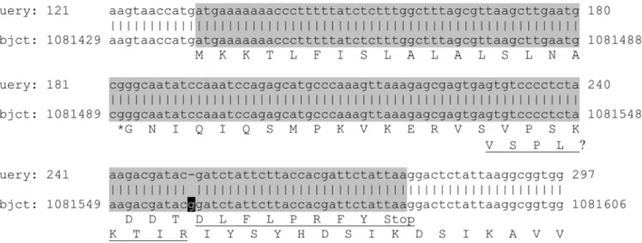
peptide, in contrast to HtrA ofE. coli. The genehp1019has an N-terminal overlap with the adjoining gene hp1018, which is 147 bases long and in a different reading frame. It has been suggested before that those genes might belong together [30]. Thus, we re-sequenced the gene hp1018 and aligned it to the published genomic data of H. pylori Hp26695 (Figure 3). Here, we demonstrate thathp1018reveals a wrongly sequenced guanidine at position 1081558 of the published genome of H. pyloristrain 26695. We conclude from our data that the translation of Hp1018 actually contains a signal peptide-like sequence (SignalP
score.0.99) at its N-terminus, and it is most likely that Hp1018 represents the N-terminal part of Hp1019 resulting in a new sequence with 475 amino acids.
To prove proteolytic activity of Hp1018/19 for the first time, we fused thehp1018/19gene lacking the putative signal peptide to the glutathione-S-transferase (gst) gene and transformed the construct intoE. coliBL21 to express the recombinant protein (Figure 4A). Both, induction and enrichment of GST-Hp1018/19Dsp protein were analyzed by Coomassie-stained SDS PAGEs (Figure 4B). During GST-Hp1018/19Dsp preparation, contaminating proteins were co-purified, which were identified by MALDI-TOF-MS as glutathione-S-transferase and degradation products of HtrA. Accordingly, it had been demonstrated thatE. coliencoded HtrA is an endopeptidase [31]. To remove the GST tag from the fusion protein, GST-Hp1018/19Dsp coupled to GST sepharose was incubated with PreScission protease resulting in the release of Hp1018/19Dsp protein (Figure 4B, lane 6).
Purified proteins were then probed for proteolytic activity (Figure 4C). The GST-Hp1018/19Dsp proteins were bound to GST sepharose, washed and eluated using reduced glutathione. As a control, we cloned and purified the Hp1018/19DspS205Aprotein in which serine-205 was mutated to alanine in the presumable active center of HtrA. As expected, we observed casein degradation by GST-Hp1018/19Dsp protein (Figure 4C, lane 3), but not by the GST-Hp1018/19DspS205A(Figure 4C, lanes 1– 2). This finding demonstrates that H. pylori HtrA actually is an active protease, which can be inactivated by mutation of serine-205. In parallel, we cloned and purified Hp0506, Hp0657, Hp1012, Hp1037, Hp1543, and Hp0169, which previously had been described as a collagenase [13]. With the exception of Hp1019, we did not detect any proteolytical activities using casein as a substrate in zymography studies (data not shown). Therefore, we conclude that the observed caseinolytic activities were actually mediated by Hp1018/19.
Table 2.Proteins that were identified by mass-spectrometry (cf.Figure 2B).
Band Accession number Protein name Sample
Number of matched peptides
MASCOT Score
Sequence coverage
1 gi|19338960 urease B subunit [H.pylori] Aa 9 69b 25%
B 8 98 20%
2 n.d.c
3 gi|15645633 Serine protease (htra) [H.pylori 26696] A 10 91 30%
B 12 88 29%
4 n.d.
5 n.d.
6 gi|15645633 Serine protease (htra) [H.pylori 26696] A 10 84 30%
B 10 88 25%
aresults of two independently processed samples;bprotein score was below the level that indicates ap-value of
,0,05,cnot determined. doi:10.1371/journal.pone.0003510.t002
Figure 2. Identification of H. pylori proteases. (A) For a preparative analyses, 186109 bacteria were lysed and analyzed by zymography. The upper (1) and lower (2) negatively stained protease bands were excised, proteins were eluated and separated by SDS PAGE and Coomassie staining(B). Indicated protein bands were analyzed by mass-spectrometry.
doi:10.1371/journal.pone.0003510.g002
Secreted Proteases ofH.pylori
As shown my mass-spectrometry, we also co-purified processed HtrA variants with GST-Hp1018/19Dsp (Figure 4B). We detected proteolytic activity of these proteins in casein zymography (Figure 4C). We therefore assume that processed variants of HtrA formed multimers with GST-Hp1018/19Dsp during the purifica-tion steps. This suggespurifica-tion is supported by the finding that removal of the GST tag from GST-Hp1018/19Dsp protein led to the formation of the 170 kDa protease (Figure 4C, lane 4), which was not detected after purification of Hp1018/19DspS205A(Figure 4C, lane 2). Together with our analysis showing that HtrA was present in the upper and lower protein bands (Figure 2), we conclude from our data that HtrA might also be active as a multimer.
Conclusions
The complex mechanisms how H. pylori strongly induce inflammatory responses and invasive growth leading to the disruption of the human epithelium are still unclear. Although exported proteases of pathogens represent extensively studied virulence factors, not much is known about their involvement in
H. pylori-associated pathogenesis. This comprehensive analysis of the
H. pylori strain 26695 genome by sequence analysis and activity prediction methods revealed several genes coding for putative proteases. Among those, we identified the HtrA fromH. pylorias a secreted enzyme exhibiting proteolytic activity. We also found that HtrA forms proteolytically active multimers, which is consistent with an earlier report of Windle and colleagues who demonstrated thatH. pylorisecretes a metalloprotease with a native molecular size of approximately 200 kDa and speculated whether this metallo-protease activity may be involved in proteolysis of a variety of host
proteinsin vivoand thereby contributes to gastric pathology [12]. The E. coli homologue HtrA functions as a heat shock protein, although it cannot be excluded that Hp1019 represents a so-called ‘‘moonlighting’’ protein [32], serving a function both in the periplasm in heat shock degradation and the extracellular matrix as a virulence factor. In fact, a secreted collagenase Hp0169 was identified as an important virulence factor forH. pyloricolonization [13]. Although the biological function of Hp0169 and the recently detected extracellular proteases Hp0657, Hp1012, and Hp1350 [29] are unknown, it underscores the potential importance of secreted bacterial proteases in H. pylori mediated pathogenesis, which represent attractive vaccine and drug target candidates.
Materials and Methods Homology search
Proteases were compiled for the reference data set by selecting all entries from the UniProtKB/SwissProt database (version 6.7) [18] containing the keyword ‘‘protease’’, but lacking the phrases ‘‘inhibitor*’’, ‘‘probable*’’, ‘‘fragment*’’, ‘‘hypothetical*’’, ‘‘puta-tive*’’, ‘‘possible*’’ or ‘‘predicted*’’ in the keyword and description fields, where the asterisk is a wildcard for any arbitrary suffix. The NCBI BLAST package was employed for pairwise sequence alignment [19]. The Blastp program was used for protein-protein comparison. Tblastn was used to compare the whole DNA sequence of H. pylori strain 26695 with the protease sequence set. The substitution matrices PAM30, PAM70, PAM250, BLOSUM45, BLOSUM62 and BLOSUM80 were used with default parameter settings (e.g.scoring penalties, window size).
Figure 3. Blastn alignment of the re-sequenced nucleotide sequence (query) with the original genomic sequence (subject) of
hp1019.The annotated genehp1018is marked in grey. The letter ‘r’ represents (‘a’ OR ‘g’), while the letter ‘k’ represents (‘t’ OR ‘g’). The inserted guanidine is printed white on black. Numbers give residue positions. The amino acid translation is given in single letter code for Hp1018, starting at position 1081440, and for Hp1019, starting at position 1081537. The predicted most likely signal peptidase cleavage site between the amino acids LNA and GNI is marked with an asterisk. The underlined part of the amino acid sequences will not be part of the translation if the marked guanidine is removed.
Bacteria
H. pylori wildtype strains 26695 and P12, its isogenic mutant strains DVacA, and DPAI had been described before [11,33]. Bacteria were grown in protein-free liquid brain heart infusion (BHI) medium (Merck, Darmstadt, Germany) supplemented with
b-cyclodextrin for 48 hours, which has been previously optimized for minimal autolysis ofH. pyloricells [14]. Lysates ofH. pyloriwere obtained by sonification in PBS containing 0.1% Triton X-100. Supernatants ofH. pyloriBHI cultures were sterilized by filtration (pore size 0.22mm).
Amplification and sequencing of hp1018
The genehp1018was amplified from the genomic DNA ofH. pylori strain 26695 by standard PCR using the Pfx DNA-polymerase (Invitrogen, Karlsruhe, Germany). The following primers were used: hp1018for: 59-GGC TAT GGA TAA GGA TCA ACG C-39, hp1018rev: 59-CCA CCG CCT TAA TAG AGT CCT T-39. The PCR product, having a calculated length of 333 bases, was submitted to a commercial provider (GENterprise, Mainz, Germany) for sequencing.
Cloning, mutation and purification of HtrA
The construct Hp1018/19Dsp was amplified from genomic DNA of H. pyloristrain 26695 using the primers 59 -aaggatccgg-caatatccaaatccagagcatg-39 and 59 -aagaattcgacccacccctatcatttcacc-39 with Pfx DNA polymerase in supplied buffer with 26PCR
Enhancer (Invitrogen). The amplified BamH1/EcoR1 flanked PCR product was then ligated into the pGEM-T Easy plasmid (Promega), subcloned into the pGEX-6P-1 plasmid DNA (GE Healthcare Life Sciences) and transformed inE. coliBL21. The construction of the protease-inactive Hp1018/19DspS205Aprotein, serine 205 was mutated to alanine using the QuikChangeH
Lightning Site-Directed Mutagenesis Kit (Stratagene) according to the manufacturer’s instructions. For heterologous overexpression and purification of GST-Hp1018/19Dsp, transformedE. coliwas grown in 500 ml TB medium to an OD550 of 0.6 and the
expression was induced by the addition of 0.1 mM isopropylthio-galactosid (IPTG). The bacterial culture was pelleted at 40006g
for 30 minutes and lysed in 25 ml PBS by sonification. The lysate was cleared by centrifugation and the supernatant was incubated with glutathione sepharose (GE Healthcare Life Sciences) at 4uC over night. The fusion protein was either eluted with 10 mM reduced glutathione for 10 minutes at room temperature or cleaved with 180 U Prescission Protease for 16 h at 4uC (GE Healthcare Life Sciences). Elution and cleavage products were analyzed by SDS PAGE and zymography.
Zymography and protein eluation
Undiluted aliquots were loaded onto 8% SDS-PAGE contain-ing 0.1% casein (Invitrogen, Germany) and separated by electrophoresis. After separation, the gel was re-naturated in 2.5% Triton X-100 solution at room temperature for 60 min with
Figure 4. Proteolytic activity of the Hp1018/19 protein. (A)For the construction of the GST-Hp1018/19Dsp fusion protein, the re-sequenced Hp1018/19 gene was amplified without the putative signal peptide and cloned into the pGEX-6P-1 vector.(B)Thegst-hp1018/19Dspconstruct was transformed inE. colifor overexpression and total protein extracts from untreated (lane 1) and IPTG-inducedE. coli(lane 2) were separated by SDS PAGE. Overexpressed GST-Hp1018/19Dsp was precipitated using glutathione sepharose and released by three eluation steps (lanes 3–5). To remove the GST tag, GST-Hp1018/19Dsp bound to glutathione sepharose were treated with PreScission protease and 30mg protein of the supernatant containing the Hp1018/19 (lane 6) were loaded on a SDS PAGE followed by Coomassie staining.(C)Threemg of purified GST-Hp1018/19DspS205A
(lane 1), GST-Hp1018/19Dsp (lane 3), PreScission protease-treated Hp1018/19DspS205A(lane 2) and Hp1018/19Dsp (lane 4) were analyzed by casein zymography for proteolytic activity.
doi:10.1371/journal.pone.0003510.g004
Secreted Proteases ofH.pylori
gentle agitation, equilibrated in developing buffer (50 mM Tris-HCl, pH 7.4; 200 mM NaCl, 5 mM CaCl2, 0.02% Brij35) at
room temperature for 30 min with gentle agitation, and incubated overnight at 37uC in fresh developing buffer. Transparent bands of caseinolytic activity were visualized by staining with 0.5% Coomassie Blue R250. For identification of proteases present in zymographies, negatively stained bands were excised from a preparative casein zymogram and proteins were eluted twice for 6 hours via D-TubeTM Dialyzers Maxi MWCO 6–8 kDa (Novagene). Eluated proteins were desalted and concentrated using Vivaspin columns from Sartorius (Germany).
Mass spectrometry
Eluated proteins from zymograms were separated by means of SDS-PAGE and stained with Coomassie for subsequent MS analysis in two independent experiments (A+B). In gel digestion was performed as previously described with several minor modifications [34]. Peptide mixtures were additionally purified and concentrated by using ZipTipC-18 tips (Millipore) according
to the manufacturers’ instructions. The identity of HtrA and urease B was proven by mass spectrometry as described [35,36]. Briefly, Samples were mixed with peptide standard (peptide standard II, Bruker) and matrix (a saturated solution of HCCA in 50% ACN+0,5%TFA) at a ratio of 1:1:2; v:v:v), and with matrix only at a ratio of 1:1; v:v, and transferred on a ground steel target. Mass analysis was done on a Bruker Reflex II mass spectrometer with predefined default instrument settings. Proteins were identified by running MASCOT (http://www.matrixscience. com) against the entire NCBI database. Peptide tolerance was set to 50 ppm and a maximum of one missed cleavage site was allowed. A hit was considered as significant at a probability value ofp,0.05.
Author Contributions
Conceived and designed the experiments: ML ASP SW GS. Performed the experiments: ML CW AR. Analyzed the data: ML CW DM AR ASP SW GS. Contributed reagents/materials/analysis tools: DM AR. Wrote the paper: ML SW GS.
References
1. Blaser MJ, Atherton JC (2004) Helicobacter pylori persistence: biology and disease. J Clin Invest 113: 321–333.
2. Peek Jr RM, Crabtree JE (2006) Helicobacter infection and gastric neoplasia. J Pathol 208: 233–248.
3. Rieder G, Fischer W, Haas R (2005) Interaction of Helicobacter pylori with host cells: function of secreted and translocated molecules. Curr Opin Microbiol 8: 67–73.
4. Backert S, Feller SM, Wessler S (2008) Emerging roles of Abl family tyrosine kinases in microbial pathogenesis. Trends Biochem Sci 33: 80–90.
5. Hatakeyama M (2006) Helicobacter pylori CagA - a bacterial intruder conspiring gastric carcinogenesis. Int J Cancer 119: 1217–1223.
6. Okamoto T, Akaike T, Suga M, Tanase S, Horie H, et al. (1997) Activation of human matrix metalloproteinases by various bacterial proteinases. J Biol Chem 272: 6059–6066.
7. Min D, Moore AG, Bain MA, Breit SN, Lyons JG (2002) Activation of macrophage promatrix metalloproteinase-9 by lipopolysaccharide-associated proteinases. J Immunol 168: 2449–2455.
8. Oggioni MR, Memmi G, Maggi T, Chiavolini D, Iannelli F, et al. (2003) Pneumococcal zinc metalloproteinase ZmpC cleaves human matrix metallo-proteinase 9 and is a virulence factor in experimental pneumonia. Mol Microbiol 49: 795–805.
9. Gooz M, Gooz P, Smolka AJ (2001) Epithelial and bacterial metalloproteinases and their inhibitors in H. pylori infection of human gastric cells. Am J Physiol Gastrointest Liver Physiol 281: G823–G832.
10. Piotrowski J, Slomiany A, Slomiany BL (1997) Suppression of Helicobacter pylori protease activity towards growth factors by sulglycotide. J Physiol Pharmacol 48: 345–351.
11. Schmitt W, Haas R (1994) Genetic analysis of the Helicobacter pylori vacuolating cytotoxin: structural similarities with the IgA protease type of exported protein. Mol Microbiol 12: 307–319.
12. Windle HJ, Kelleher D (1997) Identification and characterization of a metalloprotease activity from Helicobacter pylori. Infect Immu 65: 3132–3137. 13. Kavermann H, Burns BP, Angermuller K, Odenbreit S, Fischer W, et al. (2003) Identification and characterization of Helicobacter pylori genes essential for gastric colonization. J Exp Med 197: 813–822.
14. Bumann D, Aksu S, Wendland M, Janek K, Zimny-Arndt U, et al. (2002) Proteome analysis of secreted proteins of the gastric pathogen Helicobacter pylori. Infect Immun 70: 3396–3403.
15. Tomb JF, White O, Kerlavage AR, Clayton RA, Sutton GG, et al. (1997) The complete genome sequence of the gastric pathogen Helicobacter pylori. Nature 388: 539–547.
16. Pruitt KD, Tatusova T, Maglott DR (2005) NCBI Reference Sequence (RefSeq): a curated non-redundant sequence database of genomes, transcripts and proteins. Nucl Acids Res 33: D501–D504.
17. Krojer T, Garrido-Franco M, Huber R, Ehrmann M, Clausen T (2002) Crystal structure of DegP (HtrA) reveals a new protease-chaperone machine. Nature 416: 455–459.
18. Bairoch A, Apweiler R, Wu CH, Barker WC, Boeckmann B, et al. (2005) The Universal Protein Resource (UniProt). Nucl Acids Res 33: D154–D159.
19. Altschul SF, Madden TL, Schaffer AA, Zhang J, Zhang Z, et al. (1997) Gapped BLAST and PSI-BLAST: a new generation of protein database search programs. Nucl Acids Res 25: 3389–3402.
20. Bendtsen JD, Kiemer L, Fausboll A, Brunak S (2005) Non-classical protein secretion in bacteria. BMC Microbiol 5: 58.
21. Bendtsen JD, Nielsen H, von Heijne G, Brunak S (2004) Improved prediction of signal peptides: SignalP 3.0. J Mol Biol 340: 783–795.
22. Kall L, Krogh A, Sonnhammer EL (2004) A combined transmembrane topology and signal peptide prediction method. J Mol Biol 338: 1027–1036.
23. Yu CS, Lin CJ, Hwang JK (2004) Predicting subcellular localization of proteins for Gram-negative bacteria by support vector machines based on n-peptide compositions. Prot Sci 13: 1402–1406.
24. Lu Z, Szafron D, Greiner R, Lu P, Wishart DS, et al. (2004) Predicting subcellular localization of proteins using machine-learned classifiers. Bioinfor-matics 20: 547–556.
25. Gardy JL, Laird MR, Chen F, Rey S, Walsh CJ, et al. (2005) PSORTb v.2.0: expanded prediction of bacterial protein subcellular localization and insights gained from comparative proteome analysis. Bioinformatics 21: 617–623. 26. Cover TL, Blanke SR (2005) Helicobacter pylori VacA, a paradigm for toxin
multifunctionality. Nat Rev Microbiol 3: 320–332.
27. Henderson IR, Navarro-Garcia F, Nataro JP (1998) The great escape: structure and function of the autotransporter proteins. Trends Microbiol 6: 370–378. 28. Abdullah KM, Lo RY, Mellors A (1991) Cloning, nucleotide sequence, and
expression of the Pasteurella haemolytica A1 glycoprotease gene. J Bacteriol 173: 5597–5603.
29. Smith TG, Lim JM, Weinberg MV, Wells L, Hoover TR (2007) Direct analysis of the extracellular proteome from two strains of Helicobacter pylori. Proteomics 7: 2240–2245.
30. Pflock M, Dietz P, Scha¨r J, Beier D (2004) Genetic evidence for histidine kinase HP165 being an acid sensor of Helicobacter pylori. FEMS Micro Letters 234: 51–61.
31. Lipinska B, Zylicz M, Georgopoulos C (1990) The HtrA (DegP) protein, essential for Escherichia coli survival at high temperatures, is an endopeptidase. J Bacteriol 172: 1791–1797.
32. Jeffery CJ (2003) Moonlighting proteins: old proteins learning new tricks. Trends Genet 19: 415–417.
33. Wessler S, Hocker M, Fischer W, Wang TC, Rosewicz S, et al. (2000) Helicobacter pylori activates the histidine decarboxylase promoter through a mitogen-activated protein kinase pathway independent of pathogenicity island-encoded virulence factors. J Biol Chem 275: 3629–3636.
34. Shevchenko A, Wilm M, Vorm O, Mann M (1996) Mass spectrometric sequencing of proteins silver-stained polyacrylamide gels. Anal Chem 68: 850–858.
35. Hillenkamp F, Karas M (1990) Mass spectrometry of peptides and proteins by matrix-assisted ultraviolet laser desorption/ionization. Methods Enzymol 193: 280–295.