Submitted1 September 2016 Accepted 15 November 2016 Published20 December 2016
Corresponding author
Simon Swift, [email protected]
Academic editor Nancy Keller
Additional Information and Declarations can be found on page 10
DOI10.7717/peerj.2795
Copyright 2016 Robertson et al.
Distributed under
Creative Commons CC-BY 4.0
OPEN ACCESS
The tuberculocidal activity of polyaniline
and functionalised polyanilines
Julia Robertson1, James Dalton1, Siouxsie Wiles1,2, Marija Gizdavic-Nikolaidis3
and Simon Swift1
1Department of Molecular Medicine and Pathology, University of Auckland, Auckland, New Zealand 2Bioluminescent Superbugs Lab, Department of Molecular Medicine and Pathology, University of Auckland,
Auckland, New Zealand
3School of Chemistry, University of Auckland, Auckland, New Zealand
ABSTRACT
Tuberculosis is considered a leading cause of death worldwide. More than 95% of cases and deaths occur in low- and middle-income countries. In resource-limited countries, hospitals often lack adequate facilities to manage and isolate patients with infectious tuberculosis (TB), relying instead on personal protective equipment, such as facemasks, to reduce nosocomial transmission of the disease. Facemasks impregnated with an antimicrobial agent may be a cost-effective way of adding an extra level of protection against the spread of TB by reducing the risk of disease transmission. Conducting polymers, such as polyaniline (PANI), and their functionalised derivatives are a novel class of antimicrobial agents with potential as non-leaching additives to provide contamination resistant surfaces. We have investigated the antimicrobial action of PANI and a functionalised derivative, poly-3-aminobenzoic acid (P3ABA), against mycobacteria and have determined the optimal treatment time and concentration to achieve significant knockdown of Mycobacterium smegmatis andMycobacterium tuberculosison an agar surface. Results indicated that P3ABA is a potential candidate for use as an anti-tuberculoid agent in facemasks to reduce TB transmission.
SubjectsMicrobiology, Infectious Diseases, Public Health, Respiratory Medicine
Keywords Tuberculosis, Infection control, Antimicrobial
INTRODUCTION
MDR-TB is difficult to cure as treatment is required for longer periods of time with more costly drugs with toxic side effects (WHO, 2015).
Conducting polymers (CPs) and their functionalised derivatives are a novel class of antimicrobial agents that may be used as an additive to provide contamination resistant surfaces. PANI and its functionalised derivatives (fPANIs) comprise a widely studied class of conducting polymers (Dhand et al., 2011). Utilisation of PANI for potential applications is restricted because of its insolubility in common solvents, which renders it difficult to process (Gizdavic-Nikolaidis et al., 2011b;Pandey, Annapoorni & Malhotra, 1993). fPANIs are easily and inexpensively synthesised using substituted aniline monomers, which improves the solubility, and thus processability, of the resulting polymer (Gizdavic-Nikolaidis et al., 2011b;Pandey, Annapoorni & Malhotra, 1993). PANI and fPANIs have good thermal stability and have been incorporated as melt blends in plastics (Nand et al., 2013) and electrospun in nanofibers (Gizdavic-Nikolaidis et al., 2010a) where antimicrobial activity is retained. These novel antimicrobial agents have broad spectrum activity against gram-negative and gram-positive bacteria, but activity against mycobacteria has not been reported (Gizdavic-Nikolaidis et al., 2011a; Shi et al., 2006). Preliminary studies have suggested fPANIs have a multifunctional mechanism of action that act, in part, via perturbation of aerobic energy metabolism to cause lethal oxidative damage (Gizdavic-Nikolaidis et al., 2011a). We hypothesise that there will be activity against mycobacteria given that they are obligate aerobes and are sensitive to agents that interfere with aerobic energy metabolism (Hards et al., 2015;Leibert, Danckers & Rom, 2014;Koul et al., 2014). Any role for conduction in antimicrobial activity is unclear, P3ABA is 103–104less conductive than PANI (Gizdavic-Nikolaidis et al., 2010a;Gizdavic-Nikolaidis et al., 2010b).
In 2006, the WHO developed the ‘Stop TB strategy’ with the aim of diminishing the global TB burden (WHO, 2015). One vital component of this strategy is improved infection control measures in health facilities to reduce transmission, where effective use of personal protective equipment (PPE) is necessary (WHO, 2015;Menon, 2013;WHO, 2014;Buregyeya et al., 2013). However, healthcare facilities in LMICs rarely implement recommended TB infection control measures due to financial and resource constraints (Menon, 2013;
Gonzalez-Angulo et al., 2013;Buregyeya et al., 2013;Kompala, Shenoi & Friedland, 2013) and health care workers (HCWs) are at greater risk of contracting drug-resistant TB as a result (O’Donnell et al., 2010;Joshi et al., 2006).
Facemasks fabricated from materials impregnated with an antimicrobial agent are a potential cost-effective strategy to protect against the spread of TB (Yang et al., 2011). Antimicrobial facemasks may decrease the risk of TB transmission that occurs due to reuse of contaminated facemasks or reuse of facemasks that have been structurally compromised during decontamination (Menon, 2013;Yang et al., 2011;Chughtai et al., 2015). Contamination resistant facemasks are a realistic infection control measure for resource-constrained settings as they would not need to be replaced as frequently as disposable masks. This would reduce the overall expense of providing facemasks to HCWs and could increase facemask availability.
cellular viability. Bioluminescence is the production of light via a luciferase catalysed reaction (Andreu, Zelmer & Wiles, 2011). As tagged cells only produce a signal when alive, bioluminescence is an excellent reporter to rapidly assay for antimicrobial compounds, non-destructively and in real-time, in microtitre plate formats using a luminometer, or in vivousing sensitive imaging equipment (Andreu et al., 2013;Andreu et al., 2012). We determined the optimal treatment time and concentration to achieve a >103-fold knockdown for bioluminescently-tagged strains of M. smegmatis andM. tuberculosis inoculated onto a solid surface. Results indicated that P3ABA is a potential candidate for use as an antimicrobial agent incorporated into materials and PPE in the patient environment to reduce TB transmission.
METHODS AND MATERIALS
Bacterial strains and growth conditions
M. smegmatis ATCC 700084 was tagged with an integrating plasmid (pMVhspLux-ABG13CDE) containing the bacterial luciferase (lux) operon and designated BSG200 using a standard method (Wiles et al., 2005;Andreu et al., 2010).M. tuberculosisreference strain ATCC 27294 andM. tuberculosisclinical isolate Rangipo N were similarly tagged and designated BSG001 and BSG002, respectively (Colangeli et al., 2014;Wang et al., 2016). All strains were grown at 37 ◦C, with 200 rpm agitation where appropriate. The University
of Auckland Institutional Biological Safety Committee approved the construction and use of genetically modified risk group 2 mycobacteria (GMO11-UA007). The New Zealand Environmental Protection Agency approved the construction and use of genetically modified risk group 3Mycobacterium tuberculosis(APP201346).
Media and chemicals
All strains were cultured in Middlebrook 7H9 broth supplemented with 10% Albumin Dextrose Catalase (ADC) and 0.5% glycerol (subsequently referred to as supplemented 7H9) or on Middlebrook 7H11 agar supplemented with 10% Oleic Albumin Dextrose Catalase (OADC) and 5% glycerol (subsequently referred to as supplemented 7H11). Bac-terial growth media were obtained from Fort Richard, Auckland. For the starting inocula, 0.05% Tween 80 (Sigma Aldrich) was added to the supplemented 7H9 to avoid cellular agglomeration. To generate the experimental inocula, cells were diluted in supplemented 7H9 without tween 80. PANI and P3ABA were synthesised via chemical oxidation of aniline and 3-aminobenzoic acid monomers, respectively (Gizdavic-Nikolaidis et al., 2011a).
Antibacterial action of surface incorporated PANI and P3ABA against
M. smegmatis
Aliquots (200µl) of supplemented 7H11 agar containing 8% PANI, 10% PANI, 1% P3ABA, 2% P3ABA or 3.5% P3ABA were added to wells of a black 96-well plate (Perkin Elmer) and allowed to set. In each well the surface of the agar was inoculated with∼104CFUM. smegmatisBSG200 in 10µl and incubated at 37 ◦C. At 15 min, 30 min and 120 minM.
This plate was incubated at 37 ◦C and bioluminescence was measured using a VictorTMX
light luminescence plate reader (2030-0010) (Perkin Elmer) at 0 h and 24 h incubation. Uninoculated wells were measured to generate background luminescence readings. The experiment was performed in triplicate using independent cultures.
Antibacterial action of surface incorporated PANI and P3ABA against
M. tuberculosis
The method developed forM. smegmatiswas adapted to examine the tuberculocidal action of surface incorporated PANI and P3ABA. M. tuberculosisBSG001 or M. tuberculosis BSG002 were inoculated onto agar in black 96-well plates and rescued at the stated time points. The recovered cells were incubated at 37 ◦C . The level of bioluminescence
was measured after approximately 21 days using a VictorTM X light luminescence plate reader and growth was assessed visually. The experiments were done in triplicate using independent cultures.
Statistical analyses
Statistical analysis was performed using GraphPad Prism software version 6 (GraphPad Software, Inc.). Analysis of the effect of PANI and P3ABA treatment was based on the bioluminescence measurement 24 h post-rescue forM. smegmatisand 21 days post-rescue for M. tuberculosis. The Friedman test was used to detect differences in antimicrobial treatments utilising a 5% level of statistical significance. To determine which PANI or P3ABA concentration could achieve significant knockdown with the shortest exposure time, where 15, 30 and 120 min challenges were made, Dunn’s multiple comparison post-hoc test was used.
RESULTS
Antibacterial action of surface incorporated PANI and P3ABA against
M. smegmatisBSG200
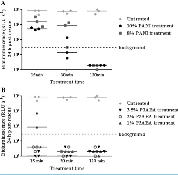
To investigate the activity of PANI and P3ABA against M. tuberculosis, the agar surface decontamination assay was first optimised using M. smegmatis BSG200. M. smegmatisis used as a safer and faster growing surrogate for the highly pathogenicM. tuberculosis(Chaturvedi et al., 2007). The surface of agar with 10% and 8% PANI reduced bioluminescence measurements fromM. smegmatisBSG200 to background levels after 30 min and 120 min exposure times, respectively (Fig. 1A). The activity of PANI was statistically significant (Friedman test, P value: 0.0033), with both 10% and 8% PANI treatment differing significantly from untreated cells at the 120 min time point (Dunn’s multiple comparison post-hoc test).
Figure 1 Antimicrobial action of surface incorporated PANI and P3ABA againstM. smegmatis
BSG200.(A)M. smegmatisBSG200 cells were exposed to the surface of agar containing 10% PANI (closed circles) and 8% PANI (squares). (B)M. smegmatisBSG200 cells were exposed to the surface of agar containing 3.5% P3ABA (downward triangles), 2% P3ABA (open circles) and 1% P3ABA (upward triangles). Following 15 min, 30 min and 120 min treatments,M. smegmatisBSG200 cells were recovered by addition of supplemented 7H9 and incubation at 37 ◦C in a fresh 96-well plate. The vertical axis represents the median bioluminescence measurements (given as relative light units [RLU] per second) from recovered cells grown for 24 h. The dashed line denotes background luminescence of uninoculated wells.
the significant activity of 3.5% P3ABA in a 15 min treatment time. Having established the protocol for fast-growing, non-pathogenic mycobacteria, testing was extended to the slow-growing human pathogenM. tuberculosis.
Antibacterial action of surface incorporated PANI and P3ABA against
M. tuberculosis BSG001
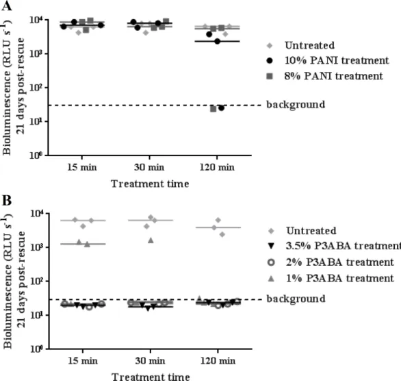
Figure 2 Antimicrobial action of surface incorporated PANI and P3ABA againstM. tuberculosis
BSG001.(A)M. tuberculosisBSG001 cells were exposed to the surface of agar containing 10% PANI (closed circles) and 8% PANI (squares). (B)M. tuberculosisBSG001 cells were exposed to the surface of agar containing 3.5% P3ABA (downward triangles), 2% P3ABA (open circles) and 1% P3ABA (upward triangles). Following 15 min, 30 min and 120 min treatments,M. tuberculosisBSG001 cells were recovered by addition of supplemented 7H9 and incubation at 37 ◦C in a fresh 96-well plate. The vertical axis represents the median bioluminescence measurements (given as relative light units [RLU] per second) from recovered cells grown for approximately 21 days. The dashed line denotes background luminescence of uninoculated wells.
Figure 3 Antimicrobial action of surface incorporated PANI and P3ABA againstM. tuberculosis
BSG002.(A)M. tuberculosisBSG002 cells were exposed to the surface of agar containing 10% PANI (closed circles) and 8% PANI (squares). (B)M. tuberculosisBSG002 cells were exposed to the surface of agar containing 3.5% P3ABA (downward triangles), 2% P3ABA (open circles) and 1% P3ABA (upward triangles). Following 15 min, 30 min and 120 min treatments,M. tuberculosisBSG002 cells were recovered by addition of supplemented 7H9 and incubation at 37 ◦C in a fresh 96-well plate. The vertical axis represents the median bioluminescence measurements (given as relative light units [RLU] per second) from recovered cells grown for approximately 21 days. The dashed line denotes background luminescence of uninoculated wells.
Antibacterial action of surface incorporated PANI and P3ABA against
the clinical isolate M. tuberculosis BSG002
M. tuberculosisBSG002 was tested using the surface decontamination assay to ascertain the activity of PANI and P3ABA against a clinical isolate. Similar to the activity against M. tuberculosisBSG001, both 10% and 8% PANI incorporated into agar did not mediate any meaningful knockdown ofM. tuberculosisBSG002 (Fig. 3A).
down to slightly above background levels after a 30 min treatment (Fig. 3B). Treatment of M. tuberculosisBSG002 with surface incorporated 3.5% P3ABA was statistically significant (Friedman test,Pvalue: 0.0042). Dunn’s multiple comparison test confirmed the significant activity of 3.5% P3ABA in a 15 min treatment time.
DISCUSSION
PANI and P3ABA have previously been shown to have broad spectrum activity against a range of gram-positive and gram-negative bacteria (Gizdavic-Nikolaidis et al., 2011a;Shi et al., 2006). The results presented here demonstrate the previously untested activity of these compounds against mycobacteria. Only a 15 min exposure to 2% P3ABA was required to mediate knockdown of M. smegmatisandM. tuberculosis. This time interval was the shortest that could be tested due to the time constraints involved in the local Standard Operating Procedures forM. tuberculosislaboratory work. It is possible that P3ABA can decontaminate mycobacteria laden surfaces in less than a 15 min exposure.
The WHO recommends that HCWs in high incidence TB areas wear respirators to protect against TB transmission (WHO, 2015). More than 50% of new MDR-TB cases occur within hospitals and communities among people that haven’t been previously treated for TB, which highlights the lack of adequate infection control measures in high MDR-TB incidence areas (Menon, 2013;WHO, 2014;Kompala, Shenoi & Friedland, 2013). Improved TB infection control is essential to curtail the development and spread of drug resistance (Menon, 2013). There are not many studies that address the efficacy of infection control measures on reducing transmission; however, there is limited evidence which suggests that the incidence of TB infection decreases after execution of control measures (Joshi et al., 2006).
Respirators are intended to protect the wearer from inhaling infectious particles by filtering small infectious droplets (Coia et al., 2013;Cleveland, Robison & Panlilio, 2009). This type of PPE is effective at preventing inhalation of infectious particles; however, the cost of the product renders respirators unobtainable for healthcare facilities in LMICs, which decreases the level of protection offered (Menon, 2013;Coia et al., 2013). Efforts to curtail the spread of MDR-TB is hampered by insufficient funding (WHO, 2015). The funding gaps associated with a full response to the global TB epidemic in LMICs has been steadily increasing, amounting to US$ 1.4 billion in 2015 (WHO, 2015).
Facemasks are worn over nose and mouth to establish a barrier between the respiratory tract and splashes and droplets in the external environment (Coia et al., 2013; Cleveland, Robison & Panlilio, 2009). Facemasks offer less protection againstM. tuberculosistransmission as they have a lower filtration efficiency and a higher degree of face seal leakage than respirators (Coia et al., 2013;Cleveland, Robison & Panlilio, 2009;
2013). The greater affordability of facemasks is associated with fewer availability issues than respirators (Menon, 2013).
Facemasks provide important protection in resource-limited settings where recommended infection control measures are not feasible. However, healthcare facilities in these areas are not able to reliably provide HCWs with sterile facemasks, increasing the risk of TB transmission in high bacterial load situations, such as an infectious patient in a poorly ventilated room (Luksamijarulkul, Aiempradit & Vatanasomboon, 2014;Brosseau, McCullough & Vesley, 1997;Rengasamy, Zhuang & Berryann, 2004). Used contaminated facemasks can be a source of infection either due to penetration of particles through the facemask into the respiratory tract of the wearer or release of aerosols back into the air (Luksamijarulkul, Aiempradit & Vatanasomboon, 2014; Brosseau, McCullough & Vesley, 1997). Used facemasks isolated from a hospital have been shown to have bacterial contamination on the outside of the mask (Luksamijarulkul, Aiempradit & Vatanasomboon, 2014).M. abscessus, used as a surrogate forM. tuberculosis, is able to survive on respiratory PPE for 5 days and can be transferred to gloves during handling (Brosseau, McCullough & Vesley, 1997). Decontamination of facemasks by HCWs may also cause reaerosolisation of TB bacilli (Rengasamy, Zhuang & Berryann, 2004). A facemask impregnated with an antimicrobial agent would ameliorate the risk of TB transmission associated with these non-recommended practices. Bacteria that come into contact with the facemask surface would be killed, facilitating safer reuse and disposal as well as eliminating the need to decontaminate (Chellamani, Veerasubramanian & Balaji, 2013).
There are several commercially available decontaminating facemasks; however, there is limited published work on the efficacy of these facemasks. Two notable examples include silver-based facemasks and quaternary ammonium compound (QAC) based facemasks (Li et al., 2006;Tseng, Pan & Chang, 2016). The cell wall ofM. tuberculosisis protective against penetration of silver nanoparticles into the cytoplasm as disruption with chloroform was required for inhibitory activity (Praba et al., 2013).Mycobacterium avium, a pathogenic mycobacterium, was reduced slightly in number after a 24–48 h treatment with silver nanoparticles, which suggests that the bactericidal activity of silver nanoparticles against M. tuberculosisin a relevant time frame (15 min) would be negligible (Islam et al., 2013;
Miyamoto, Yamaguchi & Sasatsu, 2000). Furthermore, the potential for development of resistance to silver and cross-resistance to antibiotics reduces the suitability of silver as an antimycobacterial agent. Following a single exposure to silver nanoparticles,M. smegmatis developed resistance to silver nanoparticles, silver nitrate and the antibiotic isoniazid (Larimer et al., 2014). QACs have been demonstrated to be active against hydrophilic bacteria that have a negatively charged cell surface, including LPS-expressing gram-negative bacteria and teichoic-acid containing gram-positive bacteria; however, the efficacy against hydrophobic mycobacteria is limited (Russell, 1996;Gottenbos et al., 2002).
activity of fPANI may confer protection against other respiratory pathogens that also have a high burden in LMICs, such as bacterially derived pneumonia (Macintyre et al., 2014).
Challenges remain for researchers; the first is to demonstrate tuberculocidal activity of P3ABA in facemask material. We believe this is likely as melt blends of PANI in low density polyethylene (Nand et al., 2013) and P3ABA electrospun in nanofibers of poly(lactic acid) (Gizdavic-Nikolaidis et al., 2011b;Gizdavic-Nikolaidis et al., 2010b) retain antimicrobial activity. Assuming an effective facemask can be fabricated it will be important to establish that it is effective in reducing TB transmission and to determine the expected usage lifetime to give guidelines as to when to discard a used mask. As users may be tempted to reuse masks, irrespective of mask age, for the same reasons that they currently reuse infected masks; thought will still need to be given to removing the reasons for extended use and to enforcing expiration guidelines.
CONCLUSION
A surface self-decontamination assay was established and used to test the activity of two novel antimicrobial agents, PANI and P3ABA, against mycobacteria. PANI is active againstM. smegmatisBSG200 after 120 min exposure; however, it did not mediate surface self-sanitisation ofM. tuberculosisBSG001 orM. tuberculosisBSG002. A 15 min exposure to a surface containing 3.5% P3ABA was sufficient to kill all three strains tested. Therefore, P3ABA has potential to be used to create anti-tubercular facemasks, which would serve as a cost-effective TB infection control measure in low-resource, high TB burden areas.
ACKNOWLEDGEMENTS
The authors thank Sudip Ray, Adeline Le Cocq, Chris Wilcox and Walt Wheelwright for purified PANI and P3ABA.
ADDITIONAL INFORMATION AND DECLARATIONS
Funding
The authors received research funding from both the New Zealand Ministry of Business, Innovation and Employment (MBIE) for research programmes UOAX0812 and UOAX1410, and the University of Auckland’s Vice Chancellors Strategic Development Fund, grant number 23563. The funders had no role in study design, data collection and analysis, decision to publish, or preparation of the manuscript.
Grant Disclosures
The following grant information was disclosed by the authors:
New Zealand Ministry of Business, Innovation and Employment (MBIE) for research programmes: UOAX0812, UOAX1410.
University of Auckland’s Vice Chancellors Strategic Development Fund: 23563.
Competing Interests
Author Contributions
• Julia Robertson conceived and designed the experiments, performed the experiments, analyzed the data, wrote the paper, prepared figures and/or tables, reviewed drafts of the paper.
• James Dalton conceived and designed the experiments, performed the experiments, analyzed the data, contributed reagents/materials/analysis tools, wrote the paper, reviewed drafts of the paper.
• Siouxsie Wiles conceived and designed the experiments, analyzed the data, contributed reagents/materials/analysis tools, wrote the paper, reviewed drafts of the paper.
• Marija Gizdavic-Nikolaidis conceived and designed the experiments, contributed reagents/materials/analysis tools, wrote the paper, reviewed drafts of the paper.
• Simon Swift conceived and designed the experiments, analyzed the data, wrote the paper, reviewed drafts of the paper.
Ethics
The following information was supplied relating to ethical approvals (i.e., approving body and any reference numbers):
The University of Auckland Institutional Biological Safety Committee approved the construction and use of genetically modified risk group 2 mycobacteria (GMO11-UA007). The New Zealand Environmental Protection Agency approved the construction and use of genetically modified risk group 3Mycobacterium tuberculosis(APP201346).
Data Availability
The following information was supplied regarding data availability: The raw data has been supplied as aData S1.
Supplemental Information
Supplemental information for this article can be found online athttp://dx.doi.org/10.7717/ peerj.2795#supplemental-information.
REFERENCES
Andreu N, Fletcher T, Krishnan N, Wiles S, Robertson BD. 2012.Rapid measurement
of antituberculosis drug activityin vitroand in macrophages using bioluminescence. Journal of Antimicrobial Chemotherapy67:404–414DOI 10.1093/jac/dkr472.
Andreu N, Zelmer A, Fletcher T, Elkington PT, Ward TH, Ripoll J, Parish T, Bancroft
GJ, Schaible U, Robertson BD, Wiles S. 2010.Optimisation of bioluminescent
reporters for use with mycobacteria.PLoS ONE5:e10777 DOI 10.1371/journal.pone.0010777.
Andreu N, Zelmer A, Sampson SL, Ikeh M, Bancroft GJ, Schaible UE, Wiles S,
Robert-son BD. 2013.Rapidin vivoassessment of drug efficacy againstMycobacterium
tu-berculosisusing an improved firefly luciferase.Journal of Antimicrobial Chemotherapy
Andreu N, Zelmer A, Wiles S. 2011.Noninvasive biophotonic imaging for studies of infectious disease.FEMS Microbiology Reviews35:360–394
DOI 10.1111/j.1574-6976.2010.00252.x.
Brosseau LM, McCullough NV, Vesley D. 1997.Bacterial survival on respirator filters
and surgical masks.Journal of the American Biological Safety Association2:32–43.
Buregyeya E, Nuwaha F, Verver S, Criel B, Colebunders R, Wanyenze R, Kalyango JN,
Katamba A, Mitchell EM. 2013.Implementation of tuberculosis infection control
in health facilities in Mukono and Wakiso districts, Uganda.BMC Infectious Diseases
13:360 DOI 10.1186/1471-2334-13-360.
Chaturvedi V, Dwivedi N, Tripathi RP, Sinha S. 2007.Evaluation of Mycobacterium
smegmatis as a possible surrogate screen for selecting molecules active against multi-drug resistantMycobacterium tuberculosis.Journal of General and Applied Microbiology53:333–337DOI 10.2323/jgam.53.333.
Chellamani KP, Veerasubramanian D, Balaji RSV. 2013.Surgical face masks:
manu-facturing methods and classification.Journal of Academia and Industrial Research
2:320–324.
Chughtai AA, Macintyre CR, Zheng Y, Wang Q, Toor ZI, Dung TC, Hien NT, Seale H.
2015.Examining the policies and guidelines around the use of masks and respirators
by healthcare workers in China, Pakistan and Vietnam.Journal of Infection Preven-tion16:68–74DOI 10.1177/1757177414560251.
Cleveland JL, Robison VA, Panlilio AL. 2009.Tuberculosis epidemiology, diagnosis
and infection control recommendations for dental settings.Journal of the American Dental Association140:1092–1099DOI 10.14219/jada.archive.2009.0335.
Coia JE, Ritchie L, Adisesh A, Makison Booth C, Bradley C, Bunyan D, Carson G, Fry C, Hoffman P, Jenkins D, Phin N, Taylor B, Nguyen-Van-Tam JS, Zuckerman M.
2013.Guidance on the use of respiratory and facial protection equipment.Journal of
Hospital Infection85:170–182 DOI 10.1016/j.jhin.2013.06.020.
Colangeli R, Arcus VL, Cursons RT, Ruthe A, Karalus N, Coley K, Manning SD, Kim
S, Marchiano E, Alland D. 2014.Whole genome sequencing ofMycobacterium
tuberculosisreveals slow growth and low mutation rates during latent infections in humans.PLoS ONE9:e91024DOI 10.1371/journal.pone.0091024.
Dhand C, Das M, Datta M, Malhotra BD. 2011.Recent advances in polyaniline based
biosensors.Biosensors and Bioelectronics26:2811–2821 DOI 10.1016/j.bios.2010.10.017.
Gizdavic-Nikolaidis MR, Bennett JR, Swift S, Easteal AJ, Ambrose M. 2011a.Broad
spectrum antimicrobial activity of functionalized polyanilines.Acta Biomater
7:4204–4209DOI 10.1016/j.actbio.2011.07.018.
Gizdavic-Nikolaidis MR, Ray S, Bennett JR, Easteal AJ, Cooney RP. 2010a.Electrospun
functionalized polyaniline copolymer-based nanofibers with potential application in tissue engineering.Macromolecular Bioscience 10:1424–1431
DOI 10.1002/mabi.201000237.
Gizdavic-Nikolaidis MR, Ray S, Bennett JR, Swift S, Bowmaker GA, Easteal AJ. 2011b.
and their potential in biomedical applications.American Journal of Infection Control
49:4902–4910DOI 10.1002/pola.24946.
Gizdavic-Nikolaidis MR, Zujovic ZD, Ray S, Easteal AJ, Bowmaker GA. 2010b.
Chemical synthesis and characterization of poly (aniline- co -ethyl 3-aminobenzoate) copolymers.Journal of Polymer Science Part A: Polymer Chemistry48:1339–1347 DOI 10.1002/pola.23895.
Gonzalez-Angulo Y, Geldenhuys H, Van As D, Buckerfield N, Shea J, Mahomed H,
Hanekom W, Hatherill M. 2013.Knowledge and acceptability of patient-specific
infection control measures for pulmonary tuberculosis.American Journal of Infection Control41:717–722DOI 10.1016/j.ajic.2012.10.003.
Gottenbos B, Van der Mei HC, Klatter F, Nieuwenhuis P, Busscher HJ. 2002.In vitro
andin vivoantimicrobial activity of covalently coupled quaternary ammonium silane coatings on silicone rubber.Biomaterials23:1417–1423
DOI 10.1016/S0142-9612(01)00263-0.
Hards KJ, Robson JR, Berney M, Shaw L, Bald D, Koul A, Andries K, Cook GM. 2015.
Bactericidal mode of action of bedaquiline.Journal of Antimicrobial Chemotherapy
70:2028–2037DOI 10.1093/jac/dkv054.
Islam MS, Larimer C, Ojha A, Nettleship I. 2013.Antimycobacterial efficacy of silver
nanoparticles as deposited on porous membrane filters.Materials Science and Engineering: C 33:4575–4581DOI 10.1016/j.msec.2013.07.013.
Joshi R, Reingold AL, Menzies D, Pai M. 2006.Tuberculosis among health-care workers
in low- and middle-income countries: a systematic review.PLoS Medicine3:e494 DOI 10.1371/journal.pmed.0030494.
Kompala T, Shenoi SV, Friedland G. 2013.Transmission of tuberculosis in
resource-limited settings.Current HIV/AIDS Reports10:264–272 DOI 10.1007/s11904-013-0164-x.
Koul A, Vranckx L, Dhar N, Göhlmann HWH, Özdemir E, Neefs J-M, Schulz M, Lu
P, Mørtz E, McKinney JD, Andries K, Bald D. 2014.Delayed bactericidal response
ofMycobacterium tuberculosisto bedaquiline involves remodelling of bacterial metabolism.Nature Communications5:3369DOI 10.1038/ncomms4369.
Larimer C, Islam MS, Ojha A, Nettleship I. 2014.Mutation of environmental
mycobac-teria to resist silver nanoparticles also confers resistance to a common antibiotic. BioMetals27:695–702DOI 10.1007/s10534-014-9761-4.
Leibert E, Danckers M, Rom WN. 2014.New drugs to treat multidrug-resistant
tubercu-losis: the case for bedaquiline.Journal of Therapeutics and Clinical Risk Management
10:597–602.
Li Y, Leung P, Yao L, Song QW, Newton E. 2006.Antimicrobial effect of surgical masks
coated with nanoparticles.Journal of Hospital Infection62:58–63 DOI 10.1016/j.jhin.2005.04.015.
Luksamijarulkul P, Aiempradit N, Vatanasomboon P. 2014.Microbial contamination
on used surgical masks among hospital personnel and microbial air quality in their working wards: a hospital in Bangkok.Oman Medical Journal29:346–350
Macintyre CR, Wang Q, Rahman B, Seale H, Ridda I, Gao Z, Yang P, Shi W, Pang
X, Zhang Y, Moa A, Dwyer DE. 2014.Efficacy of face masks and respirators in
preventing upper respiratory tract bacterial colonization and co-infection in hospital healthcare workers.Preventive Medicine62:1–7DOI 10.1016/j.ypmed.2014.01.015.
Menon S. 2013.Preventing nosocomial MDR TB transmission in sub Saharan Africa:
where are we at?Global Journal of Health Science5:200–210.
Miyamoto M, Yamaguchi Y, Sasatsu M. 2000.Disinfectant effects of hot water,
ultra-violet light, silver ions and chlorine on strains of Legionella and nontuberculous mycobacteria.Microbios101:7–13.
Nand AV, Swift S, Uy B, Kilmartin PA. 2013.Evaluation of antioxidant and
antimicro-bial properties of biocompatible low density polyethylene/polyaniline blends.Journal of Food Engineering 116:422–429DOI 10.1016/j.jfoodeng.2012.11.023.
Nicas M. 1995.Respiratory protection and the risk ofMycobacterium tuberculosis
infection.American Journal of Industrial Medicine27:317–333 DOI 10.1002/ajim.4700270302.
O’Donnell MR, Jarand J, Loveday M, Padayatchi N, Zelnick J, Werner L, Naidoo K, Master I, Osburn G, Kvasnovsky C, Shean K, Pai M, Van der Walt M, Horsburgh
CR, Dheda K. 2010.High incidence of hospital admissions with multidrug-resistant
and extensively drug-resistant tuberculosis among South African health care workers.Annals of Internal Medicine153:516–522
DOI 10.7326/0003-4819-153-8-201010190-00008.
Pandey S, Annapoorni S, Malhotra BD. 1993.Synthesis and characterization of
poly(aniline-co-o-anisidine): a processable conducting copolymer.Macromolecules
26:3190–3193DOI 10.1021/ma00064a032.
Praba VL, Kathirvel M, Vallayyachari K, Surendar K, Muthuraj M, Jesuraj PJ,
Govindarajan S, Raman KV. 2013.Bactericidal effect of silver
nanoparti-cles againstMycobacterium tuberculosis.Journal of Bionanoscience7:282–287 DOI 10.1166/jbns.2013.1138.
Rengasamy A, Zhuang Z, Berryann R. 2004.Respiratory protection against bioaerosols:
literature review and research needs.American Journal of Infection Control
32:345–354DOI 10.1016/j.ajic.2004.04.199.
Russell AD. 1996.Activity of biocides against mycobacteria.Society for Applied
Bacteriol-ogy Symposium Series25:87S–101SDOI 10.1111/j.1365-2672.1996.tb04602.x.
Shi N, Guo X, Jing H, Gong J, Sun C, Yang K. 2006.Antibacterial effect of the
conduct-ing polyaniline.Journal of Materials Science & Technology22:289–290.
Tseng C-C, Pan Z-M, Chang C-H. 2016.Application of a quaternary ammonium agent
on surgical face masks before use for pre-decontamination of nosocomial infection-related bioaerosols.Aerosol Science and Technology50:199–210
DOI 10.1080/02786826.2016.1140895.
Wang J, Pearce AN, Chan STS, Taylor RB, Page MJ, Valentin A, Bourguet-Kondracki
M-L, Dalton JP, Wiles S, Copp BR. 2016.Biologically active acetylenic amino
the New Zealand Ascidian Pseudodistoma opacum.Journal of Natural Products
79:607–610DOI 10.1021/acs.jnatprod.5b00770.
WHO. 2014.Drug-resistant TB surveillance & response supplement: global tuberculosis
report 2014 (WHO/HQ/TB/2014.12).Available athttp:// apps.who.int/ iris/ bitstream/ 10665/ 137094/ 1/ 9789241564809_eng.pdf.
WHO. 2015.Global tuberculosis report 2015 (WHO/HTM/TB/2015.22).Available at
http:// apps.who.int/ iris/ bitstream/ 10665/ 191102/ 1/ 9789241565059_eng.pdf.
Wiles S, Ferguson K, Stefanidou M, Young DB, Robertson BD. 2005.Alternative
luciferase for monitoring bacterial cells under adverse conditions.Applied and Environmental Microbiology71:3427–3432DOI 10.1128/AEM.71.7.3427-3432.2005.
Yang P, Seale H, Macintyre CR, Zhang H, Zhang Z, Zhang Y, Wang X, Li X, Pang X,
Wang Q. 2011.Mask-wearing and respiratory infection in healthcare workers in