Axial Ligation and Redox Changes at the
Cobalt Ion in Cobalamin Bound to Corrinoid
Iron-Sulfur Protein (CoFeSP) or in Solution
Characterized by XAS and DFT
Peer Schrapers1, Stefan Mebs1, Sebastian Goetzl2, Sandra E. Hennig2, Holger Dau1, Holger Dobbek2, Michael Haumann1*
1Freie Universität Berlin, Department of Physics, 14195, Berlin, Germany,2Humboldt-Universität zu Berlin, Department of Biology, 10115, Berlin, Germany
Abstract
A cobalamin (Cbl) cofactor in corrinoid iron-sulfur protein (CoFeSP) is the primary methyl group donor and acceptor in biological carbon oxide conversion along the reductive acetyl-CoA pathway. Changes of the axial coordination of the cobalt ion within the corrin macro-cycle upon redox transitions in aqua-, methyl-, and cyano-Cbl bound to CoFeSP or in solu-tion were studied using X-ray absorpsolu-tion spectroscopy (XAS) at the Co K-edge in
combination with density functional theory (DFT) calculations, supported by metal content and cobalt redox level quantification with further spectroscopic methods. Calculation of the highly variable pre-edge X-ray absorption features due to core-to-valence (ctv) electronic transitions, XANES shape analysis, and cobalt-ligand bond lengths determination from EXAFS has yielded models for the molecular and electronic structures of the cobalt sites.
This suggested the absence of a ligand at cobalt in CoFeSP inα-position where the
dimethylbenzimidazole (dmb) base of the cofactor is bound in Cbl in solution. As main spe-cies, (dmb)CoIII(OH2), (dmb)CoII(OH2), and (dmb)CoIII(CH3) sites for solution Cbl and
CoIII(OH2), CoII(OH2), and CoIII(CH3) sites in CoFeSP-Cbl were identified. Our data support
binding of a serine residue from the reductive-activator protein (RACo) of CoFeSP to the
cobalt ion in the CoFeSP-RACo protein complex that stabilizes Co(II). The absence of anα
-ligand at cobalt not only tunes the redox potential of the cobalamin cofactor into the physio-logical range, but is also important for CoFeSP reactivation.
Introduction
The cobalamin cofactor (Cbl, also denoted vitamin B12) since its discovery in 1925 has attracted much research interest [1–4]. Cbl is essential for all mammals [5] and in bacteria it is involved in carbon oxide (COx) conversion pathways related to potential renewable energy applications [6,7]. Anaerobic CO2reduction along the bacterial Wood-Ljungdahl pathway includes several unique enzymes [8,9]. The corrinoid iron-sulfur protein (CoFeSP) carries a Cbl cofactor [10,11] and shuttles a methyl group from transferase bound
methyl-a11111
OPEN ACCESS
Citation:Schrapers P, Mebs S, Goetzl S, Hennig SE, Dau H, Dobbek H, et al. (2016) Axial Ligation and Redox Changes at the Cobalt Ion in Cobalamin Bound to Corrinoid Iron-Sulfur Protein (CoFeSP) or in Solution Characterized by XAS and DFT. PLoS ONE 11(7): e0158681. doi:10.1371/journal.pone.0158681
Editor:Maxim I. Boyanov, Argonne National Laboratory, UNITED STATES
Received:April 12, 2016
Accepted:June 20, 2016
Published:July 6, 2016
Copyright:© 2016 Schrapers et al. This is an open access article distributed under the terms of the Creative Commons Attribution License, which permits unrestricted use, distribution, and reproduction in any medium, provided the original author and source are credited.
Data Availability Statement:All relevant data are within the paper and its Supporting Information files.
Funding:This work was funded by Deutsche Forschungsgemeinschaft (http://www.dfg.de/), Berlin Cluster of Excellence“Unifying Concepts in Catalysis —UniCat”(EXC 314), to HDo and HDa; Deutsche Forschungsgemeinschaft (http://www.dfg.de/), grants Ha3265/2-1 and Ha3265/6-1 and Bundesministerium für Bildung und Forschung (http://pt.desy.de/), grant 05K14KE1, to MH; and Deutsche
tetrahydrofolate to acetyl-CoA synthase. The latter enzyme, after receiving a CO group derived from CO2reduction by carbon monoxide dehydrogenase, synthesizes acetyl-CoA for many metabolic reactions [12]. CoFeSP alternates in the methyl transfer cycle between Co(III)-CH3 and Co(I) states [13]. The Co(I) state is prone to oxidative inactivation generating Co(II), which can be reductively reactivated in an ATP-dependent reaction catalyzed by the reductive-activator protein (RACo) [14–16]. Redox and ligation changes at cobalt in Cbl in the CoFe-SP-RACo system thus are essential in the COxconversion pathway.
Cobalamin is among the most complex non-polymeric compounds in nature and consists of a unique corrin hetero-macrocycle binding a central cobalt ion by four equatorial nitrogen ligands [17]. Two axial cobalt ligands (αand ß) may be bound in addition. Theα-ligand in Cbl
in solution or in prototypic Cbl-proteins in the so-called base-on configuration is the nitrogen atom of a dimethylbenzimidazole (dmb) group connected to the corrin ring (Fig 1). Replace-ment of the dmb ligand (base-off) by a water species or by other amino acids occurs in many proteins [1–3]. Crystal structures of isolated CoFeSP and of the protein in complex with RACo or methyl transferase have been reported [12,14,16,18,19]. In all CoFeSP structures, the dmb group is folded away from the corrin so that theα-site apparently is vacant (Fig 1). However, it
could also be occupied by a crystallographically less visible (disordered) water species or even by the hydroxyl group of a nearby threonine residue modeled at about 3.5 Å to cobalt in the structures. The ß-site in CoFeSP-Cbl can be occupied by a water species (AqCbl), a methyl group (MeCbl) [20], or may be vacant (Fig 1). In the CoFeSP-RACo protein complex, binding of the hydroxyl group of a serine (Ser 398) of RACo to cobalt at the ß-position has been shown [14–16].
Binding of the axial ligands is closely related to the cobalt oxidation state [4]. In Cbl, both in solution and bound to proteins, the formal Co(I), Co(II), and Co(III) states are associated with low-spin (3d8, 3d7, 3d6) valence electron configurations [22
–24]. Only Co(II) thus is EPR active. A decrease of the oxidation state may be accompanied by a decreasing number of axial ligands, meaning that (L = ligand) (Lα)CoIII(Lß), CoII(Lß) or (Lα)CoII, and CoIspecies may
prevail [24,25], but in protein environments deviations from such configurations may occur. Control of the axial cobalt ligation in group-transferring Cbl-enzymes such as CoFeSP is important in the reactions. However, relating the redox state to the axial ligation of cobalt can be difficult both by crystallography and spectroscopy. For example, in solution Cbl mixtures of base-on/off states may occur, in crystal structures of Cbl-proteins axial ligands may be unre-solved, or certain spectroscopic methods do not provide structural and electronic parameters or only for selected cobalt redox states. Further insight in the cobalt site structures in the CoFeSP-Cbl-RACo system is required to understand the interplay of protein-protein interac-tions, redox transiinterac-tions, axial ligand exchange, and methyl group transfer.
Here, we employed X-ray absorption spectroscopy (XAS) at the Co K-edge in combination with density functional theory (DFT) to study redox and coordination changes at cobalt in CoFeSP-Cbl in comparison to Cbl in solution. XAS in principle facilitates oxidation state, metal-ligand bond lengths, and site symmetry determination for solution and protein systems and can be applied to all spin and oxidation states of metal sites [26–29]. In particular the XAS features due to resonant 1selectron excitation into unoccupied valence levels (for example with Co(3d) character) in the so-called pre-edge absorption spectral region (core-to-valence transi-tions, ctv), which can be calculated by DFT [30–33], are sensitive to the molecular and elec-tronic structure of the Cbl cofactor [34–39]. Pronounced alterations of the ctv spectra upon changes at cobalt were observed, which were reproduced by the computational approach. Combination of experimental and theoretical analyses has established relations between the X-ray spectroscopic features and the redox state and axial ligation at the cobalt centers, thereby design, data collection and analysis, decision to
publish, or preparation of the manuscript.
Competing Interests:The authors have declared that no competing interests exist.
providing structural models for the AqCbl and MeCbl cofactors in CoFeSP and in the CoFe-SP-RACo protein complex.
Materials and Methods
Sample preparation
CoFeSP and RACo proteins fromCarboxydothermus hydrogenoformanswere heterologously overexpressed inEscherichia colifollowing previously established protocols [15,16,19] and protein purification and biochemical treatments were performed under anoxic conditions in 95% N2and 5% H2atmosphere at room temperature in a glove box. Synthetic cobalamin (denoted AqCblox, CNCblox, and MeCblox) containing Co(III) was purchased from Sigma-Aldrich, all chemicals were at least analysis grade. Purified CoFeSP (25μM) was reconstituted
in 20 mM TRIS-HCl buffer (pH 8.0) with synthetic AqCbloxor MeCblox(40
μM) by overnight
incubation at 25°C, subsequently unbound cofactor was removed and the protein concentrated using Vivaspin 500 concentrators (10 kDa cut-off). CoFeSP-AqCbloxand CoFeSP-MeCblox samples for XAS contained 1.0±0.1 mM protein as determined by the Bradford method [40]. A glass-forming agent was not present in the XAS samples. Titanium(III)-citrate (2 mM) was added to CoFeSP-Cbloxsamples for cofactor reduction (red). The CoFeSP-AqCbl-RACo pro-tein complex was prepared according to the previously reported protocol [14–16]. Solution samples of synthetic cobalamins (7 mM) were prepared by solvation of AqCblox, CNCblox, and MeCbloxpowders in 20 mM TRIS-HCl buffer (pH 8.0) and chemical reduction was achieved by addition of sodium dithionite (100 mM) to obtain CNCblredor titanium-citrate (40 mM) to Fig 1. Cobalamin crystal structures.(a) Structure of the Cbl cofactor in CoFeSP enzyme (PDB entry 2H9A, 1.9Åresolution [12]) showing a base-off configuration (dmb ligand not bound to cobalt inα-position). Ligand X at the ß-position (light green) at cobalt can be absent or can be a water species, a methyl group, or an oxygen from the side chain of RACo-Ser398 in the CoFeSP-RACo complex [14]; red balls show resolved water molecules. (b) Structure of Cbl in base-on configuration [21]; X can be a water, cyanide, or methyl species. Color code: magenta, Co; blue, N; red, O; grey, C; dark green, P; protons were omitted for clarity.
obtain AqCblredsamples. Aliquots of samples (50
μL) were loaded into Kapton-covered acrylic
glass holders for XAS and frozen in liquid nitrogen. Optical absorption spectra of samples using 2μl aliquots were recorded on a Specord 50 Plus instrument (Analytik Jena, Germany).
Total reflection X-ray fluorescence analysis
TXRF [41] was performed for metal content determination in protein samples using a PicoFox instrument (Bruker, Berlin, Germany). Protein samples were mixed (v/v 2:1) with a gallium concentration standard (Sigma, 50 mg/L) and three measurements were carried out per sample using 5μl aliquots.
X-ray absorption spectroscopy
XAS at the Co K-edge was performed at the bending-magnet beamline KMC-1 at BESSY (Helmholtz-Center for Materials and Energy Berlin) with the storage ring operated in top-up mode (250 mA). The excitation energy was tuned by a Si[111] double-crystal monochromator. Kα-fluorescence-detected XAS spectra were collected using an energy-resolving 13-element
germanium detector (Canberra) on samples held in a liquid-helium cryostat (Oxford) at 20 K. The detector was shielded against scattered X-rays by a 10μm iron foil. The K-edge inflection
point at 7709 eV of a simultaneously measured cobalt metal foil was used for calibration of the energy axis. Detector deadtime corrected XAS spectra (scan duration ~30 min) were averaged (up to 9 scans, 2 scans per sample spot) for signal-to-noise ratio improvement. No radiation induced spectral changes (i.e. in the XANES) were observed for increasing XAS scan numbers on single sample spots. XAS data processing was carried out as previously described [27] to yield normalized XANES and EXAFS spectra. Simulation ofk3-weighted EXAFS spectra in k-space was carried out using the in-house software SimX and phase functions calculated with FEFF7.0 (S02= 0.85) [42,43]. In the fits the number of C-atoms was set to the values corre-sponding to the corrin ring. Fourier-transforms of EXAFS spectra were calculated fork= 1.8– 12.2 Å-1using cosine windows extending over 10% of bothk-range ends. The XANES pre-edge features were extracted by polynomial spline subtraction with the program XANDA [44]. Mul-tiple-scattering theory simulations of K-edge spectra were performed with the FEFF9.0 code [45] using model structures based on the cobalamin crystal structure in CoFeSP (PDB entry 2H9A, 1.9 Å resolution [12]); for details see the Supporting Information (Fig C inS1 File).
Density functional theory calculations
functions (FWHM 2.5 eV), 158.7 eV shifted on the energy axis, and their amplitudes were scaled (x900) for comparison with experimental ctv spectra.
Results
Cofactor content and oxidation state
Cobalamin (Cbl) species were investigated when bound to the CoFeSP enzyme, in the CoFe-SP-RACo protein complex, and in solution samples serving as reference materials. The expected axial cobalt ligations in the samples included dimethylbenzimidazole (dmb) or water (Aq) species at theαposition (occupied by the dmb ligand in crystalline Cbl) or Aq, cyanide
(CN), or methyl (Me) group species at the ß position (opposite to the dmb ligand). CoFeSP-Cbl and solution Cbl samples in the oxidized state (ox) and after chemical reduction (red) were compared. We denote the various redox and axial ligation species of cobalt as (α-ligand)Cox
(ß-ligand) (x = valence state) in the following.
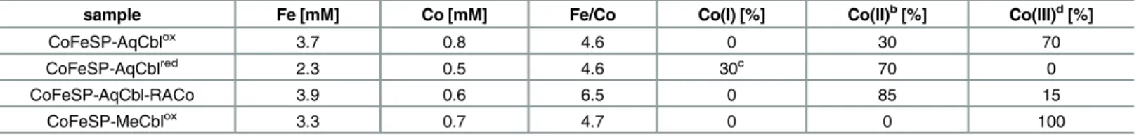
The cobalt concentrations in the Cbl-reconstituted protein samples were determined by TXRF, which on average yielded 0.8±0.1 Co ions per CoFeSP containing AqCbl or MeCbl (Table 1). This suggested close to stoichiometric reconstitution of CoFeSP with the cofactors. The mean amount of 3.5±0.5 Fe ions per CoFeSP protein was in reasonable agreement with the near-quantitative presence of the [4Fe4S] cluster in CoFeSP. The mean Fe to Co ratio was 4.6 ±0.2, which for 4 Fe ions in the [4Fe4S] cluster per protein, suggested ~0.85 Co ions per CoFeSP, in agreement with the protein to cobalt ratios. The increased Fe to Co ratio of 6.5 in the CoFeSP-AqCbl-RACo sample was in good agreement with two additional Fe ions in the sample compared to CoFeSP-AqCbl, due to the presence of close to one RACo protein contain-ing a [2Fe2S] cluster per CoFeSP.
Optical absorption spectra (Fig A inS1 File) of the solution Cbl samples confirmed the expected quantitative presence of Co(III) in AqCblox, CNCblox, and MeCblox, and showed mostly Co(II) in AqCblredand a Co(II) species in CNCblred. For the protein samples, the absorption spectra (Fig A inS1 File) indicated the expected Co(III) in the cofactor in CoFeSP--MeCblox, suggested dominance of Co(III) in oxidized CoFeSP-AqCbl and of Co(II) in CoFe-SP-AqCbl-RACo, and showed preferentially Co(II) in CoFeSP-AqCblredwith minor (~30%) Co(I) amounts only in this sample. Electron paramagnetic resonance spectroscopy (EPR) detecting only the Co(II)-containing cofactor was used to quantify the relative Co(II) contents in the protein samples (Fig B inS1 File). This showed that CoFeSP-AqCbloxcontained ~30% Co(II) and, considering also the optical spectra, ~70% Co(III), CoFeSP-AqCblredcontained ~70% Co(II), and CoFeSP-AqCbl-RACo near-quantitative amounts of Co(II) (~85%) (Table 1). The altered EPR signal shape of CoFeSP-AqCbl-RACo (Fig B inS1 File) also
Table 1. Metal content and cobalt oxidation state in the CoFeSP samplesa.
sample Fe [mM] Co [mM] Fe/Co Co(I) [%] Co(II)b[%] Co(III)d[%]
CoFeSP-AqCblox 3.7 0.8 4.6 0 30 70
CoFeSP-AqCblred 2.3 0.5 4.6 30c 70 0
CoFeSP-AqCbl-RACo 3.9 0.6 6.5 0 85 15
CoFeSP-MeCblox 3.3 0.7 4.7 0 0 100
aProtein concentrations for CoFeSP-AqCbloxand -MeCbloxwere 1.0±0.1 mM. Metal concentrations were determined by TXRF (error±0.1 mM) bCo(II) contents were determined by EPR (Fig B inS1 File, error±10%)
cCo(I) contents were estimated from optical absorption spectra (Fig A inS1 File) dCo(III) contents agree with Co(II)/Co(I) contents and data in Figs A and B inS1 File.
suggested near-stoichiometric RACo binding to CoFeSP [15,19], in agreement with the TXRF data.
EXAFS on the Cbl systems
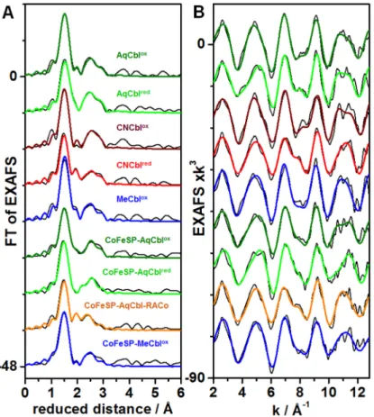
Simulation of EXAFS spectra facilitates determination of interatomic distances such as the cobalt-ligand bond lengths with ~0.02 Å precision in favorable cases. Visual inspection of the EXAFS spectra of the Cbl and CoFeSP-Cbl samples revealed a dominant Fourier-transform (FT) peak due to Co-C/N/O bonds from the corrin ring and the axial ligands and smaller fea-tures at larger distances mostly due to second-sphere Co-Ccorrininteractions (Fig 2). The fit analysis (Table 2) revealed typical bond lengths (~1.87 Å) of the equatorial Co-Ncorrinligands in the AqCblox, CNCblox, and MeCbloxsolution samples, which were only ~0.02 Å elongated in AqCblredand CNCblred. The second-sphere EXAFS features were well described by a mean Co-Ccorrindistance of ~2.9 Å and a multiple-scattering contribution with an apparent N-C dis-tance in the corrin ring of ~1.4 Å. These Co-N/Ccorrindistances are in agreement with Cbl crys-tal structures [54–57] and earlier XAS data [34,36]. Axial cobalt ligands also were discernable in the EXAFS. For AqCblox, the dmb (α) and water (ß) ligands showed relatively similar (1.96
±0.08 Å) bond lengths at cobalt, attributed to a slightly longer Co-N and shorter Co-O bond [56]. Both bonds were ~0.3 Å elongated in AqCblred(Table 2). For CNCblox, the Co-C bond was ~0.04 Å shorter than the Co-Ncorrinbonds as in crystalline CNCblox[55] and the Co-Ndmb
Fig 2. EXAFS spectra of cobalamin systems.Panel (A) shows Fourier-transforms (FTs) of the EXAFS oscillations in panel (B) for indicated solution Cbl or CoFeSP-Cbl samples. Black lines, experimental data; coloured lines, simulations with parameters inTable 2(fits 2, 5, 7, 10, 12, 14, 16, 19, 21); spectra in (A) and (B) were vertically shifted for comparison.
bond was similar to AqCblox. Lower coordination numbers and elongated axial bonds (~2.13 Å) in CNCblredsuggested that one ligand possibly was detached. For MeCblox, the longer and shorter axial bonds likely were attributed to the dmb (~2.20 Å) and CH3(~1.95 Å) ligands [54]. Overall, the fit results showed that axial cobalt ligation changes dominated the EXAFS spectral variations.
The CoFeSP-Cbl samples showed similar Co-N/Ccorrindistances as found for solution Cbl in the EXAFS fits (Table 2), revealing the integrity of the base-off cofactor in the reconstituted protein [18,19]. For CoFeSP-AqCblox, lower coordination numbers of the axial ligands com-pared to solution AqCbloxsuggested only one axial ligand. Two detectable Co-O bond lengths were attributed to a larger contribution (~2.0 Å) from 5-coordinated Co(III) and a smaller con-tribution (~2.3 Å) from 5-coordinated Co(II). CoFeSP-AqCblredshowed significantly (~0.02 Å) shorter Co-Ncorrinbonds compared to CoFeSP-AqCblox, presumably due to the minor Co Table 2. EXAFS simulation parametersa.
sample fit Co-N Co-C/N/O Co-C RF
N / R / 2σ2 N / R / 2σ2 N / R / 2σ2 [%]
AqCblox 1 4*/ 1.87 / 3* 1.8 / 1.96 / 3* 11*/ 2.88 (1.44) / 11# 8.1
2 4*/ 1.88 / 3* 0.9 / 1.92 / 3*0.8 / 1.97 / 3* 11*/ 2.89 (1.42) / 10# 8.0
AqCblred 3 4
*/ 1.89 / 3* 11*/ 2.90 (1.34) / 8# 17.1
4 4*/ 1.89 / 3* 0.9 / 2.29 / 3* 11*/ 2.91 (1.37) / 8# 13.6
5 4*/ 1.89 / 3* 1.1 / 2.30 / 3*1.1 / 2.47 / 3* 11*/ 2.91 (1.41) / 8# 8.4
CNCblox 6 4*/ 1.87 / 3* 1.5 / 1.97 / 3* 11*/ 2.87 (1.38) / 10# 16.5
7 4*/ 1.89 / 3* 0.8 / 1.85 / 3*1.2 / 2.05 / 3* 11*/ 2.88 (1.39) / 9# 12.1
CNCblred 8 4
*/ 1.87 / 3* 11*/ 2.90 (1.40) / 12# 19.6
9 4*/ 1.88 / 3* 0.9 / 2.14 / 3* 11*/ 2.91 (1.39) / 12# 12.3
10 4*/ 1.88 / 3* 1.0 / 2.14 / 3*0.5 / 2.54 / 3* 11*/ 2.91 (1.41) / 13# 11.6
MeCblox 11 4
*/ 1.89 / 3* 1.3 / 1.93 / 3* 11*/ 2.89 (1.43) / 11# 10.5
12 4*/ 1.89 / 3* 1.2 / 1.95 / 3*0.7 / 2.21 / 3* 11*/ 2.90 (1.42) / 10# 7.3
CoFeSP-AqCblox 13 4
*/ 1.88 / 3* 0.7 / 2.01 / 3* 11*/ 2.91 (1.41) / 12# 10.4
14 4*/ 1.88 / 3* 0.6 / 2.02 / 3*0.4 / 2.29 / 3* 11*/ 2.91 (1.41) / 11# 8.5
CoFeSP-AqCblred 15 4
*/ 1.85 / 3* 11*/ 2.88 (1.44) / 16# 19.0
16 4*/ 1.86 / 3* 0.6 / 2.31 / 3* 11*/ 2.88 (1.44) / 15# 14.4
17 4*/ 1.86 / 3* 0.3 / 1.96 / 3*0.8 / 2.33 / 3* 11*/ 2.89 (1.45) / 14# 12.8
CoFeSP-AqCbl-RACo 18 4*/ 1.88 / 3* 0.8 / 2.12 / 3* 11*/ 2.89 (1.34) / 13# 19.2 19 4*/ 1.88 / 3* 0.8 / 2.10 / 3*0.4 / 2.53 / 3* 11*/ 2.87 (1.41) / 19# 8.4
CoFeSP-MeCblox 20 4
*/ 1.87 / 3* 1.2 / 2.00 / 3* 11*/ 2.90 (1.42) / 15# 14.5
21 4*/ 1.87 / 3* 1.2 / 2.01 / 3*0.2 / 2.50 / 3* 11*/ 2.89 (1.44) / 16# 12.5 aData refer to EXAFS spectra inFig 2.N, coordination number per Co ion;R, interatomic distance inÅ(i.e. cobalt-ligand bond length); 2σ2, Debye-Waller
parameter in x10-3Å2;R
F,fit error sum (calculated for reduced distances of 1–3Å[27], RFrepresents the mean root square deviation in % between the
experimental Fourier-isolated k-space EXAFS spectrum in the given reduced-distance range of thefit and thefit curve)
*parameters that werefixed at given physically reasonable values in thefits
#2σ2was coupled to yield the same values for the ~2.9ÅCo-C shell (N
Co-Cwas set to the crystallographic distances in the ~2.9–3.3Årange, the
Debye-Waller factor reflects this distance distribution with more emphasis on the 8 shorter Co-C distances).
A further Co-N-C multiple-scattering shell with the sameNand 2σ2values as for the Co-C shell was included in thefits (apparent N-C distances given in parenthesis). The 2σ2values for the Co-N and Co-C/N/O shells were chosen to provide bestfit results. Two lines for a given coordination shell mean that both distances were included in the respectivefit. We note that splitting of the axial ligation shells in thefit procedure is tentative due to the ~0.1Ådistance discrimination limit of ourk= 13Å-1EXAFS data [58]. We note that the smallN-values of the second Co-C/N/O shell with relatively long distances for
CoFeSP-MeCbl (fit 19) and CoFeSP-RACo (fit 21) may not be significant and suggest dominance of 5-coordinated cobalt sites (see Fig F inS1 File).
(I) contribution, and predominance of one axial ~2.3 Å bond, attributed to a water species at Co(II). CoFeSP-AqCbl-RACo revealed only one significant short axial ligand bond (~2.1 Å); a longer interaction (~2.5 Å) showed a small and possibly insignificant coordination number (Fig F inS1 File). The short bond may reflect the Co-OSerinteraction in the CoFeSP-RACo complex. CoFeSP-MeCbloxrevealed only one axial ligand (~2.0 Å) due to the Co(III)-CH3 interaction, which was slightly longer than in solution MeCblox(Table 2).
XANES spectral analysis
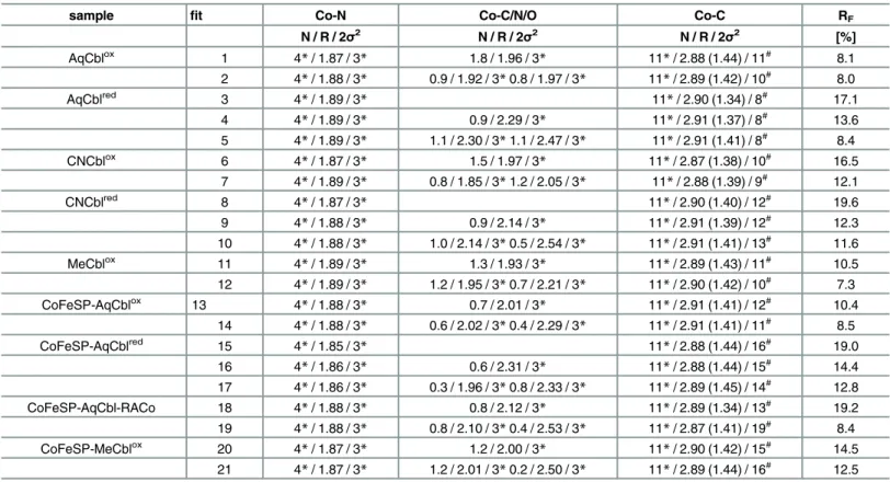
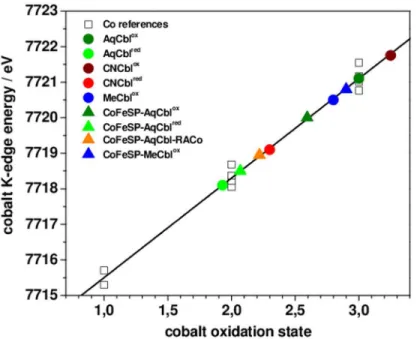
The XANES spectrum is sensitive to the spin and oxidation state of the metal, as well as to the chemical nature and symmetry of its ligands. The XANES of the solution Cbl and CoFeSP-Cbl samples revealed overall similar shapes (Fig 3), as explained by the spectral dominance of the equatorial Ncorrinligands at cobalt. The significantly different K-edge energies (Fig 4) thus likely were related to the cobalt redox and axial ligation changes. Reference K-edge energies for the cobalt redox states species were derived from synthetic complexes (Fig C inS1 File) and were determined as ~7715.5 eV for Co(I), ~7718.3 eV for Co(II), and ~7721.1 eV for Co(III) species, revealing a ~2.8 eV edge energy increase per single-electron cobalt oxidation (Fig 4). All Cbl and CoFeSP-Cbl samples showed K-edge energies in the Co(II) to Co(III) region, well above the Co(I) level (Fig 4). The K-edge energies for MeCblox, AqCblox, and CNCbloxwere centered around Co(III), with AqCbloxlocated at the mean Co(III) energy and a ~1 eV differ-ence between CNCbloxand MeCblox. The ~2.8 eV lower K-edge energies for AqCblredand CNCblredsugegsted near-quantitative Co(II) contents. For the protein samples, the K-edge energy of CoFeSP-MeCbloxwas closest to the Co(III) level, but the edge shape differed strongly from solution MeCblox(Figs3and4). The edge energy for CoFeSP-AqCbloxwas lower than the mean Co(III) level due to Co(II) admixture and a ~1.5 eV lower edge energy for CoFe-SP-AqCblredreflected the increased Co(II) content. The K-edge energy for CoFeSP-AqCbl-RACo was close to the Co(II) level, but the different edge shape compared to CoFeSP-AqCblred suggested a coordination change at cobalt.
Qualitative multiple-scattering K-edge simulations on structural models for the cobalt sites (Fig D inS1 File) fairly reproduced the experimental K-edge shape and energy differences between AqCblox, CNCblox, and MeCblox(base-on octahedral cobalt sites) (Fig E inS1 File). Simulations for the base-off sites in CoFeSP-Cbl showed that replacement of the dmb by a water ligand in 6-coordinated sites results in lower edge energies compared to the base-on structures for AqCbloxand MeCbloxsimilar to the experimental data,α-ligand removal decreased the edge energy compared to the 6-coordinated sites by ~1 eV, elongation of the axial bond as in CoFeSP-AqCblredresulted in a small (~0.5 eV) edge energy decrease, and the edge energy of a square-planar cobalt site was close to the Co(II) level. These results suggested that the lowered edge energies in the CoFsSP-Cbloxcompared to the solution Cbloxsamples at least in part were explained by loss of one axial ligand and the K-edges of CoFeSP-AqCblred and the CoFeSP-Cbl-RACo complex reflected a different axial ligand.
The XANES pre-edge feature
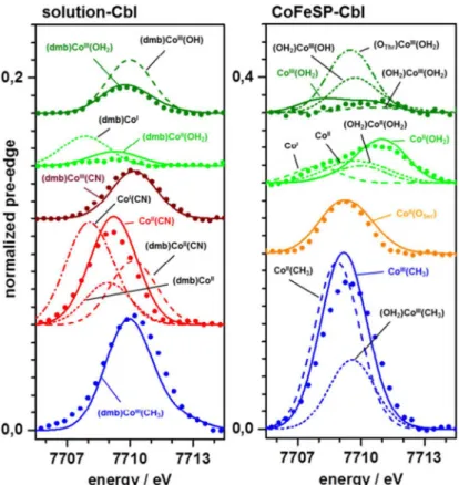
broader envelope of the ctv feature. Also CoFeSP-AqCbl-RACo showed a ctv amplitude increase, but a shift to lower energies. CoFeSP-MeCbloxexhibited by far the largest ctv feature, exceeding that of solution MeCblox.Density functional theory (DFT) was employed to generate geometry-optimized model structures of the cobalt sites and to calculate ctv features on their basis (Fig 5). The (dmb)CoIII(OH2) site from DFT showed metal-ligand bond lengths in agree-ment with crystal structures and our EXAFS data (Table 3). This structure also reproduced the small ctv feature of AqCblox, whereas an OH-ligand yielded a too large ctv amplitude (Fig 5). The diminished ctv amplitude in AqCblredwas best reproduced using a (dmb)CoII(OH2) site. Co(II) or Co(I) sites in which the water ligand, the dmb ligand, or both ligands were absent yielded larger ctv amplitudes and/or lower or higher peak energies disagreeing with the experi-mental data (Fig 5). A (dmb)CoIII(CN) site reproduced the ctv feature of CNCbloxwell. For CNCblred, however, the increased ctv amplitude was only calculated for a CoII(CN) site (base-off), whereas (dmb)CoII(CN) or (dmb)CoIIsites yielded too small and shifted ctv features. The large ctv feature for MeCbloxwas reproduced by the expected (dmb)CoIII(CH3) geometry. This shows that the ctv feature is a specific indicator of cobalt redox and ligation changes.
The small ctv feature of CoFeSP-AqCbloxwas seemingly described by a 6-coordinated (OH2)CoIII(OH2) site (Fig 5). However, the experimental ctv feature likely was increased by a Fig 3. Cobalt XANES spectra.(A) Indicated Cbl solution samples, (B) CoFeSP-Cbl samples. Dotted lines mark edge half-height. Spectra of CoIII2O3(solid black line) and CoIIO (dashed black line) are shown for
comparison in (A) and (B). Inset: Isolated pre-edge (core-to-valence, ctv) absorption features after subtraction of a smooth edge rise background (not shown) from the XANES spectra. For XANES spectra of further cobalt reference compounds see Fig C inS1 File.
Co(II) admixture so that a 5-coordinated CoIII(OH2) site with a weak ctv feature at lower ener-gies accounted equally well for the CoFeSP-AqCbloxspectrum. The broader and larger ctv fea-ture of CoFeSP-AqCblredwas best explained by dominance of the large ctv feature of a 5-coordinated CoII(OH2) site and minor contributions of weak ctv features from a Co(I) site without axial ligands (Fig 5). The large ctv peak at lower energies of CoFeSP-AqCbl-RACo was well reproduced assuming a CoII(OSer) site, i.e. binding of the hydroxyl group of the serine of the RACo protein to cobalt at ß-position in the absence of anα-ligand, in agreement with the
CoFeSP-RACo crystal structure [16]. Amplitude and energy of the largest ctv feature of CoFeSP-MeCbloxwere reasonably reproduced only by a 5-coordinated CoIII(CH3) site, whereas a (OH2)CoIII(CH3) site showed a much too small ctv feature (Fig 5).
Molecular structures of the cobalt sites
The analysis of the EXAFS, XANES, and ctv spectra using DFT (and multiple-scattering) calcu-lations, as well as the TXRF, optical absorption, and EPR data, converged towards consistent cobalt site assignments (Fig 6). Solution Aq/CN/MeCbl samples showed the expected octahe-dral base-on (dmb)CoIII(ß-ligand) configurations. AqCblredlikely contained a (dmb)
CoII(OH2) species with a weak water ligand (~2.5 Å) whereas CNCblredseemingly preferred a base-off CoII(CN) configuration with an elongated (~2.1 Å) Co-CN bond under our condi-tions. Co(III) species thus were generally 6-coordinated and Co(II) species preferred 6- or 5-coordinated geometries in solution Cbl.
The cobalt ion in CoFeSP-Cbl protein showed a tendency towards lower coordination num-bers compared to the same metal oxidation state in solution Cbl. CoFeSP-AqCbl containing Co(III) presumably contained a 5-coordinated CoIII(OH2) site as main species. Contributions from octahedral (OH2)CoIII(OH2) sites, however, were not excluded. Single-electron reduction Fig 4. Cobalt K-edge energies.Shown are K-edge energies (at 50% level) of XANES spectra inFig 3of Cbl and CoFeSP-Cbl samples (colored symbols) and of cobalt reference compounds (Fig C inS1 File) containing Co(I), Co(II), or Co(III) (open squares). Black line, linear regression to the reference data (EK-edge= 7712.77
eV + 2.76 eV*x, x = cobalt oxidation state); data points for solution Cbl and CoFeSP-Cbl were placed on the fit curve according to their K-edge energies. (For K-edge energies from XANES simulations see Figs D and E inS1 File.)
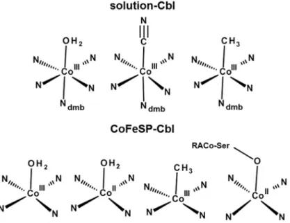
likely resulted in a CoII(OH2) site (Fig 6). CoFeSP-MeCbloxshowed a clearly 5-coordinated CoIII(CH3) site, meaning that water species at theα-position were undetectable. The cobalt spectral changes in the CoFeSP-RACo protein complex supported binding of the serine side chain of RACo to Co(II) at the ß-position (Fig 6).
Electronic structure considerations
Calculated ctv spectra for the relevant low-spin cobalt site species were analyzed in terms of the electric dipole and quadrupole contributions to the underlying electronic transitions and of the metal/ligand characters of the target MOs. The ctv spectra were dominated (>75%) by formally selection-rule forbidden electric dipole transitions in 6-coordinated base-on and 5-coordinated base-off Co(III) and Co(II) sites with water ligands (Table 4). Increased contributions from allowed quadrupole transitions in the base-off sites lead to increased ctv intensities. Increased quadrupole contributions (up to ~50%) for Co(III) and Co(II) sites with two water ligands account for non-negligible ctv intensities in these symmetric structures. The ctv spectra of CH3-, CN-, or OSer-ligand containing Co(III) sites showed almost exclusive dipole transitions, their more intense ctv features resulted from increased ligand characters of target MOs (Table 4). Dominating corrin character of target MOs for the corresponding Co(II) sites explained their more intense ctv features. The small contributions (<15%) of water ligands to target MOs generally exceeded those of dmb, but influenced the ctv intensities only moderately. Fig 5. Comparison of DFT calculated and experimental ctv features.Lines, spectra from DFT; dots, experimental data (Fig 3); spectra were vertically shifted for comparison; note the doubled y-scale in (B). Calculated spectra represent the indicated model structures; solid lines and coloured annotations denote calculated spectra for the indicated structures, which show superior agreement with the experimental data (broken lines show calculation results less in agreement with the experimental data).
However, twice as large ctv contributions from the methyl ligand and increased corrin contri-butions accounted for the large ctv features of the CoIII(CH3)-containing sites. Similarly large corrin and weaker axial ligand contributions for the Co(II)(CN)/(OSer) sites explained their smaller ctv features.
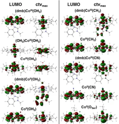
The LUMO, corresponding to the lowest-energy ctv transition, and the target MO for the maximal-intensity ctv transition were compared for the main cobalt site species (Fig 7). For (dmb)CoIII(OH2) the LUMO was delocalized on the corrin ring and the highest-intensity tar-get MO showed predominant Co-3d(z2) character oriented along the axial ligands. These MO locations were reversed when dmb was replaced by water. Enhanced delocalization of both orbitals over the corrin ring occurred in the absence of theα-ligand. Loss of theα-ligand
fur-ther caused a 1–2 eV decrease of the HOMO and LUMO energies and a ~50% decrease of the LUMO–HOMO energy gap (ΔE) from ~3 eV in (dmb)CoIII(OH2) to ~2 eV in CoIII(OH2),
mostly due to a larger relative E(LUMO) drop (Table 5). These energy changes are expected to facilitate reduction of CoIII(OH2) at more positive potentials than (dmb)CoIII(OH2). Exchange or loss of the Co(III)α-ligand further caused a cobalt charge increase by a factor up to ~1.5.
For Co(II) species, less pronounced changes and LUMO delocalization onto the corrin rather independent of theα-ligand and more delocalized MOs with Co(d) character were found.
However, a ~50% decreasedΔE compared to (dmb)CoII(OH2) was observed only for (OH2)
CoII(OH2), due to a larger relative E(HOMO) drop, whereas CoII(OH2) showed an even slightly increasedΔE (Table 5). Compared to (dmb)CoII(OH2), (OH2)CoII(OH2) may thus be
Table 3. Cobalt-ligand bond lengths from crystallography, EXAFS, and DFT.
species bond length [Å]
sample (Lα)Cox(Lß) Co-Ncorrinmean Co-Lα Co-Lß
crystal EXAFS DFT crystal EXAFS DFT crystal EXAFS DFT
AqCblox (dmb)CoIII(OH
2) 1.89a 1.88 1.92 1.92a 1.92 1.96 1.95a 1.97 2.11
AqCblred (dmb)CoII(OH
2) - 1.89 1.92 - 2.30 2.36 - 2.47 2.96
(dmb)CoII - 1.89 1.92 - 2.30 2.30 - -
-CNCblox (dmb)CoII(CN) 1.91b 1.89 1.92 2.04b 2.05 2.15 1.87b 1.85 1.88
CNCblred CoII(CN) - 1.88 1.92 - - 2.14 2.10
(dmb)CoII - 1.88 1.92 - 2.54 2.30 - -
-MeCblox (dmb)CoIII(CH
3) 1.90c 1.89 1.92 2.16c 2.21 2.36 1.99c 1.95 1.97
CoFeSP-AqCblox (OH2)CoIII(OH2) - 1.88 1.92 - 2.02 1.99 - 2.02 1.99
CoIII(OH
2) - 1.88 1.91 - - - - 2.02 1.97
CoFeSP-AqCblred (OH
2)CoII(OH2) - 1.96 1.92 - 2.33 2.53 - 2.33 2.54
CoII(OH
2) 1.90d 1.86 1.91 - - - 2.55d 2.33
-CoFeSP-AqCbl-RACo CoII(OSer) 1.90e 1.88 1.92 - - - 2.40e 2.10 2.08
CoFeSP-MeCblox (OH
2)CoIII(CH3) - 1.87 1.91 - 2.50 2.46 - 2.00 1.96
CoIII(CH
3) 1.90f 1.87 1.91 - - - 2.00f 2.00 1.96
Crystal data for Cbl and CoFeSP-Cbl species were derived from refs.
a[56] b[55] c[54] d[12,19] e[14] f[18]
DFT data refer to geometry-optimized model structures with the indicated cobalt oxidation states and axial ligations; bond lengths from EXAFS (Table 2) were placed in the table to match the other data best and facilitate species comparison.
harder to reduce, but CoII(OH2) may be reduced at most positive potentials. The charge on cobalt for most Co(II) species was even slightly more positive compared to the Co(III) sites and the surplus negative charge was thus mostly located on the corrin ring.
For MeCbl, loss of the dmb ligand left the LUMO delocalization almost unchanged, but increased the valence level delocalization onto the corrin (Fig 7). Loss of theα-ligand rather
increasedΔE due to a smaller relative E(LUMO) drop in CoIII(CH3) compared to (dmb)
CoIII(CH3) (Table 5), making the CoIII(CH3) species easier to reduce. In addition, the charges Fig 6. Cobalt coordination models.Shown structures represent most prominent species identified in the solution Cbl (top) and CoFeSP-Cbl (bottom) samples. The dmb ligand binds inα-position at cobalt; a water
ligand in ß-position in Co(III)-containing CoFeSP-AqCbl cannot be fully excluded; RACo-Ser398 binds at Co (II) in the CoFeSP-RACo complex.
doi:10.1371/journal.pone.0158681.g006
Table 4. Core-to-valence electronic transition characters.
core-to-valence transition characters
electric contribution [%]a metal/ligand contribution [%]b
cobalt site dipole quadrupole Co corrin Lα Lß
(dmb)CoIII(OH
2) 85.0 11.3 58.3 25.8 4.2 11.7
(OH2)CoIII(OH2) 61.1 38.1 46.5 47.9 2.8 2.8
CoIII(OH
2) 80.8 16.8 53.2 43.4 - 3.4
(dmb)CoII(OH2) 77.0 20.4 24.3 55.5 8.1 12.1
CoII(OH
2) 74.8 23.5 26.9 58.4 - 14.7
(dmb)CoIII(CH
3) 96.5 0.3 28.0 24.2 15.5 32.3
CoIII(CH
3) 98.0 1.8 37.3 40.7 - 22.0
(dmb)CoIII(CN) 88.7 9.8 35.5 49.4 7.2 7.9
CoII(CN) 93.7 5.6 40.3 53.8 - 5.9
CoII(O
Ser) 93.1 6.1 41.9 54.4 - 3.7
Data represents the summed relative contributions to the respective DFT-calculated stick spectra underlying the ctv spectra inFig 5.
adifference to 100% = magnetic pole contribution.
bMetal/ligand contributions (Lα, Lß = axial cobalt ligands) denote respective characters of ctv target molecular orbitals.
on the Co(III) center and in particular on the corrin in the MeCbl species were lowered com-pared to AqCbl, with little charge located on the methyl group. The MO configurations were Fig 7. Molecular orbitals in Cbl model structures from DFT.LUMO, lowest unoccupied MO
corresponding to the lowest energy core-to-valence electronic transition in the pre-edge absorption X-ray spectral region; ctvmax, MO corresponding to the highest-intensity ctv transition of the pre-edge absorption.
Cobalt oxidation state and axial ligation are indicated.
doi:10.1371/journal.pone.0158681.g007
Table 5. HOMO and LUMO energies and natural population analysis charges from DFTa.
cobalt site energy [eV] ΔE [eV] NPA charge [e]
HOMO LUMO Co corrin Lα Lß
(dmb)CoIII(OH
2) -11.4 -8.3 3.1 0.39 0.90 0.49 0.23
(OH2)CoIII(OH2) -11.8 -8.8 3.0 0.45 0.95 0.30 0.30
CoIII(OH
2) -12.3 -10.2 2.1 0.56 1.15 - 0.28
(dmb)CoII(OH2) -7.9 (-7.9)b -4.5 (-4.5)b 3.4 (3.4)b 0.58 0.26 0.14 0.03
CoII(OH
2) -8.4 (-8.4)b -4.8 (-4.8)b 3.6 (3.6)b 0.61 0.30 - 0.08
(dmb)CoIII(CH
3) -7.9 -4.6 3.3 0.21 0.59 0.20 0.00
CoIII(CH
3) -8.7 -5.0 3.7 0.30 0.65 - 0.05
(dmb)CoIII(CN) -8.2 -4.9 3.3 0.04 0.85 0.29 -0.18
CoII(CN) -4.2 (-5.1)b -1.8 (-1.8)b 2.4 (3.3)b 0.28 0.27 - -0.55
CoII(O
Ser) -3.8 (-3.7)b -1.6 (-1.6)b 2.2 (2.1)b 0.50 0.17 - -0.67
aValues correspond to model structures with the indicated cobalt oxidation states and axial ligations in low-spin species
ΔE = E(LUMO)–E(HOMO); Lα, Lß = axial cobalt ligands
benergies of
“up" and“down”(in parenthesis) -spin MOs are given for the Co(II) species (and have the same energy in the Co(I) and Co(III) species).
remarkably similar in CoII(CN) and CoII(OSer) (Fig 7), as were the similar E(HOMO/LUMO) andΔE values. Significantly less positive charges on cobalt and corrin in CoII(CN) and in
par-ticular in CoII(OSer) compared to the other Co(II) species were accompanied by strongly nega-tive charges on the CN or OSerligands. The relatively higher E(LUMO) compared, e.g., to CoII(OH2) suggested metal reduction at more negative potentials in CoII(OSer), meaning that RACo binding to CoFeSP was expected to stabilize Co(II).
Discussion
Molecular and electronic structures of cobalamin species bound to CoFeSP or in solution were characterized using XAS in combination with DFT calculations. The observed K-edge energies are affected both by axial coordination and formal cobalt oxidation state changes, in agreement with earlier studies [38,39,59]. Transition from octahedral cobalt sites in solution Cbl and ref-erence compounds to square-pyramidal sites in CoFeSP-Cbl leads to relatively lower edge ener-gies and significant shape changes, although the Co-Ncorrinbond length shows only minor changes due to redox and geometry changes at cobalt. The EXAFS spectra were dominated by the Co-Ncorrinbonds, but facilitated estimation of the axial ligand bond lengths, which were elongated for Co(II) species as in crystal and DFT structures.
The pre-edge absorption due to core-to-valence electronic excitations (ctv) revealed pro-nounced spectral variations in response to cobalt redox and site geometry changes. Interpreta-tion of the ctv spectra in terms of resonant electronic excitaInterpreta-tion of a 1score electron into unoccupied valence levels with variable metal/ligand characters was achieved using DFT. Good agreement between experimental and calculated ctv spectra was obtained for the solution Cbl and CoFeSP-Cbl systems, as previously found for other metal complexes (see, e.g., [30–33,60– 63]). The ctv intensity variations were consistently explained by changes in the cobalt/ligand character ratio of the target MOs and, to a lesser extent, by electric dipole/quadrupole contribu-tion variacontribu-tions of the underlying electronic transicontribu-tions due to axial ligacontribu-tion changes. This showed for example that the intense ctv features of cobalt sites with a methyl ligand are related to significant CH3character of the target MOs, thus unambiguously establishing a CoIII(CH3) site in CoFeSP-MeCbl. The ctv-XAS/DFT combination appears to be viable for redox state and ligation geometry assignment of cobalt sites in cobalamin.
The discriminated cofactor species revealed a trend for fewer ligands at cobalt in CoFeSP-Cbl compared to solution Cbl for the same oxidation state. Octahedral (dmb)
CoIII(OH2) and (dmb)CoII(OH2) sites were dominant in oxidized and reduced solution AqCbl. XAS and DFT showed a tendency for detachment of the water species from the Co(II) ion. However, the transition from (dmb)CoIII(CN) to CoII(CN) species suggested preference for detachment of the weaker dmb ligand upon cobalt reduction for CNCbl in solution. MeCbl in solution showed the anticipated (dmb)CoIII(CH3) structure. The spectroscopic and theoretical data converged to the same cobalt site structures in solution Cbl, corroborating the adequacy of the applied theory level (B3LYP/TZVP) for cobalamin structure description.
Crystal structures of CoFeSP-Cbl have shown the dmb group in base-off configuration in the protein [12,14,18,19]. The crystal data furthermore were interpreted as showing the absence also of water species at theα-position. The oxygen of a threonine side chain (Thr374)
at theαside was modeled at 3.2–4.6 Å to cobalt in different crystals (Fig 1), suggesting the
absence of a Co-OThrbond. However, the ß-ligand bond length at cobalt also varied consider-ably or a ß-ligand was not assigned [12,14,18,19]. These results could be related to site hetero-geneity in the crystals, which may render detection of axial cobalt ligands difficult.
presence or absence of a water ligand in theα-position at Co(III) in CoFeSP-AqCbl might be
related for example to redox state variations of the [4Fe4S] cluster bound to the large subunit (CfsA) of CoFeSP [64–67]. Taking into account also the crystallographic data, we consider a CoIII(OH2) site as more likely. Direct binding of Thr374 at theα-position at Co(III) was
seem-ingly excluded, corroborating the crystallographic assignment. Single-electron reduction of CoFeSP-AqCbl results in formation of a CoII(OH2) site with a weaker water-cobalt interaction. Cobalt sites lacking aα-ligand were clearly identified in CoFeSP-MeCbl containing Co(III) and
in CoFeSP-AqCbl-RACo containing Co(II) [18]. Our interpretation that a serine residue of RACo coordinates to Co(II) in CoFeSP-AqCbl-RACo is in agreement with the crystal structure [16] and previous spectroscopic data [15]. Serine binding at Co(II) likely is a prerequisite for ATP-induced CoFeSP activation and reaction with methyl-tetrahydrofolate, resulting in methyl group binding at cobalt [13,14], as supported by our detection of a CoIII(CH3) site in CoFeSP-MeCbl. In an earlier study of CoFeSP from another organism, a MeCbl species with a water (α) and a methyl (ß) ligand at Co(III) has been proposed; the water-cobalt bond,
how-ever, presumably was considerably elongated [65]. Our data favor a CoIII(CH3) site in theC. hydrogenoformansenzyme under our conditions. We cannot fully exclude a remote water ligand, which might have escaped detection in the XAS analysis, but consider it as unlikely.
Our DFT calculations suggest that Co(III) and Co(II) reduction in base-off CoFeSP-AqCbl presumably occurs at more positive potentials compared to base-on AqCbl or CoFeSP-AqCbl with two water ligands. The determined reduction potentials of the Co(II/III) and Co(I/II) cou-ples in CoFeSP-AqCbl (about +350 mV and -500 mV) indeed are more positive compared to the values of AqCbl in solution (about +200 mV and -600 mV) [4,66,68]. The absence of aα-ligand
at cobalt thus may tune the Cbl reduction potential in CoFeSP into the physiological range for Co(I) formation prior to methylation [13,18,65]. However, our DFT results and redox titrations [14] suggest stabilization of the Co(II) state when Ser398 of RACo is bound to the metal. The resulting apparent disabling of Co(I) formation likely is overcome by ATP binding to the CoFe-SP-RACo complex, inducing electron transfer from the [2Fe2S] cluster in RACo to the Co(II) site by a yet unresolved mechanism [14]. ATP binding could be accompanied by loss of the serine ligand at Co(II) to facilitate Co(I) formation by destabilizing Co(II), such that the Co(I/II) mid-point potential approaches the one of the [2Fe2S] cluster (-340 mV [14]). The square-planar Co (I) [36] then binds the methyl group to form CoIII(CH3) and transfer of the methyl cation to ace-tyl-CoA synthase is facilitated by a more positive reduction potential, compared, e.g., to base-on MeCbl, of the CoIII(CH3) site in CoFeSP. Control of the axial cobalt ligation therefore may play an important role both in methyl group transfer and reductive activation of CoFeSP.
Conclusions
Analysis of cobalt K-edge XAS spectra in combination with DFT calculations of pre-edge absorp-tion features facilitates determinaabsorp-tion of axial ligaabsorp-tion and redox state of cobalt in soluabsorp-tion Cbl and CoFeSP-Cbl. This supports the likely absence of aα-ligand in base-off CoFeSP-AqCbl and
-MeCbl compared to base-on solution AqCbl and MeCbl, in agreement with earlier crystallo-graphic and spectroscopic data. Coordination of a serine side chain from RACo to Co(II) in the CoFeSP-RACo protein complex is in agreement with our analysis. Control of the axial cobalt liga-tion may tune the redox potential of the cobalamin cofactor into the range of its electron transfer partners and likely is important for reductive activation of CoFeSP and methyl group shuttling.
Supporting Information
multiple scattering calculations of cobalamin XANES spectra (Fig D), K-edge energies from XANES simulations (Fig E), correlation of EXAFS fit parameters (Fig F), supporting refer-ences.
(PDF)
Acknowledgments
We thank R. Kositzki for help in XAS data collection, the group of F. Schäfers at KMC-1 of BESSY (Helmholtz Zentrum für Materialien und Energie Berlin) for technical assistance, and C. Teutloff (FU-Berlin) for generous EPR support.
Author Contributions
Conceived and designed the experiments: MH H. Dobbek. Performed the experiments: PS SM SG SEH. Analyzed the data: PS SM MH. Contributed reagents/materials/analysis tools: SG H. Dau H. Dobbek. Wrote the paper: PS MH H. Dobbek. Provided access to XAS equipment: H. Dau.
References
1. Giedyk M, Goliszewska K, Gryko D. Vitamin B12 catalysed reactions. Chem Soc Rev. 2015; 44 (11):3391–404. doi:10.1039/c5cs00165jPMID:25945462
2. Brown KL. Chemistry and enzymology of vitamin B12. Chem Rev. 2005; 105(6):2075–149. PMID: 15941210
3. Banerjee R, Ragsdale SW. The many faces of vitamin B12: catalysis by cobalamin-dependent enzymes. Annu Rev Biochem. 2003; 72:209–47. PMID:14527323
4. Dereven'kov IA, Salnikov DS, Silaghi-Dumitrescu R, Makarov SV, Koifman OI. Redox chemistry of cobalamin and its derivatives. Coord Chem Rev. 2016; 309:68–83.
5. Herrmann W, Obeid R. Cobalamin deficiency. Subcell Biochem. 2012; 56:301–22. doi: 10.1007/978-94-007-2199-9_16PMID:22116706
6. Bengelsdorf FR, Straub M, Durre P. Bacterial synthesis gas (syngas) fermentation. Environ Technol. 2013; 34(13–16):1639–51. PMID:24350425
7. Appel AM, Bercaw JE, Bocarsly AB, Dobbek H, DuBois DL, Dupuis M, et al. Frontiers, opportunities, and challenges in biochemical and chemical catalysis of CO2fixation. Chem Rev. 2013; 113(8):6621–
58. doi:10.1021/cr300463yPMID:23767781
8. Ragsdale SW. Enzymology of the Wood-Ljungdahl pathway of acetogenesis. Ann N Y Acad Sci. 2008; 1125:129–36. doi:10.1196/annals.1419.015PMID:18378591
9. Jeoung JH, Goetzl S, Hennig SE, Fesseler J, Wormann C, Dendra J, et al. The extended reductive ace-tyl-CoA pathway: ATPases in metal cluster maturation and reductive activation. Biol Chem. 2014; 395 (5):545–58. doi:10.1515/hsz-2013-0290PMID:24477517
10. Bender G, Pierce E, Hill JA, Darty JE, Ragsdale SW. Metal centers in the anaerobic microbial metabo-lism of CO and CO2. Metallomics. 2011; 3(8):797–815. doi:10.1039/c1mt00042jPMID:21647480
11. Matthews RG, Koutmos M, Datta S. Cobalamin-dependent and cobamide-dependent methyltrans-ferases. Curr Opin Struct Biol. 2008; 18(6):658–66. doi:10.1016/j.sbi.2008.11.005PMID:19059104
12. Svetlitchnaia T, Svetlitchnyi V, Meyer O, Dobbek H. Structural insights into methyltransfer reactions of a corrinoid iron-sulfur protein involved in acetyl-CoA synthesis. Proc Natl Acad Sci U S A. 2006; 103 (39):14331–6. PMID:16983091
13. Seravalli J, Zhao S, Ragsdale SW. Mechanism of transfer of the methyl group from (6S)-methyltetrahy-drofolate to the corrinoid/iron-sulfur protein catalyzed by the methyltransferase fromClostridium ther-moaceticum: a key step in the Wood-Ljungdahl pathway of acetyl-CoA synthesis. Biochemistry. 1999; 38(18):5728–35. PMID:10231523
14. Hennig SE, Goetzl S, Jeoung JH, Bommer M, Lendzian F, Hildebrandt P, et al. ATP-induced electron transfer by redox-selective partner recognition. Nat Commun. 2014; 5:4626. doi:10.1038/
15. Meister W, Hennig SE, Jeoung JH, Lendzian F, Dobbek H, Hildebrandt P. Complex formation with the activator RACo affects the corrinoid structure of CoFeSP. Biochemistry. 2012; 51(36):7040–2. PMID: 22924695
16. Hennig SE, Jeoung JH, Goetzl S, Dobbek H. Redox-dependent complex formation by an ATP-depen-dent activator of the corrinoid/iron-sulfur protein. Proc Natl Acad Sci U S A. 2012; 109(14):5235–40. doi:10.1073/pnas.1117126109PMID:22431597
17. Abeles RH, Dolphin D. Vitamin-B12 coenzyme. Accounts Chem Res. 1976; 9(3):114–120.
18. Kung Y, Ando N, Doukov TI, Blasiak LC, Bender G, Seravalli J, et al. Visualizing molecular juggling within a B12-dependent methyltransferase complex. Nature. 2012; 484(7393):265–9. doi:10.1038/ nature10916PMID:22419154
19. Goetzl S, Jeoung JH, Hennig SE, Dobbek H. Structural basis for electron and methyl-group transfer in a methyltransferase system operating in the reductive acetyl-CoA pathway. J Mol Biol. 2011; 411 (1):96–109. doi:10.1016/j.jmb.2011.05.025PMID:21640123
20. Krautler B. Vitamin B12: chemistry and biochemistry. Biochem Soc Trans. 2005; 33(Pt 4):806–10. PMID:16042603
21. Dittrich B, Koritsanszky T, Volkov A, Mebs S, Luger P. Novel approaches to the experimental charge density of vitamin B12. Angew Chem Int Ed Engl. 2007; 46(16):2935–8. PMID:17342782
22. Stich TA, Brooks AJ, Buan NR, Brunold TC. Spectroscopic and computational studies of Co3+-corri-noids: Spectral and electronic properties of the B-12 cofactors and biologically relevant precursors. J Am Chem Soc. 2003; 125(19):5897–5914. PMID:12733931
23. Jensen KP, Ryde U. Cobalamins uncovered by modern electronic structure calculations. Coord Chem Rev. 2009; 253:769–778.
24. Gherasim C, Lofgren M, Banerjee R. Navigating the B-12 road: assimilation, delivery, and disorders of cobalamin. J Biol Chem. 2013; 288(19):13186–13193. doi:10.1074/jbc.R113.458810PMID:23539619
25. Lexa D, Saveant JM. The electrochemistry of vitamin-B12. Accounts Chem Res. 1983; 16(7):235–243.
26. Dau H, Haumann M. The manganese complex of photosystem II in its reaction cycle—Basic framework and possible realization at the atomic level. Coord Chem Rev. 2008; 252:273–295.
27. Dau H, Liebisch P, Haumann M. X-ray absorption spectroscopy to analyze nuclear geometry and elec-tronic structure of biological metal centers—Potential and questions examined with special focus on the tetra-nuclear manganese complex of oxygenic photosynthesis. Anal Bioanal Chem. 2003; 376 (5):562–583. PMID:12802563
28. Yachandra VK. X-ray absorption spectroscopy and applications in structural biology. In: Sauer K, edi-tor. Methods in Enzymology—Biochemical Spectroscopy. 246. New York: Academic Press; 1995. p. 638–678.
29. Koningsberger DC, Prins R. X-ray absorption: principles, applications, techniques of EXAFS, SEXAFS, and XANES. New York: Wiley; 1988.
30. Chandrasekaran P, Stieber SCE, Collins TJ, Que L, Neese F, DeBeer S. Prediction of high-valent iron K-edge absorption spectra by time-dependent Density Functional Theory. Dalton Trans. 2011; 40 (42):11070–11079. doi:10.1039/c1dt11331cPMID:21956429
31. DeBeer-George SD, Petrenko T, Neese F. Prediction of íron K-edge absorption spectra using time-dependent density functional theory. J Phys Chem A. 2008; 112(50):12936–12943. doi:10.1021/ jp803174mPMID:18698746
32. Lambertz C, Chernev P, Klingan K, Leidel N, Siegfridsson KGV, Happe T, et al. Electronic and molecu-lar structures of the [2Fe] and [4Fe4S] units of the active-site H-cluster in [FeFe]-hydrogenase deter-mined by spin- and site-selective XAE and DFT. Chem Sci. 2014; 5:1187–1203.
33. Chernev P, Lambertz C, Brunje A, Leidel N, Sigfridsson KG, Kositzki R, et al. Hydride binding to the active site of [FeFe]-hydrogenase. Inorg Chem. 2014; 53(22):12164–77. doi:10.1021/ic502047q PMID:25369169
34. Wirt MD, Chance MR. Temperature dependent coordination effects in base-off adenosyl and methylco-balamin by X-ray edge spectroscopy. J Inorg Biochem. 1993; 49(4):265–73. PMID:8478624
35. Giorgetti M, Ascone I, Berrettoni M, Conti P, Zamponi S, Marassi R. In situ X-ray absorption spectro-electrochemical study of hydroxocobalamin. J Biol Inorg Chem. 2000; 5(2):156–166. PMID:10819461
36. Wirt MD, Kumar M, Wu JJ, Scheuring EM, Ragsdale SW, Chance MR. Structural and electronic factors in heterolytic cleavage: formation of the Co(I) intermediate in the corrinoid/iron-sulfur protein from Clos-tridium thermoaceticum. Biochemistry. 1995; 34(15):5269–73. PMID:7711048
38. Champloy F, Gruber K, Jogl G, Kratky C. XAS spectroscopy reveals X-ray-induced photoreduction of free and protein-bound B12 cofactors. J Synchrotron Radiat. 2000; 7(Pt 4):267–73. PMID:16609206
39. Scheuring EM, Clavin W, Wirt MD, Miller LM, Fischetti RF, Lu Y, et al. Time-resolved X-ray absorption spectroscopy of photoreduced base-off Cob(II)alamin compared to the Co(II) species inClostridium thermoaceticum. J Phys Chem-Us. 1996; 100(9):3344–3348.
40. Bradford MM. Rapid and sensitive method for quantitation of microgram quantities of protein utilizing principle of protein-dye binding. Analyt Biochem. 1976; 72(1–2):248–254.
41. Klockenkämper R. Total Reflection X-ray Fluorescence Analysis. London, UK: Wiley-VCH; 1996.
42. Rehr JJ, Albers RC. Theoretical approaches to x-ray absorption fine structure. Rev Mod Phys. 2000; 72 (3):621–654.
43. Ankudinov AL, Ravel B, Rehr JJ, Conradson SD. Real-space multiple-scattering calculation and inter-pretation of x-ray-absorption near-edge structure. Phys Rev B. 1998; 58(12):7565–7576.
44. Klementiev KV. XANES dactyloscope for Windows. freeware:wwwcellses/Beamlines/CLAESS/ software/xandahtml.
45. Rehr JJ, Kas JJ, Vila FD, Prange MP, Jorissen K. Parameter-free calculations of X-ray spectra with FEFF9. Phys Chem Chem Phys. 2010; 12(21):5503–13. doi:10.1039/b926434ePMID:20445945
46. Mebs S, Henn J, Dittrich B, Paulmann C, Luger P. Electron densities of three B-12 vitamins. J Phys Chem A. 2009; 113(29):8366–8378. doi:10.1021/jp902433xPMID:19569666
47. Frisch MJT, G. W.; Schlegel H. B.; Scuseria G. E.; Robb M. A.; Cheeseman J. R.; Scalmani G.; et al. Gaussian 09, Revision D.01. Gaussian, Inc, Wallingford CT. 2009.
48. Lee C, Yang W, Parr RG. Development of the Colle-Salvetti correlation-energy formula into a functional of the electron density. Phys Rev B Condens Matter. 1988; 37(2):785–789. PMID:9944570
49. Schafer A, Huber C, Ahlrichs R. Fully optimized contracted Gaussian-basis sets of triple zeta valence quality for atoms Li to Kr. J Chem Phys. 1994; 100(8):5829–5835.
50. Reed AE, Weinstock RB, Weinhold F. Natural-Population Analysis. J Chem Phys. 1985; 83(2):735– 746.
51. Glendening ED, Badenhoop J. K., Reed A. E., Carpenter J. E., Bohmann J. A., Morales C. M., Weinhold F. NBO-5. Theoretical Chemistry Institute, University of Wisconsin, Madison, USA. 2004.
52. Neese F. The ORCA program system. Wiley Interdisciplinary Reviews-Computational Molecular Sci-ence. 2012; 2(1):73–78.
53. Neese F. ORCA: An ab-initio, DFT, and Semiempirical Electronic Structure Package. v.2.6.35. Theoret-ical Chemistry Group. Mülheim, Germany: Max-Planck Institute for ChemTheoret-ical Energy Conversion; 2008.
54. Rossi M, Glusker JP, Randaccio L, Summers MF, Toscano PJ, Marzilli LG. The structure of a B-12 coenzyme—methylcobalamin studies by X-ray and NMR methods. J Am Chem Soc. 1985; 107 (6):1729–1738.
55. Krautler B, Konrat R, Stupperich E, Farber G, Gruber K, Kratky C. Direct evidence for the conforma-tional deformation of the corrin ring by the nucleotide base in vitamin-B12—synthesis and solution spectroscopic and crystal-structure analysis of Co-beta-cyanoimidazolylcobamide. Inorg Chem. 1994; 33(18):4128–4139.
56. Brown KL, Cheng SF, Zou X, Zubkowski JD, Valente EJ, Knapton L, et al. Cis effects in the cobalt cor-rins .1. Crystal structures of chloroaquacobalamin perchlorate, chlorocyanocobalamin, and 10-chloromethylcobalamin. Inorg Chem. 1997; 36(17):3666–3675. PMID:11670058
57. Kratky C, Kräutler B. X-ray Crystallography of B12. In: Banerjee R, editor. Chemistry and biochemistry of B12. New York: Wiley; 1999. p. 9–41.
58. Koningsberger DC, Mojet BL, van Dorssen GE, Ramaker DE. XAFS spectroscopy; fundamental princi-ples and data analysis. Topics in Catalysis. 2000; 10:143–155.
59. Giorgetti M, Ascone I, Berrettoni M, Conti P, Zamponi S, Marassi R. In situ X-ray absorption spectro-electrochemical study of hydroxocobalamin. J Biol Inorg Chem. 2000; 5(2):156–66. PMID:10819461
60. Krewald V, Retegan M, Cox N, Messinger J, Lubitz W, DeBeer S, et al. Metal oxidation states in biologi-cal water splitting. Chem Sci. 2015; 6:1676–1695
61. Leidel N, Chernev P, Havelius KG, Schwartz L, Ott S, Haumann M. Electronic structure of an [FeFe] hydrogenase model complex in solution revealed by X-ray absorption spectroscopy using narrow-band emission detection. J Am Chem Soc. 2012; 134(34):14142–57. doi:10.1021/ja304970pPMID: 22860512
63. Chandrasekaran P, Chiang KP, Nordlund D, Bergmann U, Holland PL, DeBeer S. Sensitivity of X-ray core spectroscopy to changes in metal ligation: a systematic study of low-coordinate, high-spin ferrous complexes. Inorg Chem. 2013; 52(11):6286–98. doi:10.1021/ic3021723PMID:23662855
64. Lindahl PA, Munck E, Ragsdale SW. CO dehydrogenase fromClostridium thermoaceticum. EPR and electrochemical studies in CO2and argon atmospheres. J Biol Chem. 1990; 265(7):3873–9. PMID:
2154491
65. Stich TA, Seravalli J, Venkateshrao S, Spiro TG, Ragsdale SW, Brunold TC. Spectroscopic studies of the corrinoid/iron-sulfur protein fromMoorella thermoacetica. J Am Chem Soc. 2006; 128(15):5010– 20. PMID:16608335
66. Jablonski PE, Lu WP, Ragsdale SW, Ferry JG. Characterization of the metal centers of the corrinoid/ iron-sulfur component of the CO dehydrogenase enzyme complex fromMethanosarcina thermophila by EPR spectroscopy and spectroelectrochemistry. J Biol Chem. 1993; 268(1):325–9. PMID:8380157
67. Menon S, Ragsdale SW. Role of the [4Fe-4S] cluster in reductive activation of the cobalt center of the corrinoid iron-sulfur protein fromClostridium thermoaceticumduring acetate biosynthesis. Biochemis-try. 1998; 37(16):5689–98. PMID:9548955
![Fig 1. Cobalamin crystal structures. (a) Structure of the Cbl cofactor in CoFeSP enzyme (PDB entry 2H9A, 1.9 Å resolution [12]) showing a base-off configuration (dmb ligand not bound to cobalt in α-position)](https://thumb-eu.123doks.com/thumbv2/123dok_br/16409065.194264/3.918.306.634.113.508/cobalamin-crystal-structures-structure-cofactor-resolution-configuration-position.webp)