Relationship with Invasion and Lymph Node Metastasis
in Hypopharyngeal Squamous Cell Carcinoma
Qing Yang1, Yehai Liu1*, Yang Huang1, Dake Huang2, Yifan Li1, Jing Wu1, Maoli Duan1,3*
1Department of Otorhinolaryngology-Head and Neck Surgery, the First Affiliated Hospital of Anhui Medical University, Hefei, Anhui, PR China,2Department of Microbiology and Parasitology, Anhui Medical University, Hefei, Anhui, PR China,3Department of clinical science, intervention and technology, Karolonska Institute, Stockholm, Sweden
Abstract
To assess the expression of COX-2,CD44v6 and CD147 in hypopharyngeal squamous cell carcinomas and the three biomarkers correlation with tumor invasion and lymph node metastasis of Chinese people. 101 cases of surgically excised primary tumor were included in this study, and 40 tissues of epithelium adjacent to carcinoma were used as controls. We characterized the immunohistochemical expression of COX-2, CD44v6, and CD147 in141 formalin-fixed, paraffin-embedded tissues, and measured the mean optical density (OD) of the positive area to identify the expression of the three bio-markers and relationship with tumor invasion and lymph node metastasis. Our study demonstrates that the expression of the COX-2 and CD147 were significantly increased in carcinoma tissues compared to the epithelium adjacent to carcinoma. We also observed that the expression of COX-2, CD44v6, and CD147 were significantly associated with T classification, lymph node metastasis and clinical stage. There was strong significant correlation among the three biomarkers as well. Additionally, we indicated that recurrence and $P50 level of COX-2 expression had an independent prognostic effect on prognosis. In
conclusion, the three biomarkers play important roles in tumor invasion and lymph node metastases and might be valuable indicators of tumor metastasis in hypopharyngeal squamous cell carcinoma.
Citation:Yang Q, Liu Y, Huang Y, Huang D, Li Y, et al. (2013) Expression of COX-2, CD44v6 and CD147 and Relationship with Invasion and Lymph Node Metastasis in Hypopharyngeal Squamous Cell Carcinoma. PLoS ONE 8(9): e71048. doi:10.1371/journal.pone.0071048
Editor:Zhihua Liu, Chinese Academy of Medical Sciences, China
ReceivedFebruary 12, 2013;AcceptedJune 26, 2013;PublishedSeptember 3, 2013
Copyright:ß2013 Yang et al. This is an open-access article distributed under the terms of the Creative Commons Attribution License, which permits unrestricted use, distribution, and reproduction in any medium, provided the original author and source are credited.
Funding:This research was supported by key program of Educational department, Anhui province (KJ2012A162), China and Karolinska Institutet, Stockholm, Sweden. The funders had no role in study design, data collection and analysis, decision to publish, or preparation of the manuscript.
Competing Interests:The authors have declared that no competing interests exist.
* E-mail: [email protected] (Y. Li); [email protected] (MD)
Introduction
Hypopharyngeal squamous cell carcinoma, a malignant neo-plasm arising from the mucosa of the upper aerodigestive tract, is one of the most aggressive cancers in the head and neck area. Over 50% of the patients with hypopharyngeal squamous cell carcinoma have reached stage IV at presentation because of lacks of symptoms. Even after multimodality therapy 20% of the patients have residual disease, and recurrences tend to appear in the first year and 50% of first recurrences include metastasis. Eventually, 64% of patients die of the cancer [1]. Furthermore, patients with invasion and metastasis to surrounding tissues in the early stages, usually have an unfavorable prognosis [2]. Although combined modality treatments such as chemotherapy, radiation and improved surgical techniques have been applied in clinic, these have not translated into significant improvements in survival [3]. Thus, it is necessary to find novel cancer-related molecules for diagnosis and consequently to improve prognosis of hypopharyn-geal carcinoma.
Over the last decade, it has become clear that there are a large number of biological markers associated with invasion and metastasis of hypopharyngeal carcinoma. Cyclooxygenase-2 (COX-2), one of the cyclooxygenase enzymes key to prostaglandin biosynthesis, whose expression is dramatically up-regulated after inflammation and tumors stimulation, is rapidly induced by
certain growth factors, such as inflammatory cytokines, tumor promoters and oncogenes [4]. Since the overexpression of COX-2 was reported in colorectal cancer [5], several studies have shown its overexpression in other types of cancer, especially in epithelial cancers, for example, breast, prostate and lung cancer [6–8]. This suggests that COX-2 may play an important role in tumor development and progression. Meanwhile, some studies have indicated the overexpression of COX-2 occurs in patients with head and neck cancer [9–10]. CD44v6, a variant isoformof CD44, regulates tumor invasion and metastasis formation, has been shown as a protein marker for metastatic behavior in breast, colorectal and gastric cancers [11–13]. These characteristics have made CD44v6 an attractive factor for the research of metastasis of hypopharyngeal carcinoma. CD147, also known as extracellular matrix metalloproteinase inducer (EMMPRIN), has been identi-fied as a tumor-cell membrane protein that stimulates matrix metalloproteinase (MMP) production in stromal fibroblasts [14]. As a transmembrane glycoprotein of the immunoglobulin super-family, CD147 is overexpressed in various tumor cells including those in head and neck carcinoma [15], and is also known to promote tumor invasion and lymph node metastasis [15–16].
invasion and lymph node metastasis. Furthermore, we analyze the correlation among them and figure out if they can be indicated the tumor invasion and lymph node metastasis.
Methods
Cases
The study protocol was approved by the Ethics Committee of the First Affiliated Hospital of Anhui Medical University, all procedures were performed under the Helsinki declaration, and written consent was obtained from all patients.
101 cases of surgically excised primary tumor were included in this study. Paraffin-embedded specimens of 71 patients with hypopharyngeal squamous cell carcinoma were obtained from the Department of Pathology of the first Hospital Affiliated to Anhui Medical University. These patients were treated at the department of Otorhinolaryngology, Head and Neck Surgery, during January 2004–June 2010. The other 40 cancer tissues were obtained intra-operatively from patients who were operated in the hospital during July 2010 to May 2011. Meanwhile, 40 tissues of epithelium adjacent to carcinoma obtained by the same way were used for comparison.
No patient had received any prior therapy, such as radio- or chemotherapy. All hematoxylin-eosin-stained sections were re-viewed, the quality of the material was checked, and the best section from each specimen was selected. All tumors were classified histologically according to the classification by WHO, TNM and clinical stages were identified by IUCC system.
Immunohistochemistry study
All sections were deparaffinized in xylene, sequentially rehy-drated in alcohol, and washed in phosphate-buffered saline. The sections were heated twice in a microwave oven for 5 min in citrate buffer (pH 6.0) for antigen retrieval. COX-2 monoclonal antibody (1:200; Santa Cruz Biotechnology Inc., Santa Cruz, CA), CD44v6 (ready to use; Zhongshan biologic and technical company, Beijing, China) and CD147 (1:150; Santa Cruz Biotechnology Inc., Santa Cruz, CA) were used as the primary antibody. Briefly, the incubation was at 4uC overnight, followed by washing with PBS. The sections were incubated with secondary antibody for 30 min at room temperature. Signals were developed with 3,39-diaminobenzine (DAB) for 2 min and counterstained with hematoxylin.
Quantitative analysis
The immunostaining densities of COX-2, CD44v6 and CD147 in hypopharyngeal carcinoma were quantitatively assessed with the Image-pro plus6.0 (Media Cybernetics, USA). In brief, sections were placed on a microscope (Olympus CX21, Japan), and images were transferred via a digital camera (Nikon 80i, Japan) to a computer. The mean optical density (OD) of the positive areas was measured. The results were expressed as the exact value of the relative optical density units.
Statistical analysis
The SPSS software (version 17.0., SPSS Inc., Chicago, IL, USA) was used in this study. t test was applied to compare
variables. Pearson correlations between three bio-markers were estimated. Survival and recurrence were assessed with the Kaplan-Meier method and comparisons between subgroups were conducted using the log rank test. Cox’s proportional hazards regressions were used to find out the significant predictors for the survival time. Significant was at the level ofP,0.05.
Results
Clinicopathologic characteristics data
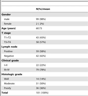
The clinicopathologic characteristics of the patients in the study are as follow: the mean age of the patients was 60.73 years, and the range was from 42 to 78. Of the patients, 99 of the patients were males and 2 were females. 59 of those (58%) presented with lymph node metastasis at diagnosis (Table 1).
Expression of COX-2, CD44v6 and CD147
In this study, diffuse cytoplasmic staining for COX-2 appeared in almost all carcinoma tissues and 70.0% (28/40) of adjacent normal epithelium tissues. However, normal epithelium presented weaker COX-2 staining (part A and B, Figure 1). The mean optical density (ODs) of COX-2 in carcinoma tissues and adjacent normal epithelium tissues were 0.2560.11 and 0.0860.06, respectively (Table 2, Table 3). COX-2 expressed in carcinoma tissues was significantly higher than that in adjacent normal tissues (P,0.001). Moreover, increased COX-2 expression was strongly
associated with lymph node metastasis (part P and Q, Figure 2;
P,0.001), T classification (P,0.001) and clinical stage (P,0.001).
No correlation was found between COX-2 and histological grades (part G, H and I, Figure 3;P= 0.956).
CD44v6 was expressed in all carcinoma tissues, mainly in the cell membrane, and staining intensity for cells in adjacent epithelium issues appeared similar to that in carcinomas (part C and D, Figure 1). There was no significant difference between carcinoma tissues and tissues adjacent to carcinoma (P= 0.668). However, CD44v6 expression was strongly correlated with lymph node metastasis (part R and S, Figure 2;P,0.001), T classification (P,0.001) and clinical stage (P,0.001), but not correlated to
histological grade (part J, K and L, Figure 3;P= 0.811).
CD147 was mainly expressed in cell membranous and cytoplasmic tissues, and also in all carcinoma tissues and adjacent normal epithelium (47.5%, 19/40) (part E and F, Figure 1). The
Table 1.Clinicopathologic characteristics data of patients.
N(%)/mean
Gender
male 99 (98%)
female 2 ( 2%)
Age (years) 60.73
T stage
T1+T2 43 (43%)
T3+T4 58 (57%)
Lymph node
Positive 59 (58%)
Negative 42 (42%)
Clinical grade
I+II 22 (22%)
III+IV 79 (78%)
Histologic grade
Well 14 (14%)
Moderate 51 (50%)
Poorly 36 (36%)
Total 101 (100%)
doi:10.1371/journal.pone.0071048.t001
mean OD measures indicate that CD147 expression in hypopha-ryngeal squamous cell carcinoma was significantly higher than that in adjacent epithelium to carcinoma (P,0.001). Although no significant difference was observed between the grade of differen-tiation of the tumors (part M, N and O, Figure 3;P= 0.630), the level of CD44v6 expression was significantly associated with the incidence of lymph node metastasis (part T and U, Figure 2;
P,0.001), T classification (P= 0.001) and clinical stage (P,0.001). Correlation analysis
Pearson correlation analyses indicate a strong significant correlation between COX-2 and CD147 (r= 0.774, P,0.001).
And there were correlations between COX-2 and CD44v6 (r= 0.473, P,0.001), as well as between CD44v6 and
CD147(r= 0.475,P,0.001).
Survival analysis
Twenty-one patients were lost during follow-up in this study, and 37 patients suffered from a recurrence. Survival analyses among 80 patients show that 1-year survival rate was 66.25%, 3-year survival rate 16.25% and 5-3-year survival 7.5%.
Results indicate that mortality of $P50 level of COX-2
expression was higher than that of , P50 level of COX-2
expression, and the difference was significant (Table 4). Survival time is significantly correlated with$P75 level of COX-2
expression,$P85 level of COX-2 expression and $P90 level of
COX-2 expression (P,0.05). Meanwhile, it is significantly
associated with$P90level of CD147 expression (P,0.05) as well.
But there is no relationship between survival time and the level of CD44v6 expression. On the basis of the above results,$P50level
of COX-2 expression and$P90level of CD147 expression were
classified as the expression of COX-2 and CD147 in hypopha-ryngeal squamous cell carcinoma for Cox’s proportional hazard
Figure 1. The three bio-markers expression by immunohistochemistry.(A) High levels of COX-2 expression in hypopharyngeal squamous cell carcinoma: diffuse cytoplasmic staining. (B) Low levels of COX-2 expression in epithelium adjacent to carcinoma. (C) High levels of CD44v6 expression in hypopharyngeal squamous cell carcinoma: cell membrane staining. (D) High levels of CD44v6 expression in epithelium adjacent to carcinoma as well as staining in carcinoma tissues. (E) High levels of CD147 expression in hypopharyngeal squamous cell carcinoma: diffuse cytoplasmic staining. (F) Low level of CD147 expression in epithelium adjacent to carcinoma.
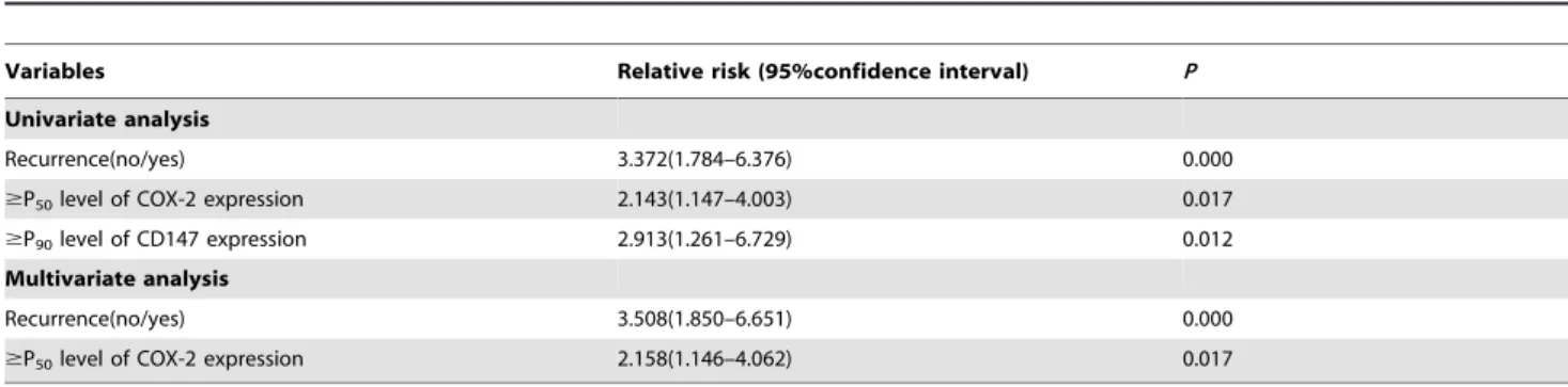
model analysis. Univariate analysis indicated that recurrence,
$P50 level of COX-2 expression and $P90 level of CD147
expression were associated significantly with a worse prognosis (Figure 4 and 5). Neither clinical stages nor node metastasis had any significant association with survival. Multivariate Cox’s proportional hazard model analysis indicates that recurrence and $P50 level of COX-2 expression had an independent
prognostic effect on prognosis (P,0.05; Table 5).
Discussion
In this study, we investigated the prognostic significance of expression of three bio-markers (COX-2, CD44v6 and CD147) in hypopharyngeal squamous cell carcinoma and epithelium adja-cent to carcinoma. The results showed that the expressions of the three bio-markers were significantly associated with tumor invasion and lymph node metastasis.
Regarding previous reports, COX-2 had an important role in invasion and metastasis of head and neck squamous cell carcinoma by a variety of pathways [17–18].To produce a marked effect, COX-2 was mediated by a series of molecules, for instance, CD44, matrix metalloproteinases and VEGF, lead to promote tumor angiogenesis and invasion, even had contribution to cell prolifer-ation and apoptosis [19]. In the present study, we suggested that COX-2 expression was significantly higher in the carcinoma samples than in the adjacent to carcinoma samples, and it was also
strongly associated with the presence of lymph node metastases and tumor invasion. Furthermore, no correlation was found between COX-2 expression and histological grades. This result is generally consistent with previous reports (9).
CD44v6 as a kind of CD44 variant isoforms, is regarded to be responsible for tumor lymphangiogenesis and lymph node metastasis. On the one hand, studies indicated that the overex-pression of CD44v6 in squamous cell carcinoma is associated with lymph node metastasis derived from skin, lung, gastric cancers as well as head and neck [20–22]. On the other hand, CD44v6 was considered as a factor of ‘‘down-regulation’’ because CD44v6 expression is weak or absent in certain samples of squamous cell carcinoma [23,24]. Therefore, the clinical significance of CD44v6 in squamous cell carcinoma remains controversial. In this study, results show that increased CD44v6 expression was strongly associated with lymph node metastasis. Our finding is similar to Guler et al. who found that the expression of CD44v6 was an indicator of malignant potential of the tumors in squamous cell carcinoma of the larynx [25]. This study also shows that the higher the grade of T classification, the higher the level of CD44v6 expression, and the difference is statistically significant. Besides, in our study, the level of CD44v6 expression in squamous cell carcinoma was comparable to that in normal squamous epithe-lium, which was similar with the result of Mack’s [26].
CD147, also known as EMMPRIN, plays a crucial role in tumor progression, invasion and metastasis in head and neck
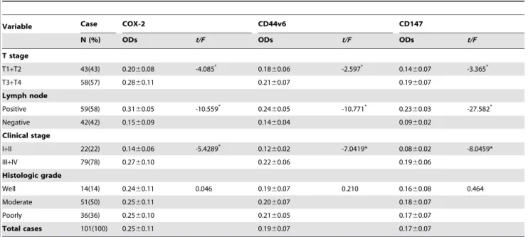
Table 2.Correlation of clinicopathologic characteristics of the patients with immunohistochemistry results.
Variable Case COX-2 CD44v6 CD147
N (%) ODs t/F ODs t/F ODs t/F
T stage
T1+T2 43(43) 0.2060.08 -4.085* 0.18
60.06 -2.597* 0.14
60.07 -3.365*
T3+T4 58(57) 0.2860.11 0.2160.07 0.1960.07
Lymph node
Positive 59(58) 0.3160.05 -10.559* 0.24
60.05 -10.771* 0.23
60.03 -27.582*
Negative 42(42) 0.1560.09 0.1460.04 0.0960.02
Clinical stage
I+II 22(22) 0.1460.06 -5.4289* 0.12
60.02 -7.0419* 0.0860.02 -8.0459*
III+IV 79(78) 0.2760.10 0.2260.06 0.1960.06
Histologic grade
Well 14(14) 0.2460.11 0.046 0.1960.07 0.210 0.1660.08 0.464
Moderate 51(50) 0.2560.11 0.2060.07 0.1860.07
Poorly 36(36) 0.2560.10 0.2160.05 0.1760.07
Total cases 101(100) 0.2560.11 0.1960.07 0.1760.07
*P,0.001
doi:10.1371/journal.pone.0071048.t002
Table 3.The three bio-markers expression in carcinoma and adjacent epithelium.
Variable Case COX-2 CD44v6 CD147
N (%) ODs t ODs t ODs t
Carcinoma tissues 101(72) 0.2560.11 9.004* 0.19
60.07 -0.429 0.1760.07 -9.450*
pithelium tissues adjacent to carcinoma 40(28) 0.0860.06 0.2060.04 0.0560.06
*P,0.001
doi:10.1371/journal.pone.0071048.t003
squamous cell carcinoma [27]. This study has demonstrated that the level of CD147 expression in hypopharyngeal carcinoma was significantly higher than that in epithelium adjacent to carcinoma. It also found that CD147 expression was significantly correlated with T classification, clinical stage and lymph node status. These findings are the same as in previous reports [16,28].
Lymph node metastasis is one of the important factors for outcome of hypopharyngeal cancer patients. There are several pathways in process of lymphatic metastasis of tumor cells, such as tumor lymphangiogenesis, migration, adhesion and proliferarion. Previous study revealed that COX-2 overexpression stimulated VEGF-C, a biomarker considered to be responsible for tumor lymphangiogenesis, up-regulated and induced the growth of new lymphatic vessels, which might be the first step for spreading of tumor cells to the lymph nodes [9]. It has also been reported that CD44v6 was associated with tumor growth and lymph node metastasis. But the relationship between CD44v6 and tumor invasion remains a controversial issue in fact. Sikorska et al [29]
reported that they found CD44v6 had no impact on tumor progression and metastasis. However, the mechanism of CD44v6 in the process of tumor metastasis has remained obscure, and CD44v6 could help tumor cells to escape identification and killing from immune system to promote lymph node metastasis [30]. CD147 can stimulate tumor cells in synthesis of MMPs and mediate the degradation of the extracellular matrix, playing an important role in tumor invasion and metastasis. CD147 can also stimulate VEGF expression to promote tumor angiogenesis by up-regulating the urokinase-type plasminogen activator system. As well as, it mediates a series of tumor promoting molecular events to facilitate tumor invasion and metastasis [31].
Our findings demonstrate a significant correlation of COX-2 and CD147 with survival, which suggests that COX-2 and CD147 are potential bio-markers for prognosis in head and neck squamous cell carcinoma. Lymph node status, T classification or clinical stage did not show any significant association with survival time, although there is a trend towards correlation of the presence
Figure 2. The three bio-markers expression in tumor tissues with and without lymph node metastasis. (P) High levels of COX-2 expression in tumor tissue with lymph node metastasis. (Q) Low levels of COX-2 expression in tumor tissue without lymph node metastasis. (R) High levels of CD44v6 expression in tumor tissue with lymph node metastasis. (S) Low levels of CD44v6 expression in tumor tissue without lymph node metastasis. (T) High levels of CD147 expression in tumor tissue with lymph node metastasis. (U) Low levels of CD147 expression in tumor tissue without lymph node metastasis.
of lymph nodes at the time of diagnosis with worse survival from the clinical standpoint.
In our study, we come to the conclusion that survival time has significant correlation with$P75level of COX-2 expression and $P90level of CD147 expression by measuring the optical density
of positive areas. In addition, we select effective percentile values to reflect the correlation between the bio-markers expression and prognosis which provides us with a novel way to predict the progression and prognosis of the tumor.
Figure 3. The three bio-markers expression in well, moderate and pooly differentiated tumor tissues.(G) COX-2 expression in well differentiated tumor tissues. (H) COX-2 expression in moderate differentiated tumor tissues. (I) COX-2 expression in poorly differentiated tumor tissues. (J) CD44v6 expression in well differentiated tumor tissues. (K) CD44v6 expression in moderate differentiated tumor tissues. (L) CD44v6 expression in poorly differentiated tumor tissues. (M) CD147 expression in well differentiated tumor tissues. (N) CD147 expression in moderate differentiated tumor tissues. (O) CD147 expression in poorly differentiated tumor tissues.
doi:10.1371/journal.pone.0071048.g003
Table 4.Kaplan-Meier survival analysis by level of the three bio-markers expression.
Variable $P50 $P75 $P85 $P90
n1 n (%) x2 n1 n (%) x2 n1 n (%) x2 n1 n (%) x2
COX-2 39 26(66.7) 6.104* 21 17(80.9) 6.870* 12 11(91.7) 8.028* 7 6(85.7) 4.725*
CD44v6 41 24(58.8) 0.752 21 12(57.1) 0.068 12 8(66.7) 0.433 7 5(71.4) 0.313
CD147 40 25(62.5) 1.629 17 11(64.7) 0.059 12 9(75.0) 0.520 8 7(87.5) 7.059*
*log rankP,0.05
n1is the number of total patients of relative percentile;
n (%) is the number of dead patients of relative percentile ( n/n1*100%) doi:10.1371/journal.pone.0071048.t004
There were some limitations in our study. Firstly, further study should focus on the relationship of the three bio-marker pathways with tumor growth, metastasis and survival, which could help to find better targets for therapeutic measures. Secondly, all the
patients were Chinese Han population, and only 2 patients were females which may be poorly representative of the whole population. Studies on multi-ethnic populations may clarify the
Figure 4. Kaplan-Meier curve for the overall survival of the patients with hypopharyngeal carcinoma, according to the expression of P50of COX-2.
doi:10.1371/journal.pone.0071048.g004
Figure 5. Kaplan-Meier curve for the overall survival of the patients with hypopharyngeal carcinoma, according to the expression of P90of CD147.
function of the three bio-markers in hypopharyngeal squamous cell carcinoma.
Acknowledgments
The authors thank Prof. Hongbo Zhang for her statistical advice and all others for their contributions.
Author Contributions
Conceived and designed the experiments: Y. Liu MD. Performed the experiments: QY YH DH. Analyzed the data: QY YH Y. Li JW. Wrote the paper: QY.
References
1. Hall SF, Groome PA, Irish J, O’Sullivan B (2008) The natural history of patients with squamous cell carcinoma of the hypopharynx. Laryngoscope118(8):1362– 1371.
2. Berrino F, Gatta G (1998) Variation in survival of patients with head and neck cancer in Europe by the site of origin of the tumours. EUROCARE Working Group. Eur J Cancer34(14):2154–2161.
3. Grandis JR, Pietenpol JA, Greenberger JS, Pelroy RA, Mohla S (2004) Head and neck cancer: meeting summary and research opportunities. Cancer Res 64(21):8126–8129.
4. Prescott SM, Fitzpatrick FA (2000) Cyclooxygenase-2 and carcinogenesis. Biochim Biophys Acta1470(2):69–78.
5. Eberhart CE, Coffey RJ, Radhika A, Giardiello FM, Ferrenbach S, et al. (1994) Up-regulation of cyclooxygenase 2 gene expression in human colorectal adenomas and adenocarcinomas. Gastroenterology 107(4):1183–1188. 6. Visscher DW, Pankratz VS, Santisteban M, Reynolds C, Ristimaki A, et al.
(2008) Association between cyclooxygenase-2 expression in atypical hyperplasia and risk of breast cancer. J Natl Cancer Inst 100(6):421–427.
7. Bin W, He W, Feng Z, Xiangdong L, Yong C, et al. (2011) Prognostic relevance of cyclooxygenase-2 (COX-2) expression in Chinese patients with prostate cancer. Acta Histochem 113(2):131–136.
8. Li F, Liu Y, Chen H, Liao D, Shen Y, et al. (2011) EGFR and COX-2 protein expression in non-small cell lung cancer and the correlation with clinical features. J Exp Clin Cancer Res30:27.
9. Kyzas PA, Stefanou D, Agnantis NJ (2005) COX-2 expression correlates with VEGF-C and lymph node metastases in patients with head and neck squamous cell carcinoma. Mod Pathol18(1):153–160.
10. Scheer M, Drebber U, Breuhahn K, Mockel C, Reuther T, et al. (2010) Expression of cyclooxygenase-2 (COX-2) in an advanced metastasized hypopharyngeal carcinoma and cultured tumor cells. Oral Maxillofac Surg 14(1):53–57.
11. Yu P, Zhou L, Ke W, Li K (2010) Clinical significance of pAKT and CD44v6 overexpression with breast cancer. J Cancer Res Clin Oncol 136(8):1283–1292. 12. Zlobec I, Gunthert U, Tornillo L, Lezzi G, Baumhoer D, et al. (2009) Systematic assessment of the prognostic impact of membranous CD44v6 protein expression in colorectal cancer. Histopathology 55(5):564–575.
13. Okayama H, Kumamoto K, Saitou K, Hayase S, Kofunato Y, et al. (2009) CD44v6, MMP-7 and nuclear Cdx2 are significant biomarkers for prediction of lymph node metastasis in primary gastric cancer. Oncol Rep 22(4):745–755. 14. Zucker S, Hymowitz M, Rollo EE, Mann R, Conner CE, et al. (2001)
Tumorigenic potential of extracellular matrix metalloproteinase inducer. Am J Pathol 158(6):1921–1928.
15. Rosenthal EL, Shreenivas S, Peters GE, Grizzle WE, Desmond R, et al. (2003) Expression of extracellular matrix metalloprotease inducer in laryngeal squamous cell carcinoma. Laryngoscope 113(8):1406–1410.
16. Huang Z, Huang H, Li H, Chen W, Pan C (2009) EMMPRIN expression in tongue squamous cell carcinoma. J Oral Pathol Med 38(6):518–523. 17. Lee DW, Sung MW, Park SW, Seong WJ, Roh JL, et al. (2002) Increased
cyclooxygenase-2 expression in human squamous cell carcinomas of the head
and neck and inhibition of proliferation by nonsteroidal anti-inflammatory drugs. Anticancer Res22(4):2089–2096.
18. Gallo O, Masini E, Bianchi B, Bruschini L, Paglierani M, et al. (2002) Prognostic significance of cyclooxygenase-2 pathway and angiogenesis in head and neck squamous cell carcinoma. Hum Pathol 33(7):708–714.
19. Dohadwala M, Luo J, Zhu L, Lin Y, Dougherty GJ, et al. (2001) Non-small cell lung cancer cyclooxygenase-2-dependent invasion is mediated by CD44. J Biol Chem 276(24):20809–20812.
20. Heider KH, Sproll M, Susani S, Patzelt E, Beaumier P, et al. (1996) Characterization of a high-affinity monoclonal antibody specific for CD44v6 as candidate for immunotherapy of squamous cell carcinomas. Cancer Immunol Immunother 43(4):245–253.
21. Nozoe T, Kohnoe S, Ezaki T, Kabashima A, Maehara Y (2004) Significance of immunohistochemical over-expression of CD44v6 as an indicator of malignant potential in esophageal squamous cell carcinoma. J Cancer Res Clin Oncol130(6):334–338.
22. Kawano T, Nakamura Y, Yanoma S, Kubota A, Furukawa M, et al (2004) Expression of E-cadherin, and CD44s and CD44v6 and its association with prognosis in head and neck cancer. Auris Nasus Larynx 31(1):35–41. 23. Kunishi M, Kayada Y, Yoshiga K (1997) Down-regulated expression of CD44
variant 6 in oral squamous cell carcinomas and its relationship to regional lymph node metastasis. Int J Oral Maxillofac Surg 26(4):280–283.
24. Rodrigo JP, Dominguez F, Alvarez C, Gonzalez MV, Herrero A, et al. (2002) Clinicopathologic significance of expression of CD44s and CD44v6 isoforms in squamous cell carcinoma of the supraglottic larynx. Am J Clin Pathol 118(1):67– 72.
25. Guler G, Sarac S, Uner A, Karabulut E, Ayhan A, et al. (2002) Prognostic value of CD44 variant 6 in laryngeal epidermoid carcinomas. Arch Otolaryngol Head Neck Surg 128(4):393–397.
26. Mack B, Gires O (2008) CD44s and CD44v6 expression in head and neck epithelia. PloS One 3(10):e3360.
27. Erdem NF, Carlson ER, Gerard DA, Ichiki AT (2007) Characterization of 3 oral squamous cell carcinoma cell lines with different invasion and/or metastatic potentials. J Oral Maxillofac Surg 65(9):1725–1733.
28. Huang C, Sun Z, Sun Y, Chen X, Zhu X, et al (2012) Association of increased ligand cyclophilin A and receptor CD147 with hypoxia, angiogenesis, metastasis and prognosis of tongue squamous cell carcinoma. Histopathology 60(5):793– 803.
29. Sikorska B, Danilewicz M, Wagrowska-Danilewicz M (2002) Prognostic significance of CD44v6 and nm23 protein immunoexpression in laryngeal squamous cell carcinoma. Pol J Pathol 53(1):17–24.
30. Kaufmann M, Heider KH, Sinn HP, von Minckwitz G, Ponta H, et al(1995)CD44 variant ex on epitopes in primary breast cancer and length of survival. Lancet 345( 8950) : 615.
31. Quemener C, Gabison EE, Naımi B, Lescaille G, Bougatef F, et al (2007) Extracellular matrix metalloproteinase inducer up-regulates the uroki-nase-type plasminogen activator system promoting tumor cell invasion. Cancer Res 67: 9– 15.
Table 5.Cox proportional hazards regression models in estimating cancer progression.
Variables Relative risk (95%confidence interval) P
Univariate analysis
Recurrence(no/yes) 3.372(1.784–6.376) 0.000
$P50level of COX-2 expression 2.143(1.147–4.003) 0.017
$P90level of CD147 expression 2.913(1.261–6.729) 0.012
Multivariate analysis
Recurrence(no/yes) 3.508(1.850–6.651) 0.000
$P50level of COX-2 expression 2.158(1.146–4.062) 0.017
*P,0.001.
doi:10.1371/journal.pone.0071048.t005