Nanostructured Grids
Maria Wolun-Cholewa1*, Krzysztof Langer2
, Krzysztof Szymanowski3, Aleksandra Glodek1, Anna Jankowska1, Wojciech Warchol4, Jerzy Langer2
1Department of Cell Biology, Poznan University of Medical Science, Poznan, Poland,2Laboratory for Materials Physicochemistry and Nanotechnology, Adam Mickiewicz University, Srem, Poland,3Department of Mother’s and Child’s Health, Poznan University of Medical Science, Poznan, Poland,4Department of Biophysics, Poznan University of Medical Science, Poznan, Poland
Abstract
Current techniques ofin vitrocell cultures are able to mimic thein vivoenvironment only to a limited extent, as they enable
cells to grow only in two dimensions. Therefore cell culture approaches should rely on scaffolds that provide support comparable to the extracellular matrix. Here we demonstrate the advantages of novel nanostructured three-dimensional grids fabricated using electro-spinning technique, as scaffolds for cultures of neoplastic cells. The results of the study show that the fibers allow for a dynamic growth of HeLa cells, which form multi-layer structures of symmetrical and spherical character. This indicates that the applied scaffolds are nontoxic and allow proper flow of oxygen, nutrients, and growth factors. In addition, grids have been proven to be useful inin situexamination of cells ultrastructure.
Citation:Wolun-Cholewa M, Langer K, Szymanowski K, Glodek A, Jankowska A, et al. (2013) An Efficient 3D Cell Culture Method on Biomimetic Nanostructured Grids. PLoS ONE 8(9): e72936. doi:10.1371/journal.pone.0072936
Editor:Bing Xu, Brandeis University, United States of America
ReceivedMay 29, 2013;AcceptedJuly 15, 2013;PublishedSeptember 2, 2013
Copyright:ß2013 Wolun-Cholewa et al. This is an open-access article distributed under the terms of the Creative Commons Attribution License, which permits unrestricted use, distribution, and reproduction in any medium, provided the original author and source are credited.
Funding:The study was supported in part by the polish National Committee for Scientific Research, grant no. NN 407 084 836. No additional external funding was received for this study. The funders had no role in study design, data collection and analysis, decision to publish, or preparation of the manuscript.
Competing Interests:The authors have declared that no competing interests exist.
* E-mail: [email protected]
Introduction
Cultures of human or animal cells in in vitro conditions are usually performed for their identification, proliferation or cell death assessment. Frequently, initial evaluations are carried out in two-dimensional (2D) cell culture systems. Despite their wide-spread use, observations made in 2D systems do not correspond to the results of in vivostudies [1]. Thus, currently, researchers are using three dimensional (3D) cultures that mimic the in vivo environment more accurately [2]. Cells grown in three-dimen-sional cultures are more valid targets for discovering and testing of new drugs for cancer treatment [3]. Contrary to 2D culture systems, 3Din vitromodels have the potential to provide insights into cellular functions such as: differentiation, migration, and gene expression in a controlled and well-defined manner [4–6]. 3D cell culture main feature is the ability to mimic the extracellular matrix (ECM) conditions. Materials used for 3D cell culture systems production include both natural and synthetic biopolymers. Lately also biodegradable materials were introduced in scaffold construc-tion, however it was proven that their stability in liquid environment is limited [5,7–14].
One of the methods for fiber-based 3D scaffolds production is electro-spinning [9]. An advantage of those nanostructured grids, which differentiates them from other types of scaffolds, is the reduced diameter of pore size [15–16]. Moreover, electrospun scaffolds are built from very small fibers which create large surface areas [1,9,13,15]. This allows for more accurate evaluation of cell proliferation [9,11,17].
In this study, electrospun nanostructured fibers in the form of spatial nanostructured 3D grids, fabricated from a polymer
mixture, including polyaniline, were examined as a potential tool for 3D culturein vitro.
Materials and Methods
Synthesis of Polyaniline
6.9 g of aniline hydrochloride was dissolved in 300 ml of 1 M HCl. The solution of aniline hydrochloride was combined with a solution of 11.4 g ammonium persulphate dissolved in 200 ml of 1 M HCl. The mixture was left at room temperature for 12 hours with continuous mixing. Next the product was filtered, washed with water, methanol and chloroform. Polyaniline (PANI) was transferred into 200 ml of chloroform and dispersed to a suspension using ultrasound bath.
Fabrication of Nanofibers using Electro-spinning
All reagents used for the synthesis of polyaniline and fabrication of nanofibers using electro-spinning were purchased from Sigma-Aldrich, St Louis, MO, USA.
Cell Culture
Human HeLa cervical epithelial cells (ATCC CCL-2) were purchased from the American Type Cell Culture (ATCC, Manassas, MA). HeLa cells were cultured in standard conditions: RPMI medium (PAN Biotech-Gmbh, Aidenbach, Germany) supplemented with 10% fetal calf serum (FCS; Sigma, St Louis, MO, USA), 2 mmol/L L-glutamine (Cambrex, Charles City, IA, USA), 100 IU/mL penicillin and streptomycin solution (Sigma-Aldrich, St Louis, MO, USA) in a 5% CO2-humidified atmosphere at 37uC.
In order to examine the usefulness of nanostructural grids as a tool for 3D cultures, the cells were seeded on grids at a density of 10 000 cells. Subsequently, the grids with cells were placed into fresh culture medium and after 24, 48 or 72 hours cells morphology and their viability were examined. Every experiment was repeated ten times.
Cell Viability Assessment
The viability of HeLa cells was analyzed using the XTT colorimetric test, based on the reduction of XTT compound (tetrazoline-2,3-bis(2-methoxy-4-nitro-5-sulphophenyl)-2H-5-car-boxyanilide, Sigma-Aldrich, St Louis, MO, USA) by living cells. Optical density of colour product was measured at the wavelength of 450 nm.
The percentage of mitochondrial activity was calculated according to the following equation: (OD of nanostructured grid with cells2OD of medium alone)/(OD of nanostructured grid only2OD medium alone)6100; where OD is optical density [18].
Statistical analysis of the results was done with the Kruskal-Wallis test with Dunn’s post-test using STATISTICA ver.5 software (Statsoft, Krakow, Poland).Pvalue less than 0.05 was considered statistically significant.
Microscopy
Fluorescence microscopy. After 24, 48 or 72 hours of culture on the grids HeLa cells were stained with 0.1mg/ml of Hoechst 33342 and 0.125mg/ml of propidium iodide (Sigma-Aldrich, St Louis, MO, USA). The presence of intact, apoptotic and/or necrotic cells was evaluated using Nicon Diaphot Eclipse TE 200 fluorescence microscope, equipped with UV-2A and FITC/FLUO-3 filters [19].
Confocal microscopy. In order to visualize the 3D construct the cells were pre-fixed for 10 minutes in 4% paraformaldehyde (Sigma-Aldrich, St Louis, MO, USA) and stained with 0.125mg/ ml of propidium iodide (Sigma-Aldrich, St Louis, MO, USA). Signals were excited at 543 nm wavelength and fluorescence emission was selected with 560 nm bandpass filter. Images of cells were represented as orthogonal projections of 20–60 optical sections in 0.3mm increments using Zeiss LSM 510 confocal microscope.
Scanning electron microscopy. After 48 hours of culture on the grids the cells were fixed in a standard way using 2.5% glutaraldehyde (at room temperature for 60 min and, then at 4uC for 24 hours). The grids with cells were washed with PBS for 30 min and the cells were gradually dehydrated in alcohols. The grids with cells were finally coated with palladium and visualized with a scanning electron microscope (Philips SEM 515).
Transmission electron microscopy. After 48 hours the nanostructured grids with the cells were put into a fixative solution containing 4% glutaraldehyde (Taab, Berkshire, UK),
buffered with 0.1 M phosphate buffer, pH 7.3 (Merck). After overnight incubation the nanostructured grids were rinsed for 60 min in 0.1 M phosphate buffer, pH 7.3, and then postfixed in 2% OsO4 in 0.1 M phosphate buffer for 2 hours (Merck). The specimens were dehydrated in ethanol, block-stained with alcoholic uranyl acetate and embedded in Spurr’s medium (Merck). After contrasting, the ultrastructure of HeLa cells was examined using JEOL 100 Transmission Electron Microscope.
Results
Morphology of Polyaniline Fibers
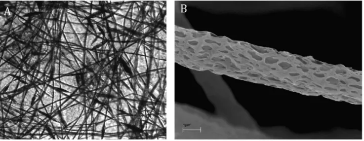
Light microscopy confirmed that the analyzed grids are transparent (Fig. 1A). Moreover, scanning electron microscopy showed that the thickness of electrospun fibers was 2000– 2500 nm, and the diameter of pores was 50–300 nm (shorter dimension). The average thickness of a grid is less than 0.5 mm (Fig. 1B).
Cell Viability
To determine whether the tested electrospun scaffolds can influence the viability of uterine cervix carcinoma cells cultured in 3D, their viability was examined using the XTT assay. Due to XTT-reducing properties of the nanostructured grids alone, the viability of cells grown on the grids were related to the results obtained for measurements of the grids alone.
As shown in Fig. 2 the grids have no cytotoxic effect on HeLa cells. After 48 hours of culture on the nanostructured grids cell viability increased compared to the evaluation made after 24 hours. However, the change was insignificant (p.0.05). Subse-quent incubation, up to 72 hours, resulted in a significant increase of cell viability, compared to the viability noted after either 24 hour (p,0.001) or 48 hour of culture (p,0.05) (Fig. 2).
Cell Morphology
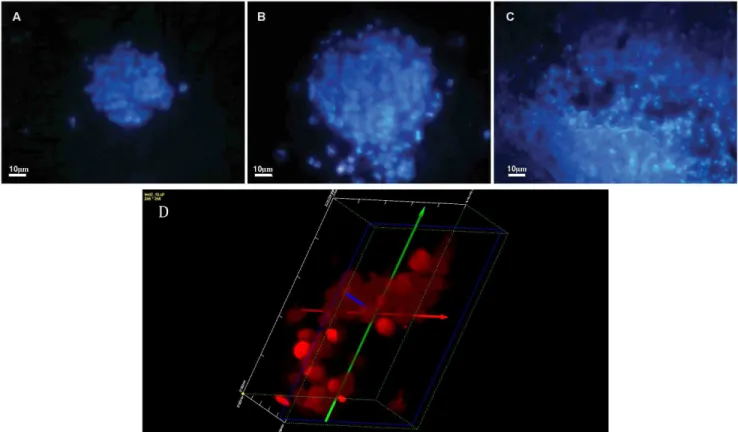
The double staining using Hoechst 33342 and propidium iodide allows distinguishing living and necrotic cells as propidium iodide penetrates only cells with a disrupted cell membrane, while Hoechst 33342 penetrates also intact cell membranes. As it was shown in Fig. 3 the nuclei of cells cultured on the nanostructured grids 24 (Fig. 3A), 48 (Fig. 3B) and 72 (Fig. 3C) hours emitted bright blue fluorescence. The staining proved that in the analyzed time intervals cells showed high viability with very low level of necrosis. Moreover, confocal microscopy confirmed that the cells cultured on the grids formed a homogenous 3D constructs (Fig. 3D).
The results of the light microscopy and scanning electron microscopy observation demonstrated that after 48 hours of culture on the nanostructured grids HeLa cells formed symmet-rical and sphesymmet-rical aggregates. The size of cells ranged between 6 and 7 micrometers (Fig. 4).
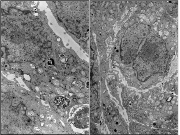
Detailed analysis of the cells grown for 48 hours on the nanostructured grids showed that there were no changes in the ultrastructure of HeLa cells. The cells were oval or elongated in shape with abundant microvilli and mostly euchromatic nuclei. The cytoplasm showed numerous intact organelles. Cisterns of the Golgi apparatus frequently contained electron-dense material in the form of laminae. The cells possessed numerous mitochondria with partially obliterated inner structure, sometimes presenting cristae. Some cells contained phagolysosomes with electron-dense content (Fig. 5).
Discussion
Three-dimensional culture scaffolds assure a clearly better growth and proliferation of cells as they mimic the in vivo conditions. Moreover the cells cultured on 3D scaffolds have been found to develop a self-specific microenvironment [2,13,24]. Culture scaffolds of such properties can be used, for instance in regenerative medicine [2,7,9,12,22–23]. However, not all three-dimensional scaffolds are adequate for cell culture, as the building material of a scaffold must fulfill certain conditions [7,10,21,25]. In
particular it should allow for attachment of cells and their undisturbed growth and proliferation.
One of the most common among technologies used in production of nano- and microfibres from polymer solutions is electro-spinning. Studies confirm that this technique can be applied to obtain spatial scaffolds for cell cultures in in vitro conditions [20–21].
The electrospun three-dimensional nanostructured grids pre-sented in this study were not only proven to be biocompatible but also suitable for cell culture. One of the advantages of these Figure 1. Light microscopy image of nanostructured grid. Original magnification x200 (A). Scanning electron microscopy image of nanostructured microfiber. Original magnification x500 (B).
doi:10.1371/journal.pone.0072936.g001
Figure 2. The viability of HeLa cells cultured on the nanostructured grid. HeLa cells were incubated for 24, 48 72 hours on the nanostructured grids and the percentage of mitochondrial activity was calculated according to the following formula: (OD of nanostructured grid with cells2OD of medium alone)/(OD of nanostructured grid only2OD medium alone)6100; where OD is optical density.
nanostructured grids is in their architecture in the form of nano-and microtubes. What is more in the process of electro-spinning these hollow fibers are being arranged randomly, forming an irregular network with micrometric distance between the tubes preserved. The electrospun capillaries possess nanopores which probably enhance the free flow of the culture medium. These features have favorable effects on cell attachment and their
viability. It has also been shown that these properties may promote migration of the proliferating cells along the fibers or between the overlaying fiber layers [1].
XTT test results have demonstrated that the described scaffold is suitable for cell culture in 3D conditions. The intense and significant increase in cell mitochondrial activity was observed particularly 48 hours after seeding of cells on the grid. The Figure 3. HeLa cells stained with Hoechst 33342 and propidium iodide after 24 (A), 48 (B) and 72 (C) hours of 3D culture on the nanostructure grids.Original magnification x200. A fragment of the spherical 3D structure of cell growing on nanostructured grids visualized after 48 hours. Original magnification x400 (D).
doi:10.1371/journal.pone.0072936.g003
Figure 4. Cells growing on the scaffold visualized using light microscopy after 48 hours of incubation.Original magnification x200 (A). Scanning electron microscopy image of HeLa cells on the nanostructured grid fibers after 48 hours of incubation. Original magnification x100 (B). doi:10.1371/journal.pone.0072936.g004
observation has been additionally supported by results from fluorescent microscopy with the use of Hoechst 33342 and propidium iodide. It was demonstrated that within 24 hours of seeding, the cells attached to nanostructured grid fibers and formed spatial agglomerates, which in the course of culture formed multilayered structures.
It can be assumed that the process of cell seeding was accompanied by the selection of particular types of cells, which were capable of proliferation on the fibers and thereby accept the new supporting material. The problem which remains to be resolved involves our inability to calculate the precise number of cells deposited on the nanostructured grid in the first day of the culture. For this reason, the obtained values of mitochondrial activity have been related to results obtained for an empty nanostructured grid and then to each other.
The analysis of results obtained using a scanning electron microscope has shown that the cells deposited on nanostructured grids form protrusions, which may either anchor the cells at the site of deposition or allow their migration along the fibers. It may be expected that the anchored cells may form a barrier preventing further amoeba-like movements of the adjacent cells. This would explain the reason of the inward growth of cells and formation of the spherical spatial structures. The observed spherical shapes of cell agglomerates formed following 24 hours after cell deposition
allow for local development of high density cultures on a limited area. Thus, the presented scaffold represents not only an excellent support for growing cells in 3D conditions but may also stabilize the environment of proliferating cells thanks to the specific microenvironment developed. Nevertheless, it should be noted that the above hypothesis requires verification using studies on interaction of cells with individual fibers.
The presence of pores in the described fibers not only promotes cell growth but also makes the grid transparent. The potential for cell observation and their growth monitoring using light micro-scope is an important advantage of the suggested method of 3D cultures. Such a feature eliminates the need for staining of cells with fluorescent markers or for their transfection using e.g. GFP. Moreover, the nanostructured grid-deposited cells may be used in studies on ultrastructure of cells inin situconditions and thus the simple technique of preparing cultures for Spurr embedding may be here applied.
Conclusions
A new solution for 3D cultures has been suggested, which is suitable for cell culture in conditions mimicking in vivo environ-ment. The relatively high surface to volume ratio permits to load such a nanostructured grid with high number of cells and to grow them in conditions of high density or even overcrowding. Figure 5. Transmission electron microscopy images of HeLa cells on the nanostructure grid fibers after 48 h culture. Original magnification x6000.
Moreover, the suggested novel 3D support in many ways surpasses other similar culture approaches: the scaffolds are transparent and thus allow observation of viable cells in time; standard fluorescence or colorimetric methods such can be used for evaluation of cells. Finally, the cell nanostructured grid can be embedded in in situ conditions for ultrastructural studies.
Author Contributions
Conceived and designed the experiments: MWC KL KS AG AJ WW JL. Performed the experiments: MWC KL AG. Analyzed the data: MWC KL WW. Contributed reagents/materials/analysis tools: MWC. Wrote the paper: MWC AJ JL.
References
1. Yamada KM, Cukierman E (2007) Modeling tissue morphogenesis and cancer in 3D. Cell 130: 601–10. 10.1016/j.cell.2007.08.006.
2. Ma PX (2008) Biomimetic materials for tissue engineering. Adv Drug Deliver Rev 60: 184–98. 10.1016/j.addr.2007.08.041.
3. Fischbach C, Chen R, Matsumoto T, Schmelzle T, Brugge JS, et al. (2007) Engineering tumors with 3D scaffolds. Nat Methods 4: 855–60. 10.1038/ nmeth1085.
4. Jang JH, Castano O, Kim HW (2009) Electrospun materials as potential platforms for bone tissue engineering. Adv Drug Deliver Rev 61: 1065–83. 10.1016/j.addr.2009.07.008.
5. Zhang S (2008) Designer self-assembling peptide nanofiber scaffolds for study of 3-D cell biology and beyond. Adv Cancer Res 99: 335–62. 10.1016/S0065-230X(07)99005-3.
6. Kraehenbuehl TP, Langer R, Ferreira LS (2011) Three-dimensional biomate-rials for the study of human pluripotent stem cells. Nat Methods 8: 731–6. 10.1038/nmeth.1671.
7. Shin H, Jo S, Mikos AG (2003) Biomimetic materials for tissue engineering. Biomaterials 24: 4353–4364. 10.1016/S0142-9612(03)00339-9.
8. Kim JH, Park CH, Lee OJ, Lee JM, Kim JW, et al. (2012) Preparation and in vivo degradation of controlled biodegradability of electrospun silk fibroin nanofiber mats. J Biomed Mater Res A 100: 3287–95. 10.1002/jbm.a.34274. 9. Jeong SI, Krebs MD, Bonino CA, Khan SA, Alsberg E (2010) Electrospun
alginate nanofibers with controlled cell adhesion for tissue engineering. Macromol Biosci 10(8): 934–43. 10.1002/mabi.201000046.
10. Jo JH, Lee EJ, Shin DS, Kim HE, Kim HW, et al. (2009) In vitro/in vivo biocompatibility and mechanical properties of bioactive glass nanofiber and poly(epsilon-caprolactone) composite materials. J Biomed Mater Res B Appl Biomater 91(1): 213–20. 10.1002/jbm.b.31392.
11. Lee JJ, Yu HS, Hong SJ, Jeong I, Jang JH, et al. (2009) Nanofibrous membrane of collagen-polycaprolactone for cell growth and tissue regeneration. J Mater Sci Mater Med 20: 1927–35. 10.1007/s10856-009-3743-z.
12. Cai YZ, Zhang GR, Wang LL, Jiang YZ, Ouyang HW,et al. (2012) Novel biodegradable three-dimensional macroporous scaffold using aligned electro-spun nanofibrous yarns for bone tissue engineering. J Biomed Mater Res A 100: 1187–94. 10.1002/jbm.a.34063.
13. Davidenko N, Campbell JJ, Thian ES, Watson CJ, Cameron RE (2010) Collagen-hyaluronic acid scaffolds for adipose tissue engineering. Acta Biomater 6: 3957–68. 10.1016/j.actbio.2010.05.005.
14. Ifkovits JL, Burdick JA (2007) Review: photopolymerizable and degradable biomaterials for tissue engineering applications. Tissue Eng 13: 2369–85. 10.1089/ten.2007.0093.
15. Rnjak-Kovacina J, Weiss AS (2011) Increasing the pore size of electrospun scaffolds. Tissue Eng Part B Rev 17: 365–72. 10.1089/ten.teb.2011.0235. 16. Nam J, Huang Y, Agarwal S, Lannutti J (2007) Improved cellular infiltration in
electrospun fiber via engineered porosity. Tissue Eng 2007;13: 2249-57. 10.1089/ten.2006.0306.
17. Kim TG, Park TG (2006): Biomimicking extracellular matrix: cell adhesive RGD peptide modified electrospun poly(D,L-lactic-co-glycolic acid) nanofiber mesh. Tissue Eng 12: 221–33. doi:10.1089/ten.2006.12.221.
18. Wołun´-Cholewa M, Szymanowski K, Nowak-Markwitz E, Warchoł W (2010) Photodiagnosis and photodynamic therapy of endometriotic epithelial cells using 5-aminolevulinic acid and steroids. Photodiagnosis Photodyn Ther 8: 58–63. 10.1016/j.pdpdt.2010.12.003.
19. Fik E, Wołun´-Cholewa M, Kistowska M, Warchoł JB, Goz´dzicka-Jo´zefiak A (2001) Effect of lectin from Chelidonium majus L. on normal and cancer cells in culture. Folia Histochem Cytobiol 39: 215–216.
20. Ahmed I, Ponery AS, Nur-E-Kamal A, Kamal J, Meshel AS, et al. (2007) Morphology, cytoskeletal organization, and myosin dynamics of mouse embryonic fibroblasts cultured on nanofibrillar surfaces. Mol Cel Biochem 301: 241–9. 10.1007/s11010-007-9417-6.
21. Rodrigues MT, Martins A, Dias IR, Viegas CA, Neves NM, et al. (2012) Synergistic effect of scaffold composition and dynamic culturing environment in multilayered systems for bone tissue engineering. J Tissue Eng Regen Med 6: e24–30. 10.1002/term.499.
22. Garg K, Bowlin GL (2011) Electrospinning jets and nanofibrous structures. Biomicrofluidics 5: 13403. 10.1063/1.3567097.
23. Sill TJ, Von Recum HA (2008) Electrospinning: applications in drug delivery and tissue engineering. Biomaterials 29: 1989–2006. 10.1016/j.biomateri-als.2008.01.011.
24. Bhattarai SR, Bhattarai N, Yi HK, Hwang PH, Cha DI, et al. (2004) Novel biodegradable electrospun membrane: scaffold for tissue engineering. Biomate-rials 25: 2595–2602. 10.1016/j.biomateBiomate-rials.2003.09.043.
25. Tuzlakoglu K, Bolgen N, Salgado AJ, Gomes ME, Piskin E, et al (2005) Nano-and micro-fiber combined scaffolds: a new architecture for bone tissue engineering. J Mater Sci Mater Med 16: 1099–104. 10.1007/s10856-005-4713-8.