Vol.57, n.5: pp. 636-643, September-October 2014 http://dx.doi.org/10.1590/S1516-8913201402165
ISSN 1516-8913 Printed in Brazil
BRAZILIAN ARCHIVES OF BIOLOGY AND TECHNOLOGY
A N I N T E R N A T I O N A L J O U R N A L
Influence of Calcium Content of Tissue on Hyperhydricity
and Shoot-Tip Necrosis of
in vitro
Regenerated Shoots of
Lavandula angustifolia
Mill.
Marília Pereira Machado
1*, André Luís Lopes da Silva
2, Luiz Antonio Biasi
1, Cícero
Deschamps
1, João Carlos Bespalhok Filho
1and Flávio Zanette
11Departamento de Fitotecnia e Fitossanitarismo; Setor de Ciências Agrárias; Universidade Federal do Paraná;
Curitiba - PR - Brasil. 2Departamento de Engenharia de Bioprocessos e Biotecnologia; Universidade Federal do Paraná; Curitiba - PR - Brasil
ABSTRACT
In the present study, the effects of two CaCl2.2H2O levels (440 and 1320 mg L-1) and two subcultures were evaluated
on in vitro shoots of Lavandula angustifolia cv. Provence Blue. Ca2+ content of the apical, middle and basal portion of shoots was determined. Increasing CaCl2.2H2O level in the culture medium increased tissue Ca2+ content and
decreased hyperhydricity. Shoot-tip necrosis also decreased with 1320 mg L-1 CaCl2.2H2O, but it did not occur in
the second subculture. The middle and basal portion had higher Ca2+ content than apical portion. In non-hyperhydric tissues, there were smaller and more juxtaposed cells. Scanning electron microscopy of the leaves demonstrated that trichomes from in vitro leaf surface occurred in smaller quantities.
Key words: True lavender, Micropropagation, CaCl2.2H2O, Leaf anatomy, Ultrastructure, Trichome
*Author for correspondence:[email protected]
INTRODUCTION
Lavandula angustifolia Mill. (syn. L. officinalis
Chaix; L. vera DC.) is a plant belonging to the Lamiaceae family. Essential oil obtained from the flowers of L. angustifolia is composed mainly of linalyl acetate, linalool, lavandulol, 1,8-cineole, lavandulyl acetate and camphor (Lis-Balchin and Hart 1999). Lavender is one of the most useful medicinal plants and has found wide application in perfumes and cosmetics. L. angustifolia may be propagated sexually or asexually. Reproduction via seeds is usually slow and the plants display a great variation in a number of characteristics such as growth rate and essential oil composition. Therefore, vegetative propagation is the most
effective to produce a large number of uniform individuals displaying a genotype of interest. Tissue culture techniques have been applied to
Lavandula plants (Quazi 1980; Calvo and Segura
1989; Jordan et al. 1990; Gras and Calvo 1996; Andrade et al. 1999; Echeverrigaray et al. 2005; Zuzarte et al. 2010; Machado et al. 2011; Silva et al. 2013). However, the performance and survival
of in vitro cultures of many plant species are often
potential of in vitro techniques for mass propagation. In general, the leaves of hyperhydric shoots are thicker, elongated, wrinkled and/or curled, and can be brittle when compared to normal leaves. Anatomical characteristics change in hyperhydric shoots and several types of abnormal structures can be recognized (Ziv 1991). Several factors can induce the hyperhydricity in explants, such as high BAP levels (Oliveira et al. 2010), cultivar (Carvalho et al. 2013), and agar concentration (Abdoli et al. 2007) but the most common cause of the hyperhydricity occurrence is the culture in liquid medium (Scheidt et al. 2011; Silva et al. 2012). Some species are so sensitive to this physiological change that even when cultivated in solid medium with a low BAP level, hyperhydricity occurs, as it is the case for L. angustifolia. In this context, one of the strategies to overcome this problem is the modification of the composition of the culture medium.
Ca2+ concentration in the culture medium has been associated with the shoot-tip necrosis, which decreases when the calcium content increases in the medium for in vitro culture of Castanea
mollissima, C. dentata and C. sativa (Qiguang et
al. 1985; Piagnani et al. 1996). Calcium is an integral component in the cell wall and plays an important role in maintaining membrane integrity (Marschner 1995; Machado et al. 2010). This study investigated the relationship between tissue Ca2+ content in the reduction of hyperhydricity and shoot-tip necrosis. The leaf anatomy in non-hyperhydric and non-hyperhydric common lavender was investigated to identify the structural alterations related with this phenomenon.
MATERIALS AND METHODS
Plant material and source of explants
Shoot-tips (approximately 5 mm) from L.
angustifolia Mill. cv. Provence Blue were surface
sterilized by immersion in 70% ethanol for 20 seconds, followed by immersion in 1% sodium hypochlorite containing two drops of Tween-20® per 100 mL for 20 min under manual agitation, followed by three rinses in distilled and autoclaved water. Explants were incubated on MS medium (Murashige and Skoog 1962) containing 3% sucrose, 0.6% agar (Vetec®) and the pH was adjusted to 5.8. After five weeks, the explants presented greening shoots. The shoot multiplication was carried out on MS medium
containing 3% sucrose, 0.6% agar (Vetec®) and supplemented with 1.0 µM 6-benzylaminopurine (BAP) at each 40 days (subcultures). The culture flasks (60 x 90 mm and 200 mL volume) were used as culture vessels and were sealed by polypropylene autoclavable lids. Each culture flask contained 20 mL culture medium. Voucher specimens (adult plant) have been deposited at the Herbarium of Federal University of Paraná (UFPR) under code numbers UPCB 61303.
Ca2+ treatments
Shoots (2.0 cm length) from in vitro multiplication (methodology described above) were used as explants. The basal medium was MS containing 3% sucrose and solidified with 0.6% agar (Vetec®). The treatments consisted of two calcium levels: 440 (MS standard concentration) and 1320 mg L-1 CaCl2.2H2O (Machado et al. 2010). The
results of two subcultures, carried out every 40 d, were compared. The shoot length (cm), number of leaves, shoot number per explant, rooting (%), apical necrosis (%) and hyperhydricity (%) were evaluated at the end of each subculture.
Determination of Ca2+ content
After second subculture, shoots were cut into apical, middle and basal portion, washed, dried and ground. Calcium was separated from the sample tissues by nitropercloric digestion and calcium content was determined by EDTA chelatometry methodology (Malavolta et al. 1997).
Histological analysis
Scanning electron microscopy
Leaf samples were collected at the second completely expanded leaves from in vitro grown (40 days-old, in the first subculture) normal and hyperhydric plants obtained from independent regeneration events. Samples were dehydrated in a routine, graded ethanol series. Leaf samples were critical-pointed-dried with CO2 using a Balzer’s
Critical Point Dryer (Bal-Tec CPD 030) and fragments were positioned on stubs prior to gold sputtering in a Sputter Coater (Balzers union FL 9496 SCD 030). The specimens were observed and micrographed using a JEOL (JSM 6360 LV) SEM at 15 kV.
Culture conditions and statistical analysis
Cultures were incubated at 25 ± 2 ºC under a light intensity of 30 µM m-2 s-1 obtained with white fluorescent lamps and a 16 h photoperiod. The experimental design was a randomized complete block in a two-way factorial arrangement (2 x 2) with four replicates of 40 explants per treatment. The data was submitted to a normality analysis by the Lilliefors´s method, followed by the analysis of variance (ANOVA), followed by the Duncan’s multiple range test with the level of significance
P ≤ 0.05. The variables from the counting were transformed to x+0.5 and variables from the
percentages were transformed to arcsin x/100.
RESULTS AND DISCUSSION
There was no interaction between the two factors, CaCl2.2H2Oconcentrations and subcultures on in
vitro multiplication of L. angustifolia cv. Provence
Blue for all the variables evaluated (Table 1). Shoot length, number of leaves, shoot number per explants and percentage of rooting were not significantly influenced by CaCl2.2H2O
concentration used. Nevertheless, shoot-tip necrosis and hyperhydricity percentage showed significant differences in the shoots cultivated with different CaCl2.2H2O concentrations. The
concentration of 1320 mg.L-1 CaCl2.2H2O was
beneficial for in vitro shoots of L. angustifolia, which decreased the occurrence of hyperhydricity and shoot-tip necrosis (Table 1). Despite high levels of the CaCl2.2H2O in MS medium, there
were leaf yellowing, weak stems and even death of tissues because of chloride toxicity (McCown and Sellmer 1987).
Table 1 - Effect of CaCl2.2H2O concentrations during two subcultures on in vitro multiplication of Lavandula
angustifolia cv. Provence Blue.
CaCl2.2H2O
(mg L-1) Subculture first Subculture second
F-test
CaCl2.2H2O Subculture
CaCl2.2H2O x
Subculture
Shoot length (cm)
440 1.9 (±0.14) 3.0 (±0.40) ns P≤ 0.05 ns
1320 2.0 (±0.53) 3.0 (±0.23)
Number of leaves
440 11.1 (±0.80) 12.6 (±1.56) ns ns ns
1320 11.0 (±1.40) 11.1 (±1.97)
Shoot number per explant
440 2.4 (±0.06) 2.4 (±0.45) ns ns ns
1320 2.2 (±0.30) 2.7 (±0.70)
Rooting (%)
440 14.2 46.5 ns P≤ 0.05 ns
1320 24.0 40.8
Apical necrosis (%)
440 21 51 P≤ 0.01 P≤ 0.05 ns
1320 10 30
Hyperhydricity (%)
440 23.0 30.0 P≤ 0.01 ns ns
1320 6.0 1.3
Significance (P-values) of the two-way ANOVA. CaCl2.2H2O concentration and subculture as the two factors. Data represent
The subcultures presented statistical differences for shoot length, rooting percentage and shoot-tip necrosis (Table 1). However, for the number of leaves, shoot number per explant and hyperhydricity percentage, there were no statistical differences (Table 1). The shoots cultured on the MS medium supplemented with 440 mg L-1 CaCl2.2H2O presented severe hyperhydricity,
exhibiting typical glassiness (Fig. 1). For both the subcultures, hyperhydricity was observed at the frequencies of 23 and 30% (440 mg L-1 CaCl2.2H2O). In contrast, only 6.0 and 1.3% of the
shoots cultured on MS with 1320 mg L-1 CaCl2.2H2O were hyperhydric (Table 1). These
results demonstrated the calcium effect to reduce the hyperhydricity in L. angustifolia shoots. Similar results were observed in the studies on calcium deficiency in tissue cultures. It was shown that an increase in calcium in the medium reduced the hyperhydricity in several plant species (Sha et al. 1985).
The shoot-tip necrosis in L. angustifolia cultivated
in vitro was characterized visually by a whiteness
color (Fig. 1). The rates of shoot-tip necrosis were also higher in the medium containing 440 mg L-1 CaCl2.2H2O (21 and 51% for the first and second
subculture, respectively) than in the shoots cultured in the presence of 1320 mg L-1 CaCl2.2H2O (10 and 30% for the first and second
subculture, respectively) (Table 1). The high relative humidity of the gaseous atmosphere is commonly observed in tissue culture vessels and this factor alone probably results in a substantial depression of transpiration and creates conditions in which calcium related disorders might tend to develop in fast growing shoot tips (Abousalim and Mantell 1994). The absorption and the translocation of Ca through the plant tissues are dependent of the mass flow and it is limited by the low transpiration of the explants cultivated in the flasks under high relative humidity, reducing the transport of ions of low mobility as the calcium (Sha et al. 1985). This symptom of shoot tip necrosis caused by calcium deficiency occurs in both ex vitroand in tissue culture (Sha et al. 1985;
Piagnani et al. 1996).
Figure 1 - Shoots of Lavandula angustifolia cultivated in vitro. (A) Hyperhydric shoots, (B) Shoots
with tip necrosis (arrow) and (C) Normal shoot. Bars: 10 mm.
Characteristic symptoms of calcium deficiency include necrosis of young meristematic regions, such as the tips of roots or young leaves, where cell division and wall formation are most rapid. Necrosis in slowly growing plants may be preceded by a general chlorosis and downward hooking of the young leaves. Young leaves may
also appear deformed. The root system of a calcium-deficient plant may appear brownish, short, and highly branched. Severe stunting may result if the meristematic regions of the plant die prematurely (Taiz and Zeiger 2006).
reactions with growth regulators and enzymatic activation (via calmodulin) (Malavolta et al. 1997). The structural function of calcium is characterized by the use in the synthesis of new cell walls, particularly the middle lamellae that separate newly divided cells (Taiz and Zeiger 2006).
Shoots of L. angustifolia cv. Provence Blue grown on 1320 mg L-1 CaCl2.2H2Ohad the highest Ca
2+
content (Fig. 2). Calcium uptake was mainly localized in the basal and middle portion of shoots (Fig. 2). The data showed that calcium had the tendency to accumulate in the basal and middle part of the shoot with a very slow movement to the apex and probably therefore, the shoots were affected by tip necrosis as a consequence of uptake deficiency.
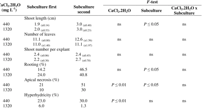
The transversal section of the non-hyperhydric and hyperhydric leaves showed uniserial epidermis with thin walls and stomata on both the surfaces. The observations revealed that the anatomy in non-hyperhydric leaves presented smaller and more juxtaposed cells and a more differentiated vascular system when compared with hyperhydric leaves (Fig. 3A-B). The leaves of hyperhydric shoots and non-hyperhydric shoots were characterized by the drastically reduced palisade
tissue. The mesophyll appeared spongy with large intercellular spaces (Fig. 3), which were similar in
the in vitro cultivated leaves of Eucalyptus
benthamii (Brondani et al. 2012).
Figure 2 - Effect of the CaCl2.2H2O concentration on
tissue Ca2+ content in different shoot portions of Lavandula angustifolia cv. Provence Blue. Means (bars) with the same letter are not different in Duncan’s test (P≤
0.05). Lowercase letters: CaCl2.2H2O
concentrations; and uppercase letters: shoot portions (tip, middle and basal parts). Bars represent the standard error of the mean.
Figure 3 - Transversal sections of leaves from shoots of Lavandula angustifolia cv. Provence Blue cultured
in vitro after 40-d. (A) Non-hyperhydric leaves (B) Hyperhydric leaves. Bars 500 µm.
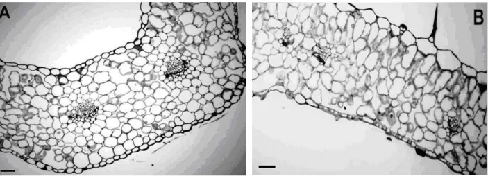
The stomata of non-hyperhydric and hyperhydric leaves displayed normal morphology (Fig. 4B-F). Louro et al. (1999) also found same basic structure (normal stomata in non-hyperhydric and hyperhydric leaves) in E. grandis W. Hill ex Maiden x Eucalyptus urophylla S. T. Blake. Due
to the high density of trichomes on the lower surface of the in vivo leaf, the detailed study of the stomata was not possible (Fig. 4A-B).
4A-F). Taha and Haron (2008) suggested that the anatomical and morphological differences observed in in vivo and in vitro leaves were due to the differences of the environment of the plants. Trichomes have a function of the defense and
protection in the plants, and, as the in vitro environment lacks predators, it is possible that in
in vitro tissues there are not signaling to produce a
high number of trichomes.
Figure 4 - Scanning electron microscopy of leaf blade surfaces of Lavandula angustifolia cv.
CONCLUSIONS
It was concluded that the increasing CaCl2.2H2O
level (1320 mg L-1) in the culture medium increased tissue Ca2+ content and decreased the hyperhydricity. Shoot-tip necrosis also decreased in the medium containing 1320 mg L-1 CaCl2.2H2O, but it did not occur in the second
subculture. The middle and basal portion tissue had higher Ca2+ content higher than apical portion. In non-hyperhydric tissues, there were smaller and more juxtaposed cells. Scanning electron microscopy of the leaves demonstrated that the quantity of trichomes on in vitro leaf surface was smaller than the quantity of trichomes on in vivo leaf surface.
REFERENCES
Abdoli M, Moieni A, Dehghani H. Effects of cultivar and agar concentration on in vitro shoot organogenesis and hyperhydricity in sunflower (Helianthus annuus L.). Pak J Bot. 2007; 39: 31-35. Abousalim A, Mantell SH. A pratical method for
alleviating shoot-tip necrosis symptoms in in vitro
shoot cultures of Pistacia vera cv. Mateur. J Hortic Sci. 1994; 69: 357-365.
Andrade LB, Echeverrigaray S, Fracaro F, Pauletti GF, Rota L. The effect of growth regulators on shoot propagation and rooting of common lavender (Lavandula vera DC). Plant Cell Tissue Org Cult.
1999; 56: 79-83.
Brondani GE, Wit Ondas HW, Baccarin FJB, Gonçalves AN, Almeida M. Micropropagation of
Eucalyptus benthamii to form a clonal micro-garden.
In Vitro Cell Dev Biol Plant. 2012; 48: 478-487. Calvo MC, Segura J. In vitro propagation of lavender.
HortScience. 1989; 24: 375-376.
Carvalho DC, Lopes da Silva AL, Schuck MR, Purcino M, Tanno GN, Biasi LA. Fox grape cv. Bordô (Vitis labrusca L.) and grapevine cv. Chardonnay (Vitis vinifera L.) cultivated in vitro under different carbohydrates, amino acids and 6-Benzylaminopurine levels. Braz Arch Biol Technol. 2013; 56: 191-201. Debergh P, Aitken-Christie J, Cohen D, Grout B, Von
Arnold S, Zimmerman R, et al. Reconsideration of the term ‘vitrification’ as used in micropropagation.
Plant Cell Tiss Organ Cult. 1992;30: 135-140. Echeverrigaray S, Basso R, Andrade LB.
Micropropagation of Lavandula dentata from axillary buds of Field-grown adult plants. Biol Plantarum.
2005; 49: 439-442.
Feder N, O´Brien TP. Plant microtechnique: some principles and new methods. Am J Bot. 1968; 55: 123-142.
Gaspar T, Kevers C, Bisbis B, Franck T, Crèvecoeur M, Greppin H, et al. Loss of plant organogenic totipotency in the course of in vitro neoplastic progression. In Vitro Cell Dev Biol Plant. 2000; 36: 171-181.
Gras MC, Calvo MC. Micropropagation of Lavandula latifolia through nodal bud culture of mature plants.
Plant Cell Tiss Organ Cult. 1996; 45: 259-261. Johansen DA. Plant Microtechnique. New York: Mac
Graw Hill Book; 1940.
Jordan AM, Calvo MC, Segura J. Morphogenesis in callus and single-cell cultures of Lavandula latifolia
Medicus. J Hortic Sci. 1990; 65: 49-53.
Lis-Balchin M, Hart S. Studies on the mode of action of the essential oil of lavender (Lavandula angustifolia
P. Miller). Phytother Res. 1999; 13: 540-542.
Louro RP, Santos AV, Machado RD. Ultrastructure of
Eucalyptus grandis x Eucalyptus urophylla. I. Shoots cultivated in vitro in multiplication and elongation-rooting media. Int J Plant Sci. 1999; 160: 217-227. Machado MP, Biasi LA, Reinhart V, Santos GD.
Redução da necrose apical e hiperidricidade de plantas de Lavandula angustifolia Mill. cultivadas in vitro. Plant Cell Cult Micropropag. 2010; 6: 18-25. Machado MP, Silva ALL, Biasi LA. Effect of plant
growth regulators on in vitro regeneration of
Lavandula dentata L. shoot tips. J Biotec Biodivers. 2011; 2: 28-31.
Malavolta E, Vitti GC, Oliveira SAD. Avaliação do estado nutricional das plantas. Piracicaba: Associação Brasileira para Pesquisa da Potassa e do Fosfato; 1997.
Marschner H. Mineral nutrition of higher plants. London: Academic Press; 1995.
McCown BH, Sellmer JC. General media and vessels suitable for woody plant culture. In: Bonga JM, Durzan DJ, editors. Cell and tissue culture in forestry. Dordrecht: Martinus Nijhoff; 1987. p 4-16.
Murashige T, Skoog F. Revised medium for rapid growth and bioassays with tobacco tissue culture.
Physiol Plant. 1962; 15: 473-497.
O’Brien TP, Feder N, McCully ME. Polychromatic staining of plant cell walls by toluidine blue.
Protoplasma. 1964; 59: 368-373.
Oliveira Y, Pinto F, Silva ALL, Guedes I, Biasi LA, Quoirin M. An efficient protocol for micropropagation of Melaleuca alternifolia Cheel. In Vitro Cell Dev Biol Plant. 2010; 46: 192-197.
Piagnani C, Zocchi G, Mignani I. Influence of Ca2+ and 6-benzyladenine on chestnut (Castanea sativa Mill.)
in vitro shoot-tip necrosis. Plant Sci. 1996; 118: 89-95.
Qiguang Q, Read PE, Fellman CD, Hosier MA. Effect of medium constituents and rooting regime on in vitro
culture of Castanea mollissima and C. dentata.
HortSci. 1985; 20: 593.
Quazi MH. In vitro multiplication of Lavandula spp.
Scheidt GN, Silva ALL, Oliveira Y, Costa JL, Biasi LA, Soccol CR. In vitro growth of Melaleuca alternifolia Cheel in bioreactor of immersion by bubbles. Pak J Bot. 2011; 43: 2937-2939.
Sha L, McCown BH, Peterson LA. Occurrence and cause of shoot-tip necrosis in shoot cultures. J Amer Soc Hort Sci. 1985; 110: 631-634.
Silva ALL, Costa JL, Alcantara GB, Carvalho DC, Schuck MR, Biasi LA, et al. Micropropagation of
Nidularium innocentii Lem. and Nidularium procerum Lindm. (Bromeliaceae). Pak J Bot. 2012; 44: 1095-1101.
Silva ALL, Rodrigues C, Costa JL, Machado MP, Penha RO, Biasi LA, et al. Gibberellic acid fermented extract obtained by solid-state fermentation using citric pulp by Fusarium moniliforme: Influence on
Lavandula angustifolia Mill. cultivated in vitro. Pak J Bot. 2013; 45: 2057-2064.
Taha RM, Haron NW. Some morphological and anatomical studies of leaves and flowers of Murraya paniculata (Jack) Linn. in vivo and in vitro. Pak J Biol Sci. 2008; 11: 1021-1026.
Taiz L, Zeiger E. Plant physiology. 3rd Edition, Sunderland MA: Sinauer Associates Inc.; 2006. Ziv M. Vitrification: morphological and physiological
disorders of in vitro plants. In: Debergh PC, Zimmerman RH, editors. Micropropagation: technology and application. Dordrecht: Kluwer Academic Publishers; 1991. p. 45-69.
Zuzarte MR, Dinis AM, Cavaleiro C, Salgueiro LR, Canhoto JM. Trichomes, essential oils and in vitro
propagation of Lavandula pedunculata (Lamiaceae).
Ind Crop Prod. 2010; 32: 580-587.