Method: The Innovation and the Results by
Light and Scanning Electron Microscopy
Aperfeiçoamento da Técnica de Embalsamamento de Cadáveres:
A Inovação da Técnica e os Resultados à Luz da Microscopia Óptica e
Electrónica de Varrimento
1. Department of Anatomy. Faculdade de Ciências Médicas. Universidade Nova de Lisboa. Lisbon. Portugal.
2. Department of Anatomy & Body Donation Office. Faculdade de Ciências Médicas. Universidade Nova de Lisboa. Lisbon. Portugal. 3. Forensic Medicine. Faculdade de Ciências Médicas. Universidade Nova de Lisboa. Lisbon. Portugal.
4. Physic Department. Faculdade de Ciências e Tecnologia. Universidade Nova de Lisboa. Lisbon. Portugal. 5. Department of Pathology. Alfredo da Costa Maternity. Lisbon. Portugal.
6. Anatomy and Otolaryngology. Faculdade de Ciências Médicas. Universidade Nova de Lisboa. Lisbon. Portugal. 7. Physic and Technological Investigation Center - CEFITEC. Lisbon. Portugal.
Recebido: 12 de Janeiro de 2013 - Aceite: 03 de Março de 2013 | Copyright © Ordem dos Médicos 2013
João GOYRI-O’NEILL1,7, Diogo PAIS2, Francisco FREIRE DE ANDRADE3, Paulo RIBEIRO4, Ana BELO5, Assunção
O’NEILL6, Samuel RAMOS1, Cláudia NEVES MARQUES1
Acta Med Port 2013 May-Jun;26(3):188-194
RESUMO
O embalsamamento é um processo químico que visa a preservação e sanitização do corpo humano por tempo indefinido. A téc-nica de embalsamamento é uma ferramenta importante no ensino e investigação em Anatomia viabilizando a conservação em boas condições de material cadavérico (minorando alterações estruturais significativas e mantendo a aparência natural). Este artigo reporta os resultados de embalsamamento de cadáveres obtidos por perfusão arterial, através da utilização de uma máquina de perfusão especialmente desenhada para o efeito. E que permite o controlo do processo de injecção de fluido de embalsamamento. A influência da técnica e a optimização dos seus parâmetros na qualidade final do embalsamamento foi avaliada através da análise histológica sequencial de tecidos cadavéricos e sua classificação por método original a partir de uma amostra de 17 cadáveres do Gabinete de Doação do Departamento de Anatomia da Faculdade de Ciências Médicas da Universidade Nova de Lisboa, sujeitos à técnica de em-balsamamento desenvolvida no Departamento. Concluímos que, com a utilização deste método, ocorre uma diminuição do processo de decomposição no momento do embalsamamento, o qual é eficaz a longo prazo (mais de um ano), exigindo apenas a manutenção do corpo a baixas temperaturas (4° C), sendo o músculo o tecido melhor preservado, com uma classificação considerada óptima.
Palavras-chave: Embalsamamento; Dissecção; Cadáver; Perfusor; Soluções; Microscopia Óptica; Microscopia Electrónica de
Var-rimento.
ABSTRACT
Embalming is a chemical process that aims the preservation and sanitization of the human body indefinitely. The technique of embalm-ing is an important tool in teachembalm-ing and research in anatomy enablembalm-ing the preservation of cadaveric material in good conditions (lessen-ing any significant structural changes and maintain(lessen-ing the natural appearance). This article presents the results of embalmed cadavers in the course of arterial perfusion, through the use of a perfusion machine, particularly designed to this objective, and which allows the control of the embalming fluid injection process. The influence of this technique and the optimization of its parameters on the final quality of embalming were evaluated by sequential histological analysis of the cadaveric tissues using an original method of classification of samples collected from 17 deceased corpses of the Corpses Donation Office of the Department of Anatomy of Faculdade de Ciências Médicas from Universidade Nova de Lisboa, subject to the embalming technique developed in the Department. We concluded that, with this method, there is a decrease of the decomposition process at the time of embalming, which is effective at long term (over a year), requiring merely the maintenance of the body at low temperatures (4° C) and it is possible to observe that the tissue best preserved over time is muscle, showing a conservation considered optimal.
Keywords: Embalming; Dissection; Cadaver; Solutions; Microscopy, Electron, Scanning.
INTRODUCTION
The high requirement level in care, technical quality, surgical and microsurgical skills, alongside with the need for a high anatomical knowledge, have recently demanded a degree of unprecedented excellence and technical mas-tery.1-3 The high risk in invasive procedure performed by inexperienced hands, often, appears as a limitation in the specialist formation because of his lack of technical exper-tise, for example, in arthroscopy or microsurgery of the tem-poral bone, rarely performed.4,5
The current surgical training programs use a variety of instruments, including unanimated models, virtual reality, live animals and human corpses, to simulate living tissue
and human anatomy and high performance of simulators for training of patient emergencies, and training in emergency teams.1,2,6,7 Although human cadavers represent currently
the most closely model to reality found in clinical practice, its cost and availability, often limited, along with the limita-tion of the tissues specificities severely reduce their utili-zation.1,8 Table 1 summarizes some of the advantages and
disadvantages of various skills training models / tools. Embalming is a chemical process that aims to preserve and sanitize the human body indefinitely.1 In most modern
ARTIGO ORIGINAL
have led to practices in which the corpse is viewed as look-ing “natural,” thus denylook-ing the reality of death. It is important to acknowledge the impact of embalming, which can also carry with it problematic psychological consequences for the family.9 From the scientific point of view, the main goal
is namely to improve the body’s resistance to extended pe-riods of exposure during anatomical dissection.1, 2, 7
The desired properties required for successful embalm-ing of cadavers for gross anatomy teachembalm-ing include a good long-term structural preservation of organs and tissues with minimal distortion, prevention of over-hardening or ap-pearance alteration, while maintaining flexibility of internal organs, prevention of desiccation and fungal or bacterial growth and spread within a specific cadaver and to other cadavers in the dissection room.10
The limitations of reproducibility of the properties of tis-sues, both in non-conserved body, with the potential risk of infection or accelerated decomposition, or in embalmed, conserved corpse, prevent the young surgeons from per-forming their learning in a safe way, without time or techni-cal limitations, such as formalin, inappropriate for observa-tion and approach of perforating vessels or delicate tissue dissection procedures.3
In 1992 was published by Walter Thiel (Graz, Austria), an embalming method with great acceptance, utilized for more than 30 years and perfected by its use in more than 977 corpses and numerous body parts. This procedure was updated in 2002.15 It’s most innovative features, compared
to previous methods2,11-13 focused on formaldehyde as a pre-servative, are almost no odor of the embalming fluid, ability to maintain a long-term conservation, and very minor mor-phological changes,2,15 of the cadaveric material and
disin-fection efficacy confirmed by bacteriological tests without release to the environment of harmful substances.2,12,17 The
previous utilization of harmful substances, such as formal-dehyde, raised important safety issues.16,17 Formaldehyde
levels should be measured periodically specially during the dissection in the anatomy laboratory, and local exhaust ven-tilation system should be installed and personal protective equipment such as safety glass and gloves should be avail-able and be used to prevent direct skin or eye contact.17
Therefore, prudent practices should seek to minimize form-aldehyde, and other harmful substances, exposure.12,16,18,19
The Thiel solution fundamental basis consists of a mix-ture of high saline components concentration causing dena-turation of proteins. The infusion is performed during three days.2,3
The physiological texture of tissue is maintained by the precipitation of the solution, without observation of tissue retraction or saturation. This solution has been modified, either the relative composition of its basic components as with the introduction of new compounds to allow a more adequacy to the particular tissue properties that we pretend to preserve. In general, an ideal result is characterized by conservation that lasts six months.2,3,6
With the Thiel’s technique, the body has a pale or red-dish skin.3 Occurs detachment of superficial epithelial layers
and nails. Compared with vivo, the skin remains smooth, oiled, hairless but firmer. The subcutaneous adipose tissue retains its yellow color and differentiation in adipose cells of different sizes. The shiny appearance and strength of fas-cias, as well as and the permeation of intermuscular spaces by vessels and neural structures is no different from living in color or texture.3
Currently, minimally invasive surgery and, specifically, the laparoscopic approach is the ‘Gold Standard’ for numer-ous surgeries (i.e., cholecystectomy, bariatric and anti-re-flux surgery).2 Its excellence requires a high degree of
dex-terity, which is directly correlated with practice and exercise. As a result, the American Society of Gastrointestinal and Endoscopic Surgeons (SAGES) and the European Associa-tion of Endoscopic Surgeons (EAES) emphasized the need
Table 1 -Types of Training instruments available skills and and their advantages, disadvantages and use situations (Adapted from Reznick R et al).
Bench Models Cheap, portable, minimal risks.
Low acceptance by trainees. Low reliability and limited basic tasks. Not allow the simulation of an entire surgery.
Acquisition of basic skills for beginners.
Improvement limited.
Live Animals
High reliability, availability, allow the practice of haemostasis and a complete surgery.
Cost, need for infrastructure ethical limitations. Use single anatomical differences.
High technical knowledge. Training situations involving haemostasis. Training dissection.
Corpses
High fidelity, currently ‘only’ true simulator, allows the realization of complete surgeries.
High cost, availability, possible use unique properties of tissues, risk of infection.
High technical knowledge. Dissection. Continuing medical education.
Simulation Models of Human Performance
Reusable, high reliability, data
to keep up with advances in technology through validated training programs, with measurement of performance, be-fore moving to real situations, reducing in this way the ad-vanced laparoscopic learning curve.1,20
In fact, the technique of embalming is an important tool, along with other conservation techniques,21 in teaching and
research in anatomy, because it enables the conservation of cadaverous material in good condition, i.e. without signifi-cant structural changes, while maintaining the natural ap-pearance22,23 andits limitations may introduce an important
bias in investigation when present.
The dissection of a human cadaver is an indispensable practice in general training of medical students, doctors in specialist training, research in fundamental anatomical and pathological phenomena and in the improvement of diag-nosis methods and therapy,23 assuming the, therefore, its
central role in teaching undergraduate and support for post-graduate teaching, the latter with great increase in recent years.2 A safe conservation technique, economic and
ac-cessible and that ensures the maintenance of lasting physi-cal properties of tissues is urgently needed, both in the con-text of pre and post graduate. Thus, the rookie running pro-cedures as demanding as the skin flap or performance of anesthetic procedure may, in the current context, be made by direct practice in patient.24,25
There are mechanisms dedicated to cadaver infusion, usually automatic or semiautomatic machines, fitted with pump units of injection to propel the solution into the em-balming cadaver26 through the arterial vasculature.
How-ever, they still lack efficiency either because there is no per-fusion technology that ensures the quality of the injection, either by unknowing the exact response of the vascular sys-tem to the injection and vascular perfusion process.23 These
machines, in particular, lack of automation, control and data acquisition modern systems, allowing the optimization of the technique, so with not suitable to scientific research. Since 2006 the Department of Anatomy, Faculty of Med-ical Sciences, in collaboration with the Center for PhysMed-ical Research and Technology at the same University, has stud-ied the project and development of machines to allow arte-rial perfusion that suppresses perfusion limitations of exist-ing systems in the market and aims to meet the needs that embalming techniques study entails.
This article reports the study of the influence of the arte-rial perfusion technique and the optimization of its param-eters in the final quality of the embalming through the anal-ysis of cadaveric tissues, through the utilization of a new machine specially designed to this intent.
MATERIAL AND METHODS
Between June 2009 and May 2010 were selected 17 ca-davers donated sequentially, through the Corpses Donation Office implemented in the Department of Anatomy of the FCM-UNL. The body is donated to this institution through a document written in life or a desire expressed before death, ethically and legally,24 as predicted in Decree-Law number
274/99 from July 22, 1999, in Portuguese Republic Diary,27
and European legal and ethical framework28,29 for body
do-nation for pre and posts graduate education and research, and integrated in this observational study. The embalming technique used was the cadaverous automated infusion of an embalming solution conceived by the Department of Anatomy and used for several years, using the automatic perfusion machine.
The embalming solution used is a combination of ali-phatic alcohols Diethylene glycol and Monoethylene glycol (90:10) optimized in order to preserve the texture, volume, color and shape of the body and its tissues as perfect as possible, in order also to allow the disinfection and sanitiza-tion of the process.
The resultant mixture solution is a clear liquid, practical-ly odorless, colorless and denser than water. These prop-erties combined with adequate hygroscopic levels, good solubility in organic acids and physiologically safe. Mono-ethylene glycol is toxic,30,31 the same being also true for diethylene glycol.32,33 Diethylene glycol can lead to serious
complications that may prove fatal when ingested.32,33 This
substance produces no toxic vapors at room temperature and isn’t harmful at touch, unless with direct contact with the product.32-34 The cadaver embalming procedure involves a closed circuit of embalming fluid perfusion, from the ma-chine itself to the closed vascular system of the preserved cadaver, without submersion of the entire body, so with no direct risk added to the practice of dissection. These char-acteristics revealed this solution as a good choice, toxico-logically comparable to glycerol,32,33 to achieve the intended
goals.
An incision was made in the right and left groin, with exposure of the femoral vessels, preparing the femoral ar-terial injection of bidirectional embalming solution in all 17 cadavers using cross-sectional or longitudinal in about 1 cm from the femoral artery without any previous conservation intervention than external washing with Chlorhexidine soap and the cooling during transport (in a temperature of ap-proximately 4 - 6° C), involved in a simple cadaver plastic bag. The injection was performed using the introduction of an appropriate size cannula at the proximal femoral artery, and a lower size cannula also appropriate in the distal femo-ral artery, at room temperature.
The injection of the embalming solution was accom-plished using a pulsed infusion at a pace between 60 to 70 pulses per minute, with the aim of mimicking normal vessels in cardiac output with recoil and variation of systolic and dia-stolic pressure, reducing the flow resistance and expanding the extent and scope of perfused tissues.35
The mean duration of cadaver vascular infusion was 30 - 45 minutes per cadaver, performed with a perfusion rate of 70 pulses / minute. The average volume of the embalming solution injected was of 7 liters per body, varying with mass and stature.
Microscopic Evaluation Morphological
Tissue biopsies were performed in all 17 corpses. The collection of tissue samples was performed in two
ent chronological phases - immediate post-embalming and more than 1 month after embalming.
The choice of material to be collected for sampling was related with the importance given to the evaluation of dif-ferent anatomical regions with very own characteristics of conservation and particular requirements. This option is intended to obtaining a better sense of the overall quality of embalming, with particular focus on tissues addressed by students and teachers during the dissections and future uses of the cadaverous material. In the immediate post-em-balming, three fragments with dimensions of 1x1 cm were picked up, respectively: skin (the anterior thigh) muscle (muscular body anterior thigh) and buccal mucosa, this col-lection was repeated one month after the embalming. In each case was also registered the cause of death and the factors of possible peri and post-embalming com-mitment of cadaverous structures, to avoid bias in the his-tological results. It was used hematoxylin eosin regular and elastin staining and an Optic Microscope for the observation of the sections of tissues.
Histological classification of methodological criteria For the stratification of the results obtained in the his-tological analysis, original criteria were established based on the principles of characterization of cellular autolysis re-ported by M. Mckenna et al.28
The established criteria also integrate assessment of
nuclear pyknosis and changes in cytoplasmic organelles, thereby enabling to determine the degree of deterioration/ breakdown of tissue samples collected. The Table 2 lists the used criteria.
Harvests were performed with the sample placed in a jar with formaldehyde, with a single random numerical iden-tification, due to the intersection of data and available only to the element that held all harvests, omitted to the patholo-gist.
After biopsies, histological blades were prepared in number of 10 for each harvest. These were stained with hematoxylin-eosin and elastin. Using this process, were ob-served 280 slides of samples.
A report was made for each tissue, establishing a rank-ing in five categories of preservation of morphological structures by microscopy (Table 3), based on the number of negative or positive levels according to the criteria of his-tological classification of methodological degree of cellular autolysis shown in Table 2.
RESULTS Macroscopy
After embalming, the body revealed a pale, occasionally with livor mortis in regions of slope. There was no change of the nail beds, scalp or in most epithelial cells of skin lay-ers, including changes in hairiness. During the final phase of embalming, occasionally was found the acquisition of
Table 2 - Histological Classification of methodological criteria of the degree of cellular autolysis.
Eosinophilia / cell hidropization 0 (absent)
1+ (< 25% cells) 2+ (25% < > 50%) 3+ (> 50%)
Nuclear pyknosis 0 (absent)
1+ (< 25% cells) 2+ (25% < > 50%) 3+ (> 50%)
Kariorexis 0 (absent)
1+ (< 25% cells) 2+ (25% < > 50%) 3+ (> 50%)
Disorganization of fibers 0 (absent) 1+ (focal)
2+ (in more than 3 filds 10x) 3+ (widespread)
Coagulation necrosis 0 (absent)
1+ (focal)
2+ (in more than 3 filds 10x) 3+ (widespread)
Table 3 - Classification Morphological microscopic tissue into 5 categories, according to their conservation status
A = 2+
Excellent conservation
2+ e = 4
Good conservation
B
C 4+ e = 6+
Satisfactory conservation
6+ e = 8+
Conservation unsatisfactory
D
E 8+
Tissue in autolysis
specific and generalized cutaneous depressions and turgor - characterize in practice as ‘orange peel aspect’.
Compared with the living, cadaveric skin in immediate post-embalming showed no increased resistance or de-tachment of skin layers or significant changes in coloration. There is, however, a slight decrease of its elasticity, objecti-fiable in the incision with facilitated removal of tissues. The maintenance of its features easily allowed the incision of tissues, mimicking the feel of the incision in living tissue. There was a slight increase in the swelling of tissues, with a slight increase in volume (eg diameter of the forearm) but only millimeters.
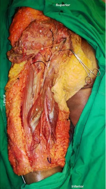
The subcutaneous tissue retained its structure and yel-low color, with slightly increased consistency, and the fascia and muscular bodies maintained their resistance, intense red color and elasticity of the pre-embalming (Fig. 1). Not observed in any of embalming commitment of muscle spac-es or mimicry of compartment syndrome by the injection of embalming solution. The joints remained movable with-out significant changes of its passive range of motion. The vessels and nerves maintained their structure and integrity, easily identifiable arteries and veins on palpation, including microns caliber vessels. The lack of color in these, due to the transparency of the embalming solution, the only dif-ficulty was its identification and distinction.
There were no significant changes between this de-scription and morphological observation after 1 month to 6 months of embalming. Fig. 1 illustrates the quality of the tissue preservation by the technique developed by the De-partment of Anatomy.
Morphological microscopy
Based on the methodological criteria of Histological Classification of the degree of cellular autolysis was pos-sible to evaluate the overall integrity of tissues.
Total of 17 corpses analyzed.
From the analysis of Fig. 2 we can see that the skin has a high rating - Excellent maintenance – in more than half the
harvest, with the rest mainly classified as good conserva-tion at the time of embalming. These results with skin are only surpassed by the muscle (Fig. 2), the tissue with the best classification of all. This classification pattern is con-firmed after more than a month of embalming.
Comparing the long-term histologic evaluation between the 17 bodies and the 3 chosen tissues, it is possible to observe that the tissue that has the larger number of level A classification is the muscle (with a total of 6 to 8 ratings with over 1 month).
Of the three tissues collected simultaneously in each body, the buccal mucosa tissue showed the worst results in microscopic evaluation - mostly classified as B, D and E at the time of embalming and maintaining, of course, a mediocre rating in the post - 1 month embalming.
It was also performed a Scanning electron microscopy
Figure 1 – Dissection of the posterior surface of the left thigh, with detail of integrity and recognition of vascular and nerve structures deep in the gluteal region in a corpse after 1 year of embalming (Images available in http://www.fcm-anatomia.com/pt/).
Figure 2 – Immediate post-embalming harvest.
Figure 3– Striated thigh Muscle collected after embalming (hema-toxylin eosin staining – Optic Microscope, 200x).
(S.E.M.) observation of some of the cadaveric tissues, veri-fying structural integrity and high morphologic correlation with living tissue.
DISCUSSION
It is possible to infer that the results are generally con-sistent in collections held over several months, showing the same diminished/absent degree of evolution of tissue au-tolysis, with excellent results in cadaveric dissection, as de-sirable for human gross anatomy dissection.10 It was found in almost all cadaver tissues similar resistance, mobility of joints in the various body segments and staining of struc-tures (including organs) to the living. These properties allow unique conservation of the embalmed corpses compared to other embalming techniques, with easy recognition and individualization of nerves, arteries and veins, even at mil-limetric levels.
This tissue integrity is currently explored in several pre-graduation courses dissection and annual regional anato-my, and in the context of post-graduated training, in surgical techniques of plastic surgery, ENT, Neurosurgery, regional anesthesia, orthopedics and urology, among others through Postgraduate Courses available through the platform of the Department of Anatomy - http://www.fcm-anatomia.com/pt/. The prolonged maintenance of the structural integrity abolishes the limitations of previous use of cadaveric mate-rial, with the completion of current perfusion techniques for research in the Department of Anatomy of the FCM-UNL, including diafinization, angiography and Scanning Electron Microscopy in corrosion molds.
CONCLUSION
Compared with the embalming method of Thiel,2 the
em-balming method developed by the Department allows a fast and effective as both macro and microscopically as shown, due to the mechanical perfusion of the body, with optimiza-tion of the procedure by its pulsed nature, allowing comple-tion of the embalming process in less than 1 hour after the start of injection. No additional submersion of the body in conservation fluid is necessary. Most of the corpses in use
with between six months and one year of embalming, pres-ent excellpres-ent texture, color and reproducibility of the tech-niques performed in vivo. This approach allows the conser-vation of superficial cutaneous plans, musculo-fascial and also endocavitary (i.e., abdominal, cranial). As mentioned in the method of Thiel,2,15 the embalming solution developed
and used by the Department of Anatomy – FCM-UNL does not present a direct risk to health during cadaveric material handling,29,30 without releasing vapors into the environment and allowing a lasting disinfection of the involved tissues. The degree of preservation obtained in skin and muscle, two of the most important tissues during the course of dis-section, identification and pedagogical use of corpse piec-es, explains the importance and scope of possible method adopted by the Department of Anatomy – FCM-UNL. Comparing the long-term histologic evaluation between the bodies 17 and the 3 tissues chosen, it is possible to observe that the tissue best preserved over time is muscle (Fig. 3), showing a conservation considered optimal (less than or equal to two criteria positive autolysis cell).
The isolated discouraging results obtained for the buc-cal mucosa at the time of embalming, imply that the change may be pre-existing. Given the average age of the cadav-ers preserved by the method of embalming - average age of 60 years - and the presence of prosthetic teeth in almost all cases it is important to future investigate the possible contribution to the further deterioration of mucosa from the post-embalming immediate and its relationship to quality of embalming.
The refinement of this technique and its perfusor has allowed the realization of human cadaver embalming in the Department of Anatomy, under the dissection courses in pre and post-graduation, some already realized and others un-derway, where the existence of well-embalmed cadaveric material differentiates this technique in a manner fully face the other, as well as the method used so far by the scientific community.
Thus, we conclude that there is an interruption of the decomposition process at the time of embalming, which is effective at long term (over a year), requiring merely the
maintenance of the body at low temperatures, while posi-tive (4° C), for optimization and perpetuation of results. This technique and machine allows the preparation of ca-daver embalming material in good condition, allowing path-ological examinations, forensic and their long-term conser-vation of the cadaveric material.
The experience has shown that the preservation ex-ceeded 5 years. We consider it important in the near future to perform tissue culture and assessment of resistance to fungal inoculation of the embalmed tissues, for additional macroscopic recognition of their conservation.
The anticipation is, in the near future, to maximize the data obtained during the embalming process, such as tem-perature and flow pulsatility already tested, to optimize the process and obtain a longer and lasting conservation, dis-rupting the tissue breakdown as early as possible.
CONFLICT OF INTERESTS None stated.
FUNDING SOURCES None stated
REFERENCES
1. Reznick R, MacRae H. Teaching surgical skills — Changes in the wind. N Engl J Med. 2006;355:2664-9.
2. Thiel W. Die Konservierung ganzer Leichen in naturlichen Farben. Ann Anat. 1992;174:185-95.
3. Wolff KD, Kesting M, Mücke T, Rau A, Hölzle F. Thiel embalming tech-nique: a valuable method for microvasular exercice and teaching of flap raising. Microsurgery. 2008; 28:273-8.
4. Oxentenko AS, Ebbert JO, Ward LE, Pankratz VS, Wood KE. A multi-dimensional workshop using human cadavers to teach bedside proce-dures. Teach Learn Med. 2003;15:127-30.
5. Tolhurst DE, Hart J. Cadaver preservation and dissection. Eur J Plast Surg. 1990;13:75-8.
6. Groscurth P, Eggli P, Kapfhammer J. Gross anatomy in the surgical curriculum in Switzerland: Improved cadaver preservation, anatomical models, and course development. Anat Rec. 2001;265:254-6. 7. Bradbury SA, Hoshino K. An improved embalming procedure for
long-lasting preservation of the cadaver for anatomical study. Acta Anat. 1978;101:97-103.
8. Macdonald G, MacGregor D. Procedures for embalming cadavers for the dissecting laboratory. Proc Soc Exp Biol Med. 1997;215:363-5. 9. Palermo G, Gumz E. The last invasion of human privacy and its
psy-chological consequences on survivors: a critique of the practice of em-balming. Theor Med. 1994;15:397-408.
10. Coleman R, Kogan I. An improved low-formaldehyde embalming fluid to preserve cadavers for anatomy teaching. J Anat. 1998;192:443-6. 11. Wineski L, English A. Phenoxyethanol as a nontoxic preservative in the
dissection laboratory. Acta Anat. 1989;136:155-8.
12. Pabst R. Exposure to formaldehyde in anatomy: an occupational health hazard? Anat Rec. 1987;219:109-12.
13. Frolich K, Andersen L, Knutsen A, Flood P. Phenoxyethanol as a non-toxic substrate for formaldehyde in long-term preservation of human anatomical specimens for dissection and demonstration purposes. Anat Rec. 1984;208:271-8.
14. Bradbury SA, Hoshino K. An improved embalming procedure for long-lasting preservation of the cadaver for anatomical study. Acta Anat. 1978;101:97-103.
15. Thiel W. Erganzung fur die Konservierung ganzer Leichen nach W. Thiel. Ann Anat. 2002;184:267-9.
16. O’Sullivan E, Mitchell BS. An improved composition for embalming fluid to preserve cadavers for anatomy teaching in the United Kingdom. J Anat. 1993;182:295-7.
17. Ohmichi K, Komiyama M, Matsuno Y, Takanashi Y, Miyamoto H, Kado-ta T, et al. Formaldehyde exposure in a gross anatomy laboratory - per-sonal exposure level is higher than indoor concentration. Environ Sci Pollut Res Int. 2006;13:120-4.
18. Whitehead M, Savoia M. Evaluation of methods to reduce formal-dehyde levels of cadavers in the dissection laboratory. Clin Anat. 2008;21:75-81.
19. Coleman R. Reducing the levels of formaldehyde exposure in gross
anatomy laboratories. Anat Rec. 1995;243:531-3.
20. Giger U, Frésard I, Häfliger A, Bergmann M, Krähenbühl L. Laparo-scopic training on Thiel human cadavers: A model to teach advanced laparoscopic procedures. Surg Endosc. 2007;22:901-6.
21. Goyri-O’Neill, J. Técnica de plastinização - sua contribuição no ensino e investigação em anatomia. Lisboa: Edição do autor;1984. 22. Mayer, RG. Embalming: History, theory and practice. Pennsylvania:
McGraw-Hill; 2006.
23. Benkhadra M, Faust A, Ladoire S. Comparison of fresh and Thiel’s embalmed cadavers according to the suitability for ultrasound-guided regional anesthesia of the cervical region. Surg Radiol Anat. 2009; 31:531-5.
24. Parecer sobre utilização de cadáveres humanos para fins de ensino médico e a sua necessidade, pertinência e legitimidade. Lisboa: Docu-mentação, CNECV. 1991-1993; 1:67-72.
25. Esperança-Pina, JA. Aspectos morfológicos gerais e actuais da micro-vascularização. Acta Med Port.1983;4:433-6.
26. McKenna MF, Goldbogen JA, St Leger J, Hildebrand JA, Cranford TW. Evaluation of postmortem changes in tissue structure in the bottlenose dolphin (Tursiops truncatus). Anat Rec. 2007;290:1023-32.
27. Decreto de Lei nº 274/99 de 22 de Julho; In: Diário da República - I Série A, Nº169 – Page 4522 to 4525—Lisbon, 1999.
28. McHanwell S, Brenner E, Chirculescu AR, Drukker J, van Mameren H, Mazzotti G, Pais D, et al. The legal and ethical framework governing Body Donation in Europe - A review of current practice and recommen-dations for good practice. Eur J Anat. 2008;12:1-24.
29. Riederer BM, Bolt S, Brenner E, Bueno-Lopez JL, Cirulescu ARM, Da-vies DC, et al. The legal and ethical framework governing Body Dona-tion in Europe – 1st update on current practice. Eur J Anat. 2012;16:1-21.
30. Brent J, McMartin K, Phillips S, Burkhart KK, Donovan JW, Wells M, et al. Fomepizole for the treatment of ethylene glycol poisoning. N Engl J Med. 1999;340:832-38.
31. Friedman EA, Greenberg JB, Merrill JP, Dammin GJ. Consequences of ethylene glycol poisoning: report of four cases and review of the literature. Am J Med.1962;32:891-902.
32. O’Brien KL, Selanikio JD, Hecdivert C, Placide M-F, Louis M, Barr DB, et al. Epidemic of pediatric deaths from acute renal failure caused by diethylene glycol poisoning. JAMA. 1998;279:1175-80.
33. Schep LJ, Slaughter RJ, Temple WA, Beasley DM. Diethylene glycol poisoning. Clin Toxicol. 2009;47:525-35.
34. Sigma-Aldrich® – Ficha de segurança Dietilenoglicol [accessed 2013
Feb 12]. Available at: http://www.sigmaaldrich.com/MSDS/MSDS/Dis-playMSDSPage.do?country=PT&language=pt&productNumber=0312 8&brand=FLUKA&PageToGoToURL=http%3A%2F%2Fwww.sigmaal-drich.com%2Fcatalog%2Fproduct%2Ffluka%2F03128%3Flang%3D pt.
35. Gabrys E, Rybaczuk M, Kedzia A. Blood flow simulation through fractal models of circulatory system. Chaos, Solitons Fractals. 2006;27:1-7.