IN VITRO BIOFILM FORMING POTENTIAL OF STREPTOCOCCUSSUIS ISOLATED FROM HUMAN AND SWINE
IN CHINA
Guo Dawei, Wang Liping*, Lu Chengping
College of Veterinary Medicine, Nanjing Agricultural University, Nanjing, 210095
Submitted: October 11, 2010; Approved: June 07, 2012.
ABSTRACT
Streptococcus suis is a swine pathogen and also a zoonotic agent. The formation of biofilms allows S. suis to become persistent colonizers and resist clearance by the host immune system and antibiotics. In this study, biofilm forming potentials of various S. suis strains were characterized by confocal laser scanning microscopy (CLSM), scanning electron microscopy (SEM) and tissue culture plates stained with crystal violet. In addition, the effects of five antimicrobial agents on biofilm formation were assayed in this study. S. suis produced biofilms on smooth and rough surface. The nutritional contents including glucose and NaCl in the growth medium modulated biofilm formation. There was a significant difference in their biofilm-forming ability among all 46 S. suis strains. The biofilm-forming potential of S. suis serotype 9 was stronger than type 2 and all other types. However, biofilm formation was inhibited by five commonly used antimicrobial agents, penicillin, erythromycin, azithromycin, ciprofloxacin, and ofloxacin at subinhibitory concentrations, among which inhibition of ciprofloxacin and ofloxacin was stronger than that of other three antimicrobial agents.Our study provides a detailed analysis of biofilm formation potential in S. suis, which is a step towards understanding its role in pathogenesis, and eventually lead to a better understanding of how to eradicate S. suis growing as biofilms with antibiotic therapy.
Key words: biofilm; Streptococcussuis; scanning electron microscopy; confocal laser scanning microscopy; microplate assay; antimicrobial agents
INTRODUCTION
Streptococcus suis is an important swine pathogen, causing a wide range of diseases in pigs, including meningitis, septicaemia, pneumonia, endocarditis, and arthritis (24). It is also a zoonotic organisms and its public health importance was highlighted by a recent large-scale outbreak of human S. suis infections in China in 2005, which resulted in 38 deaths (29).
Human can also be infected with S. suis by direct contact with pigs or its byproducts and the infection leads to development of streptococcal toxic shock syndrome (3, 4) as well as meningitis and endocarditis (2, 13).Thirty five serotypes of S. suis (types1 to 34 and type 1/2) have been described, but type 2 is considered to be the most pathogenic for both human and swine (27).
Biofilms are matrix-enclosed bacterial population adherent
to each other and/or to surfaces or interfaces. The biofilm mode of growth is widespread and is of medical and economic importance in diverse ecological niches (5). The formation of biofilm allows microorganisms to become persistent colonizers and resist clearance by the host immune system. Among pathogenic bacteria including S. suis, the formed biofilms are able to attach to the surfaces of various indwelling devices such as vascular catheters, prosthetic joints and artificial heart valves, as well as to host tissues and demonstrate superior resistance to antibiotics, which makes antibiotic therapies less effective or leads to treatment failure (9, 10). The ability of these bacteria to produce biofilms on the surfaces of biomaterials used for surgery is one of the main causes of difficult-to-cure infections. Recently, Grenier et al. (10) have investigated the ability of S. suis type 2 to form biofilms.
However, biofilm formation by other S. suis serotypeshas not been extensively examined.
In this study, we extensively investigated the biofilm-forming potential of S. suis on smooth glass coverslip and rough organic membrane by scanning electron microscopy (SEM) and confocal laser scanning microscopy (CLSM).
Additionally, the polystyrene 96-well microplate stained with crystal violet was used to analyze the factors in the growth medium contributing to the biofilm formation and biofilm-forming ability of 46 S. suis isolated from human and swine from different regions of China as well as the effect of antimicrobial agents at subinhibitory concentrations on biofilm formation.
MATERIALS AND METHODS
Bacterial strains and growth conditions
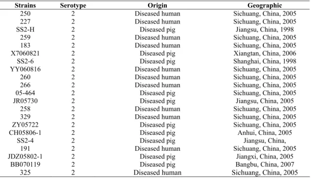
Forty six S. suis strains were isolated from human and swine from 1998 to 2008 from different regions of China except for three strains. Serotyping of S. suis was carried out by a coagglutination test, using commercial specific sera against serotype 1–28 (Statens Seruminstitut, Copenhagen, Denmark). Twenty eight of the isolates were serotype 2, 16 were serotype 9, and 2 isolates were serotype 1 and 7,
respectively (Table 1). S. suis isolates were cultured at 37℃ in
THB(Todd-Hewitt yeast Broth) containing (w/v) 0.5% beef extract, 2% peptone, 0.3% yeast extract, 0.5% glucose, 0.2% NaCl, 0.04% Na2HPO4, 0.25% Na2CO3 and 2% calf serum.
Table 1. Origins and the relevant characterstics of Streptococcus suis strains
Strains Serotype Origin Geographic
250 2 Diseased human Sichuang, China, 2005
227 2 Diseased human Sichuang, China, 2005
SS2-H 2 Diseased pig Jiangsu, China, 1998
259 2 Diseased human Sichuang, China, 2005
183 2 Diseased human Sichuang, China, 2005
X7060821 2 Diseased pig Xiangtan, China, 2006
SS2-6 2 Diseased pig Shanghai, China, 1998
YY060816 2 Diseased human Sichuang, China, 2005
260 2 Diseased human Sichuang, China, 2005
266 2 Diseased human Sichuang, China, 2005
05-464 2 Diseased pig Sichuang, China, 2005
JR05730 2 Diseased pig Jiangsu, China, 2005
258 2 Diseased human Sichuang, China, 2005
329 2 Diseased human Sichuang, China, 2005
ZY05722 2 Diseased pig Sichuang, China, 2005
CH05806-1 2 Diseased pig Anhui, China, 2005
SS2-4 2 Diseased pig Jiangsu, China,
191 2 Diseased human Sichuang, China, 2005
JDZ05802-1 2 Diseased pig Jiangxi, China, 2005
BB070119 2 Diseased pig Bangbu, China, 2007
GH05-458 2 Diseased pig Sichuang, China, 2005
719 2 Diseased pig Sichuan, China, 2005
13 2 Diseased pig Shanghai, China, 2005
T002 2 Diseased human Sichuang, China, 2005
251 2 Diseased human Sichuang, China, 2005
98012 2 Diseased human Jiangsu, China, 1998
P4254 2 Diseased human Germany
2083 9 - Denmark
NJ-2 9 Healthy carrier pig Suzhou, China, 2006
SH06 9 Diseased pig Shanghai, China, 2007
NJ-4 9 Diseased pig Yancheng, China, 2005
CZ0608 9 Healthy carrier pig Changzhou, China, 2006
55 9 Healthy carrier pig Jiangxi, China, 2005
SH26 9 Diseased pig Shanghai, China, 2007
NJ-5 9 Diseased pig Xuzhou, China, 2005
GZ0565 9 Diseased pig Guangzhou, China, 2005
SH896 9 Diseased pig Shanghai, China, 2006
27 9 Healthy carrier pig Shanghai, China, 2007
NJ-1 9 Diseased pig Nanjing, China, 2006
NJ-6 9 Diseased pig Taizhou, China, 2005
L89 9 Diseased pig Shanghai, China,2006
NJ-3 9 Healthy carrier pig Yancheng, China, 2006
40 9 Healthy carrier pig Shanghai, China,2004
SH28 1 - Canda
SH59 7 Diseased pig Shanghai, China, 2005
Scanning electron microscopy (SEM)
A mid-exponential growth culture of S. suis type 9 NJ-3 and type 2 YY060816 were diluted to an optical density of 0.1 at 600 nm (OD600) and each 200 μL were added to wells of a 6-well microplate (Cosmo) containing an 11 mm×11 mm sterilized rough organic membrane (Mosutech Co., Ltd., Shanghai, China) and a smooth glass coverslip respectively on the bottom. After incubation without shaking for 24 h at 37 oC, medium and planktonic bacteria on the organic membrane and glass coverslip were removed with sterile PBS. The biofilms and planktonic bacteria were fixed for 6 h with 4% glutaraldehyde and washed three times with 0.1 M PBS in the intervals of 10 min, then fixed to the black transparent in 2% osmium tetroxide. Samples were dehydrated and critical point dried, gold sputtered with ion sputtering instrument (current 15 mA, 2 min) and examined using a scanning electron microscopy (FEI Quanta, Netherland).
Confocal laser scanning microscopy (CLSM)
The biofilm on the glass coverslips was adapted from a
procedure as described previously by Takenaka et al. (2001). Extracellular polysaccharide in the biofilm formed by S. suis NJ-3was visualized with 50 µg/mL fluorescein
isothiocyanate-concanavalin A (FITC-ConA , Sigma , USA) which
fluoresces green. The bacterial cells in biofilms were visualized by fluorescent staining with 10 μg/mL propidium iodide (PI,
Sigma,USA) solution which fluoresces red. All confocal
images were digitized with confocal laser scanning microscope
system (CLSM,Leica TCS SP2, Mannheim, Germany) using
a 63× or 100× oil immersion objective (Leica, Mannheim, Germany). An argon laser, at 488 nm, was used as the excitation source for the fluorescent probe. A 530/30 Band Pass (BP) filter was utilized for FITC-ConA, and a 605 Long Pass (LP) filter was utilized for PI.
Analysis of factors influenicng S. suis biofilm formation
with crystal violet (TCP assay)
confirmed by SEM and CLSM. Biofilm formation of S. suis NJ-3 was determined in the presence of 0.2%, 0.5%, 1% and 2% glucose. Based on the results of this experiment, biofilm formation of S. suis NJ-3 was determined in the presence of 0.2%, 0.5%, 1% and 2% NaCl. The optimized THB medium including 1% glucose and 0.5% NaCl was used to analyze the biofilm-forming ability of 46 S. suis clinical isolates. All biofilm assays were run in triplicate and the means ± standard deviations of independent experiments were calculated.
Effect of antimicrobial agents on biofilm formation
determined by the TCP assay
Mid-exponential growth phase cultures of 46 S. suis isolates were adjusted to an optical density of 0.2 at 600 nm (OD600). One hundred μL of culture and 100 μL of antimicrobial agent solution were added to each well of a 96-well microplate and the final concentrations of each antimicrobial agent were 1 / 2 × MIC, 1 / 4 × MIC, 1 / 8 × MIC and 1 / 16 × MIC, respectively. Wells filled with only sterile growth medium were included as negative controls, and wells including 200 μL culture inoculation without antimicrobial agents were used as positive controls. After incubation without
shaking for 24 h at 37 oC, wells were subsequently rinsed and stained with crystal violet and biofilm formation was quantified as described above.
RESULTS
Figure 1. SEM observation of the biofilms formed by S suis NJ-3 and YY060816 on rough organic membrane and smooth glass coverslip. Bacteria were grown in six well tissue culture plates with organic membranes and glass coverslips at 37 oC for 24 h. The massive amounts of mucus-like extracellular materials were observed. A: Rough organic membrane; B: NJ-3 planktonic cells; C: Biofilm of NJ-3 on rough organic membrane (6000×); D: Biofilm of NJ-3 on rough organic membrane (12000×); E: Biofilm of NJ-3 on smooth glass coverslip (25000×); F: Biofilm of S. suis YY060816 on rough organic membrane.
CLSM observation of biofilm in vitro
In order to further confirm the potential of biofilm formation
in S. suis NJ-3, the extracellular polysaccharides surrounding
bacterial cells were detected by CLSM. The sterile coverslips were
not dyed either green or red fluorescence, indicating the coverslips
were clean and served as an ideal carrier for visualizing biofilms.
Planktonic S. suis NJ-3 cells cultured for 12 h stained with
FITC-ConA showed no green fluorescent materials, indicating that the
planktonic NJ-3 cells did not secrete any polysaccharides.
However, biofilm produced by S. suis NJ-3 demonstrated green
fluorescent materials dispersed among red-fluorescent cells after
Figure 2. CLSI images of S. suis NJ-3 planktonic cells and biofilms on glass coverslips. The extracellular polysaccharide in the biofilm was visualized with 50 µg/ml fluorescein isothiocyanate –concanavalin A (FITC-ConA) which fluoresces green. The bacterial cells in biofilms were visualized by staining with 10 μg/mL propidium iodide (PI) which fluoresces red. A: planktonic cells stained with PI (red); B: Polysaccharide staining of planktonic cells with FITC-ConA; C: NJ-3 cells from biofilm cultures stained with PI (red); D: Polysaccharide of biofilm of NJ-3 stained with FITC-ConA (green); E: Merged image of C and D.
Factors influencing S. suis biofilm formation
S. suis biofilm formation was also assayed by crystal violet staining under different conditions including different NaCl and glucose concentrations as well as different strains. The results demonstrated that the nutritional state of the medium had significant effect on biofilm formation. The biofilm formation can be enhanced with the increase of glucose concentration (as showed in Fig.3). Compared to THB including 0.2% glucose, biofilm formation activity significantly increased,about 6.2-fold in THB including 0.5% glucose,10.2-fold in THB including 1% glucose,and 12-fold in THB including 2% glucose. Based on the results,THB including 1% glucose was used to study the effect of NaCl concentration in medium on biofilm formation. When the final concentration of NaCl in THB was 0.5%, biofilm formation in S. suis was the strongest. However, biofilm formation
Figure 3. Effect of glucose(A and NaCl B at different concentrations (0.2%, 0.5%, 1% and 2%) on the ability of ) ( ) S. suis NJ-3 to form biofilms, as measured by CV staining. Experiments were run in triplicate and each bar represents the mean ± standard deviation
from the mean.
Figure 4. Biofilm-formation by 46 S. suis clinical isolates incubated at 37ºC for 24 h in THB including 1% glucose and 0.5% NaCl, as measured by CV staining. Experiments were performed in triplicate and each bar represents the mean ± standard deviation from the
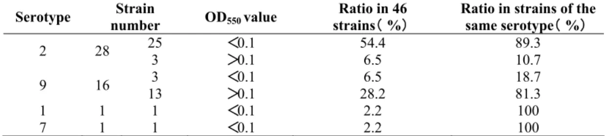
Table 2. Comparison of biofilm formation activity by S. suis of different serotypes
Serotype Strain
number OD550 value
Ratio in 46 strains(% )
Ratio in strains of the same serotype(% )
25 <0.1 54.4 89.3
2 28
3 >0.1 6.5 10.7
3 <0.1 6.5 18.7
9 16
13 >0.1 28.2 81.3
1 1 1 <0.1 2.2 100
7 1 1 <0.1 2.2 100
Effect of antimicrobial agents against biofilm formation in vitro by the TCP assay
Five antimicrobial agents commonly used for treatment of S. suis infection in clinics, include penicillin, erythromycin, azithromycin, ciprofloxacin, and ofloxacin. Their MICs against S. suis NJ-3 were 0.063, 1, 0.0039, 1 and 1mg·L-1 respectively. They inhibited biofilm formation of S. suis NJ-3 at subinhibitory concentration1 / 16 × MIC, 1 / 8 × MIC, 1 / 4 × MIC and1 / 2 × MIC. The inhibitory effect gradually increased
by increasing the concentration of antibiotics. The inhibitory effect of biofilm formation by different antimicrobial agents varied substantially. Among the five antimicrobial agents, the inhibition by ciprofloxacin and ofloxacin at 1/8 × MIC, 1/4 × MIC and1/2 × MIC was stronger than other three antimicrobial agents. The inhibition of biofilm formation by azithromycin at 1/8 × MIC and 1/4 × MIC was not stronger than that of erythromycin, while at 1/2 × MIC, the inhibitory activity of azithromycin was stronger than that of erythromycin (Fig. 5).
Figure 5. Effect of penicillin, erythromycin, azithromycin, ciprofloxacin, and ofloxacin at different concentrations on S. suis NJ-3 biofilm formation, as measured by CV staining.
DISCUSSION
A wide range of bacteria can form biofilms under various conditions. In many cases, biofilms are surface-attached
be prevalent especially in field isolates, suggesting possible role of biofilm in S. suis pathogenesis (11, 14). Early detection and management of potentially pathogenic S. suis biofilm can be one of the essential steps towards prevention and management of device-associated infections. There is also a need to evaluate a simple method for detection of biofilm forming by S. suis. Based on these objectives, we investigated S. suis adhesion and biofilm formation on rough and smooth surfaces by three different methods.
TCP assay is based on the ability of bacteria to form biofilms on the bottom of tissue culture plates, which is widely used to determine the formation of bacteria biofilms (14, 17, 20). Bacteria are cultured in the wells of tissues culture plates, and the presence or absence of a biofilm is detected by staining the wells with crystal violet. Mathur et al. (14) reported the correlation between OD values and biofilm formation. Based on this, comparison of the ability of 46 S. suis isolates to form biofilms on plastic surfaces was conducted by this method in this study.
Previous studies have indicated that the nutritional content of the growth medium can regulate biofilm formation (7, 16). In general, bacteria tend to adhere to available surfaces and form mature biofilm in environments that provide sufficient nutrients but will not adhere to surfaces in environments that are nutrients deficient. S. suis NJ-3 biofilm formation was tested in a variety of glucose concentration. The results proved that the increased glucose concentration promoted biofilm formation in S. suis (Fig. 3), which is in agreement with the results of previous studies done on other bacteria (4, 18). It is also found that 0.5% NaCl in medium is the optimal concentration of S. suis biofilm formation (Fig.3). These results support the notion that bacteria form biofilm under favorable nutrient conditions (6). Therefore, the optimized THB including 1% glucose and 0.5% NaCl was used to analyze biofilm-forming ability of different S. suis strains. The result showed that the biofilm forming ability was different in all 46 S. suis stains at 24 h (Fig.4). Grenier et al. (10) reported that 25
S. suis strains posses their ability to form biofilms, among which a strain, 95-8242, of S. suis serotype 2 isolated from a case of meningitis in pigs formed a significant biofilm. Our results showed difference with that of Grenier et al. In this study, the average biofilm-forming ability of S. suis serotype 9 stains was stronger than that of type 2 strains. The reason for this discrepancy remains to be further studied. In our study we also found that two S. suis type 2 isolates (98012 and P4254) obtained from human infected with S. suis, showed the strongest biofilm forming abilities. It is possible that the biofilm-forming ability of S. suis may be related with its virulence but possibility remains to be determined.
bacteria (red) and the production of exopolysaccharides (green) (Fig. 2). As a matter of fact, polysaccharides are belived to be a major prerequisite for the production of biofilm (2). These findings further confirm that S. suis NJ-3 can form biofilm in vitro. Although these methods have their advantages, the TCP method has a high specificity, sensitivity, and positive predictive value. Thus, it is suitable for screening biofilm formtion in large numbers of samples.
Most antibiotics of clinical relevance are derivatives of naturally occurring microbial products that probably function in microbial competition within environment niches. β-lactam antibiotics, macrolide antibiotics, and fluoroquinolone antimicrobial agents are commonly used drugs in the treatment of S. suis infection. Upon treatment with these antimicrobial agents, a fraction of S. suis is inevitably exposed to subinhibitory level of the agents. Therefore, we studied the effect of these commonly used antimicrobial agents in the subinhibitory concentrations on S. suis biofilm formation. Five antimicrobial agents, including penicillin, erythromycin, azithromycin, ciprofloxacin, and ofloxacin, can inhibit biofilm formation of S. suis NJ-3 at subinhibitory concentration, and this inhibition is a dose-dependent manner, i.e., the higher the concentration of antimicrobial agents, the stronger the inhibition of biofilm formation. In addition, the inhibition of biofilm formation varied among the antimicrobial agents. Numerous reports have described biofilm formation in the presence of subinhibitory concentrations of antimicrobial agents (3, 9, 21, 22). Yassien et al. (28) reported that subinhibitory concentrations (1/2, 1/4, and 1/8 of the MIC) of fluoroquinolones (ciprofloxacin, norfloxacin, pefloxacin, and ofloxacin) reduced the adherence of P. aeruginosa to 30 to 33, 44 to 47, and 61 to 67% of that of controls, respectively. Shibl (23) found that subinhibitory concentrations of erythromycin decreased adherence of S. aureus to tissue culture plates. Our results are in agreement with these previous findings (3, 21, 22). Although our results showed a better activity of ciprofloxacin and ofloxacin against S. suis biofilm formation, further studies should be conducted to confirm and clarify the
relationship between the relative adherence-inhibiting properties of ciprofloxacin and ofloxacin and their mechanisms. We also need to detect if the biofilm has formed, whether the common used antibiotics could killed the bacteria aggregated in biofilm or not. It will provide information for clinical use of antimicrobial agents against biofilm formed bacteria.
The study could be extrapolated on more isolates with respect to biofilm property along with analysis of the biofilm encoding genes that may lead to better understanding of the determinants involved in the adherence of the organisms growing as biofilms. Further studies are also required to define more precisely the extent to which the organism forms biofilms during colonization.
CONCLUSIONS
In summary, this study provides a detailed analysis in understanding biofilm formation potential that could be one of the important virulent and antimicrobial resistant mechanisms associated with S. suis induced pathogenesis in human and animals. This study may eventually lead to a better understanding of how to eradicate S. suis growing as biofilms with antibiotic therapy.
ACKNOWLEDGEMENTS
This work was supported by grants from the National Natural Science Foundation of China (No. 30972220), Special Funding of Public Sector Agricultural Research Project (No. 200803016) and A Project Funded by the Priority Academic Program Development of Jiangsu Higher Education Institutions (2011).
REFERENCES
1. Boyd, A.; Chakrabarty, A.M. (1995). Pseudomonas aeruginosa biofilms: role of the alginate exopolysaccharide. Indian J of Microbiol. 15:162-168.
(2007). Human meningitis caused by Streptococcus suis: the first case report from north-eastern Italy. Infez Med ,15:111-114.
3. Carsenti-Etesse, H.; Durant, J.; Entenza, J.; Mondain, V.; Pradier, C.; Bernard. E.; Dellamonica, P. (1993). Effects of subinhibitory concentrations of vancomycin and teicoplanin on adherence of
staphylococci to tissue culture plates. Antimicrob Agents and Chemother.
37:921-923.
4. Christensen, G.D.; Simpson, W.A.; Younger, J.J.; Baddour, L.M.; Barrett, F.F.; Melton, D.M. (1985). Adherence of coagulase-negative staphylococci to plastic tissue culture plates: a quantitative model for the adherence of staphylococci to medical devices. J Clinical Microbiol., 22:996–1006.
5. Costerton, J.W.; Lewandowski, Z.; Caldwell, D.E.; Korber, D.R.; Lappin-Scott, H.M. (1995). Microbial biofilms. Annual Review of
Microbiol. 49:711-745.
6. Costerton, J.W.; Stewart, P.S.; Greenberg, E.P. (1999). Bacterial biofilms: a common cause of persistent infections. Science. 284: 1318-1322.
7. Dewanti, R.; Wong, A.C. (1995). Influence of culture conditions on biofilm formation by Escherichia coli O157:H7. International J Food
Microbiol. 26: 147-164.
8. Donlan, R.M.; Murga, R.; Bell, M.; Toscano, C.M.; Carr, J.H.; Novicki, T.J. (2001). Protocol for detection of biofilms on needleless connectors attached to central venous catheters. J Clinical Microbiol. 39: 750–753. 9. Dunne, W.M.Jr. (1990). Effects of subinhibitory concentrations of
vancomycin or cefamandole on biofilm production by coagulase-negative staphylococci. Antimicrob Agents and Chemother. 34:390-393. 10. Grenier, D., Grignon, L., Gottschalk, M. (2009). Characterisation of
biofilm formation by a Streptococcus suis meningitis isolate. Vet J .179: 292-295.
11. Kay, R.; Cheng, A.F.; Tse, C.Y. (1995). Streptococcus suis infection in Hong Kong. QJM , 88: 39-47.
12. Lun, Z.R.; Wang, Q.P.; Chen, X.G.; Li, A.X.; Zhu, X.Q. (2007).
Streptococcus suis: an emerging zoonotic pathogen. The Lancet Infect
Dis, 7: 201-209.
13. Martin, C.; Heidt, W.M.; Torsten, H.; Robert, P.; Trinad, C.; Domann, E. (2005). Human infective endocarditis caused by Streptococcus suis serotype 2. J Clinical Microbiol , 43: 4898–4901.
14. Mathur, T.; Singhal, S.; Khan, S.; Upadhyay, D.J.; Fatma, T.; Rattan, A. (2006). Detection of biofilm formation among the clinical isolates of
Staphylococci: an evaluation of three different screening methods. Indian
J Medical Microbiol. 24:25–29.
15. May, T.B.; Shinabargerand, D.; Maharajand, R.; Katoand, J.; Chuand, L.; DeVault, J.D. (1991). Alginate synthesis by Pseudomonas aeruginosa: a key pathogenic factor in chronic pulmonary infections of cystic fibrosis patients. Clinical Microbiol Review. 4:191-206.
16. Oh, Y.J.; Jo, W.; Yang, Y.; Park, S. (2007). Influence of culture conditions on Escherichia coli O157:H7 biofilm formation by atomic force microscopy. Ultramicroscopy . 107: 869–874.
17. Okajima, Y.; Kobayakawa, S.; Tsuji, A.; Tochikubo, T. (2006). Biofilm formation by Staphylococcus epidermidis on intraocular lens material.
Invest Ophth and Vis Sci., 47:2971–2975.
18. O'Toole, G.A.; Kolter, R. (1998). Initiation of biofilm formation in
Pseudomonas fluorescens WCS365 proceeds via multiple, convergent
signalling pathways: a genetic analysis. Molecular Microbiol. 28:449-461.
19. Patel, R. (2005). Biofilms and antimicrobial resistance. Clinical Orthop
and Relat Res., 437:41–47.
20. Presterl, E.; Suchomel, M.; Suchomel, M.; Eder, M.; Reichmann, S.; Lassnigg, A.; Graninger, W.; Rotter, M. (2007). Effects of alcohols, povidone-iodine and hydrogen peroxide on biofilms of Staphylococcus
epidermidis. J Antimicrob Chemother, 60:417–420.
21. Rachid, S.; Ohlsen, K.; Witte, W.; Hacker, J.; Ziebuhr, W. (2000). Effect of subinhibitory antibiotic concentrations on polysaccharide intercellular adhesin expression in biofilm-forming Staphylococcus epidermidis.
Antimicro Agents and Chemother. 44: 3357-3363.
22. Rupp, M.E.; Hamer, K.E. (1998). Effect of subinhibitory concentrations of vancomycin, cefazolin, ofloxacin, L-ofloxacin and D-ofloxacin on adherence to intravascular cathetersand biofilm formation by
Staphylococcus epidermidis. J Antimicrob Chemother. 41:155-161.
23. Shibl, A.M. (1987). Influence of subinhibitory concentrations of antibiotics on virulence of Staphylococci. Rev Infec Dis. 9:704-712. 24. Staats, J.J.; Feder, I.; Okwumabua, O.; Chengappa, M.M. (1997).
Streptococcus suis: past and present. Vet Res Commun, 21: 381-407.
25. Takenaka, S.; Iwaku, M.; Hoshino, E. (2001). Artificial Pseudomonas
aeruginosa biofilms and confocal laser scanning microscopic analysis. J
Infect Chemother. 7:87-93.
26. Wei, Z.G.; Li, R.; Zhang, A.D.; He, H.K.; Hua, Y.F.; Xia, J.; Cai, X.H.; Chen, H.C.; Jin, M.L. (2009). Characterization of Streptococcus suis isolates from the diseased pigs in China between 2003 and 2007. Vet
Microbiol. 137:196–201.
27. Wisselink, H.J.; Smith, H.E.; Stockhofe-Zurwieden, N.; Peperkamp, K.; Vecht, U. (2000). Distribution of capsular types and production of muramidase-released protein (MRP) and extracellular factor (EF) of Streptococcus suis strains isolated from diseased pigs in seven European countries. Vet Microbiol.74: 237-248.
28. Yassien, M.; Khardori, N.; Ahmedy, A.; Toama, M. (1995). Modulation of biofilms of Pseudomonas aeruginosa by quinolones. Antimicrob
Agents and Chemother. 39: 2262–2268.
Ou, Y.B.; Ye, C.Y.; Jin, D.; Lu, Q.; Cui, Z.G.; Huang, Y.; Zhang, S.Y.; An, X.D.; Huang, T.; Zhou, X.Y.; Feng, L.; Pang, Q.D.; Shu, Y.L.;
Wang, Y. (2006). Human Streptococcus suis outbreak, Sichuan, China.
Emerg Infect Dis, 12:914-920.