J of Evolution of Med and Dent Sci/ eISSN- 2278-4802, pISSN- 2278-4748/ Vol. 4/ Issue 25/ Mar 26, 2015 Page 4386
A CASE OF RIGHT SIDED INFECTIVE ENDOCARDITIS WITH REACTIVE KNEE
ARTHRITIS AND ACUTE KIDNEY INJURY
Suresh Babu S1, A. K. Badrinath2, K. Suresh3
HOW TO CITE THIS ARTICLE:
Suresh Babu S, A. K. Badrinath, K. Suresh. A Case of Right Sided Infective Endocarditis with Reactive Knee Arthritis and Acute Kidney Injury. Journal of Evolution of Medical and Dental Sciences 2015; Vol. 4, Issue 25, March 26; Page: 4386-4394, DOI: 10.14260/jemds/2015/634
ABSTRACT: Right-sided infective endocarditis (RSIE) occupies 5% of infective endocarditis (IE). Here we present a 42 year old male who presented to us with the complaints of fever, bilateral knee swellings, loss of appetite, weight loss and decreased urine output for one month. Patient had pan systolic murmur in the tricuspid area with basal crepitation. 2D ECHO showed mass in the tricuspid leaflet protruding into the right atrium with severe tricuspid regurgitation. His renal parameters were elevated and blood culture was positive for coagulase negative staphylococ ci. The patient was diagnosed as right sided infective endocarditis with reactive knee arthritis and acute kidney injury.
INTRODUCTION: Infective endocarditis (IE) involves the aortic valve most commonly, the mitral valve is second valve most commonly involved while tricuspid and pulmonary valves are least commonly involved. Multiple valve involvements are usually seen in 17-22% of the patients. Infection localized to the right heart valves occurs in about 5% of cases of infective endocarditis. In the western countries this disease is seen mainly in injection drug abusers and as a complication of indwelling catheters in the subclavian vein. In spite of the high prevalence of rheumatic heart disease in our environment the right sided infective endocarditis of the heart is a rare presentation. The diagnosis is rendered more difficult when signs of left heart disease overshadow that of the right side. And also there have been only scant reports of right-sided infective endocarditis (RSIE). This article deals with the use of two dimensional echocardiography in the diagnosis of RSIE in a population in which rheumatic heart disease is more common and intravenous drug abuse uncommon.
CASE REPORT: A 42 years old gentleman, tailor presented with the complaints of high grade fever with chills and rigor for one month. He also complained of bilateral knee joint swelling with pain and restricted movements. Patient also had decreased urine output, b/l leg swellings, loss of appetite and weight loss. He had jaundice one month ago. Patient is a smoker and alcoholic, no history of drug addiction. Patient was treated as CKD at a PHC and came here for further management.
ON EXAMINATION: Patient is conscious and oriented, thin built and poorly nourished. He is pale with bilateral pitting pedal edema. Bilateral knee joint swelling with warmth and tenderness present. His pulse is 80 beats/min, normal rhythm and volume, BP normal. On auscultation S1S2 present, a pan systolic murmur was heared in the tricuspid area. The respiratory rate 20/min and there was bilateral fine basal crepitations. Abdomen soft, no organomegaly and CNS intact.
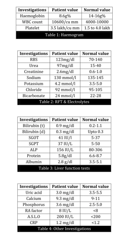
J of Evolution of Med and Dent Sci/ eISSN- 2278-4802, pISSN- 2278-4748/ Vol. 4/ Issue 25/ Mar 26, 2015 Page 4387 Investigations Patient value Normal value
Haemoglobin 8.6g% 14-16g% WBC count 10600/cu mm 4000-10000
Platelet 3.5 lakh/cu mm 1.5 to 4.0 lakh
Table 1: Haemogram
Investigations Patient value Normal value
RBS 123mg/dl 70-140
Urea 97mg/dl 15-40
Creatinine 2.6mg/dl 0.6-1.0 Sodium 130 mmol/l 135-145 Potassium 4.2 mmol/l 3.5-5.0
Chloride 92 mmol/l 95-105 Bicarbonate 24 mmol/l 22-28
Table 2: RFT & Electrolytes
Investigations Patient value Normal value Bilirubin (t) 0.9 mg/dl 0.2-1.1 Bilirubin (d) 0.3 mg/dl Upto 0.3
SGOT 41 IU/l 5-37
SGPT 37 IU/L 5-50
ALP 156 IU/L 80-306
Protein 5.8g/dl 6.6-8.7 Albumin 2.8 g/dl 3.5-5.1
Table 3: Liver function tests
Investigations Patient value Normal value Uric acid 3.0 mg/dl 3.5-5.5
Calcium 9.3 mg/dl 9-11 Phosphorus 3.6 mg/dl 2.5-5.0
RA factor 8 IU/L <8 A.S.L.O 200 IU/L <200
J of Evolution of Med and Dent Sci/ eISSN- 2278-4802, pISSN- 2278-4748/ Vol. 4/ Issue 25/ Mar 26, 2015 Page 4388 URINE EXAMINATION:
Sugar – nil. Albumin - +. Pus cells – plenty. Spot sodium - 47Meq/L. Spot protein - 46.7 mg/l.
24 hour urinary protein- 1120 mg/day.
VIRAL MARKERS: HIV I and II – negative. HbsAg – negative. HCV – negative.
SYNOVIAL FLUID: C/S – No growth.
ECG: Normal sinus rhythm, within normal limits.
USG Abdomen- Grade I medical renal disease. Rt. kidney – 12.3 X 4.8 cm.
Lt. Kidney – 12.0 X 4.5 cm.
BLOOD CULTURE:
1 – Coagulase negative staphylococci (moderate growth) 2 – Contamination
3 – Coagulase negative staphylococci (moderate growth)
HACEK/FUNGAL – No growth.
Review Blood culture – Heavy growth of staphylococcus aureus.
Figure 1: 2D –ECHO - Parasternal long axis view showing normal sized aorta, left atrium, left ventricle and normal mitral valve.
J of Evolution of Med and Dent Sci/ eISSN- 2278-4802, pISSN- 2278-4748/ Vol. 4/ Issue 25/ Mar 26, 2015 Page 4389 Figure 2: 2D-ECHO - Apical four chamber view showing dilated right atrium and right ventricle with a mass vegetation in the atrial surface of the tricuspid valve protruding into the right atrium.
Figure 3: 2D- ECHO -Apical 4chamber view with colour Doppler showing severe tricuspid regurgitation.
Figure 4: 2D- ECHO- with colour doppler showing severe tricuspid regurgitation. Figure 2
Figure 3
J of Evolution of Med and Dent Sci/ eISSN- 2278-4802, pISSN- 2278-4748/ Vol. 4/ Issue 25/ Mar 26, 2015 Page 4390 2D ECHO REPORT:
RA, RV mildly dilated;
Mass vegetation attached to the atrial surface of the tricuspid leaflet prolapsing into RA. With Severe Tricuspid Regurgitation (+).
Impaired diastolic function.
TREATMENT: The patient was treated with iv antibiotics- Inj. CEFTRIAXONE 2g iv BD for 2weeks and other supportive measures were given. The patient gradually improved and was discharged after 20days of hospitalization.
DISCUSSION: INFECTIVE ENDOCARDITIS (IE) involves the aortic valve the most commonly, the mitral valve second most commonly and infection localized to the right heart valves occurs in about 5% of cases of infective endocarditis. This disease is seen mainly in drug abusers and as a complication of indwelling catheters in the subclavian vein, and among injection drug users, commonly the tricuspid valve is involved and commonly caused by staphylococcus aureus, of which many strains are methicillin resistant.
Staphylococcus aureus has become the most common microorganism of Infective Endocarditis, while incidence of streptococcus viridians infections has reduced. Among IV drug abusers, group A streptococci have caused tricuspid valve IE similar to that noted with staphylococcus aureus. Other causative organisms are, pseudomonas aeruginosa, candida species and sporadically by unusual organisms such as bacillus, lactobacillus and corynebacterium species.
The clinical manifestations of IE in depend on the valves involved and infecting organism. The symptoms include Pleuritic chest pain, breathlessness, cough, and haemoptysis occur with tricuspid valve endocarditis, particularly when it is caused by staphylococcus aureus. In 65% to 75% of patients, chest radiograph reveals abnormalities related to septic pulmonary emboli. Murmurs of tricuspid regurgitation are noted in less than half of these patients. Infection of the aortic or mitral valve in addicts clinically resembles IE seen in patients who are not drug abusers. HIV infection has been noted in 27% to 73% of IV drug abusers with IE.
Organism
Percentage of cases- Native valve endocarditis
Percentage of cases of prosthetic valve endocarditis- months of
onset after valve surgery
Endocarditis in injection drug users Community-acquired (n-1718) Health care associated (n-788) <2months (n-144) 2-12 months (n-31) >12 months (n-194) Right sided (n-346) Left sided (n-204) Total (n-675)
Streptococci 40 9 1 9 31 5 15 12
Pneumococci 2 - - - -
Enterococci 9 13 8 12 11 2 24 9
Staph.aureus 26 53 22 12 18 77 23 57 Coagulase –ve
staphylococci 5 12 33 32 11 - - - Fastidious
gram-ve cocco Bacilli
J of Evolution of Med and Dent Sci/ eISSN- 2278-4802, pISSN- 2278-4748/ Vol. 4/ Issue 25/ Mar 26, 2015 Page 4391
Gram -ve
bacilli 1 2 13 3 6 5 13 7
Candida spp <1 2 8 12 1 - 12 4
Polymicrobial 3 4 3 6 5 8 10 7
Diphtheria - <1 6 - 3 - - 0.1
Culture –ve 9 5 5 6 8 3 3 3
Table 5
DIAGNOSIS OF INFECTIVE ENDOCARDITIS: MAJOR CRITERIA: Positive blood culture.
1) Typical microorganism for infective endocarditis from two separate blood cultures.
i) Viridians streptococci, Streptococcus gallolyticus, HACEK group, Staphylococcus aureus, or
ii) Community-acquired enterococci in the absence of a primary focus, or
Persistently positive blood culture, defined as recovery of a microorganism consistent with infective endocarditis from blood cultures drawn >12 h apart; or all of 3 or a majority of 4 separate blood cultures, with first and last drawn at least 1 h apart or single positive blood culture for Coxiella burnetii or phase I IgG antibody titer of >1:800
Evidence of endocardial involvement.
2) Positive echocardiogram - Oscillating intracardiac mass on valve or supporting structures or in the path of regurgitant jets or in implanted material, in the absence of an alternative anatomic explanation, or abscess, or new partial dehiscence of prosthetic valve, or new valvular regurgitation (increase or change in preexisting murmur not sufficient).
MINOR CRITERIA:
1) Predisposing heart condition or injection drug use. 2) Fever 38.0°C (100.4°F).
3) Vascular phenomena: major arterial emboli, septic pulmonary infarcts, mycotic aneurysm, intracranial haemorrhage, conjunctival haemorrhages, Janeway lesions.
4) Immunologic phenomena: glomerulonephritis, Osler's nodes, Roth's spots, rheumatoid factor. 5) Microbiologic evidence: positive blood culture but not meeting major criterion as noted
previously or serologic evidence of active infection with organism consistent with infective endocarditis
6) Echo: consistent with endocarditis but do not meet a major criterion as noted above.
Infective endocarditis is diagnosed by the documentation of two major criteria, or one major and three minor criteria, or five minor criteria.
INVESTIGATIONS: Blood culture.
Non- blood culture tests. a) Serologic tests.
J of Evolution of Med and Dent Sci/ eISSN- 2278-4802, pISSN- 2278-4748/ Vol. 4/ Issue 25/ Mar 26, 2015 Page 4392 c) Microscopic examination with special stains (Periodic Acid – Schiff for Tropheryma
whipplei).
d)PCR to recover unique microbial DNA.
IMAGING MODALITIES USED IN INFECTIVE ENDOCARDITIS:
Trans thoracic echocardiography (TTE) is non-invasive and exceptionally specific, it cannot image vegetations <2 mm in diameter, TTE detects vegetations in only 65% of patients with definite clinical endocarditis.
Trans esophageal echocardiography (TEE) is safe and detects vegetations in >90% of patients with definite endocarditis; TEE is the optimal method for the diagnosis of pulmonary valve endocarditis or the detection of myocardial abscess, valve perforation, or intra cardiac fistulae. Right-sided Valvular vegetations are often larger than left-sided ones.
Other investigations done are complete blood count, serum creatinine levels, liver function tests, chest radiography, erythrocyte sedimentation rate, C-reactive protein level, and circulating immune complex titre are commonly increased in endocarditis
Cardiac catheterization is useful primarily to assess coronary artery patency in older individuals who are to undergo surgery for endocarditis.
Organism (Streptococci) Drug (dose, duration)
Penicillin – susceptible streptococci, S. Gallolyticus
Penicillin G (2-3mU iv q4h for 4weeks). Ceftriaxone (2g/day iv as a single dose for
4weeks).
Vancomycin (15 mg/kg iv q12h for 4weeks). Penicillin G (2-3mU iv q4h) or Ceftriaxone (2g/day
iv). for 2weeks plus Gentamicin (3mg/kg qd iv or im as a single dose or divided into equal doses q8h for 2weeks.
Relatively penicillin-resistant streptococci
Penicillin G (4mU iv q4h0 or Ceftriaxone (2g iv qd) for 4 weeks plus Gentamicin (3mg/kg qd iv or im as a single dose or divided into equal doses q8h) for 2weeks.
Vancomycin (15 mg/kg iv q12h for 4weeks.
Streptococci- Moderately penicillin resistant, nutritionally variant organisms, or Gemella morbillorum
Penicillin G (4-5mU iv q4h) Ceftriaxone (2g iv qd) for 6weeks plus.
Gentamicin (3mg/kg qd iv or im as a single dose or divided into equal doses q8h) for 6weeks. Vancomycin as above for 4weeks.
Table 6: Treatment
Staphylococci
Methicillin-susceptible, infecting native valves
Nafcillin or oxacillin (2g iv q4h for 4-6weeks). Cefazolin (2g iv q8h for 4-6weeks).
Vancomycin (15mg/kg iv q8-12h for 4-6weeks). Methicillin-resistant,
J of Evolution of Med and Dent Sci/ eISSN- 2278-4802, pISSN- 2278-4748/ Vol. 4/ Issue 25/ Mar 26, 2015 Page 4393 Methicillin susceptible,
infecting prosthetic valves
Methicillin resistant, infecting prosthetic valves
Nafcillin or oxacillin (2g iv q4h for 4-6weeks). Gentamicin (1mg/kg im or iv q8h for 2weeks) plus
Rifampin (300mg PO q8h for 6-8weeks).
Vancomycin (15mg/kg iv q12h for 6-8weeks) plus. Gentamicin (1mg/kg iv or im q8h for 2weeks) plus
Rifampin (300mg PO q8h for 6-8weeks.
TABLE 7
Enterococci
Penicillin G (4-5 mU iv q4h) plus
Gentamicin (1mg/kg iv q8h), both for 4-6weeks Ampicillin (2g iv q4h) plus Gentamicin
(1mg/kg iv q8h), both for 4-6weeks. Vancomycin (15mg/kg iv q12h) plus
Gentamicin (1mg/kg iv q8h) both for 4-6weeks Table 8
Endocarditis, where the causative organism is Enterobacteriaceae is treated with a potent beta-lactam antibiotics plus an aminoglycoside and Corynebacterial endocarditis is treated with penicillin plus an aminoglycoside.
Therapy for Candida endocarditis consists of amphotericin B plus flucytosine and early surgery; long-term suppression with an oral azole is advised. Caspofungin treatment of Candida endocarditis has been effective in sporadic cases.
COMPLICATIONS: Cusp or leaflet rupture/flail perforation, abscess, aneurysm, fistula, dehiscence of prosthetic valve, pericardial effusion (more frequent with abscess). Hemodynamic compromise is valvular regurgitation, acute mitral regurgitation, acute aortic regurgitation, premature mitral valve closure, restrictive mitral inflow pattern, valvular stenosis and congestive heart failure, embolization into systemic, cerebral, pulmonary circulation. Tricuspid valve endocarditis causes pulmonary embolism in 69% of cases.
REFERENCES:
1. Adolf WK, Infective endocarditis, Harrison’s principles of internal medicine – 18th edition;
1052-63.
2. Shi Min Yuan, Right sided infective endocarditis recent epidemiologic changes, Int J Clin Exp Med; 2014; 7(1): 199-248.
3. Naidoo DP, Right sided endocarditis in the non-drug addict, Post Grad Med J, 1993; 69; 615-620.
J of Evolution of Med and Dent Sci/ eISSN- 2278-4802, pISSN- 2278-4748/ Vol. 4/ Issue 25/ Mar 26, 2015 Page 4394
AUTHORS:
1. Suresh Babu S. 2. A. K. Badrinath 3. K. Suresh
PARTICULARS OF CONTRIBUTORS:
1. Junior Resident, Department of General Medicine, Sri Manakula Vinayagar Medical College & Hospital, Pondicherry. 2. Professor & HOD, Department of General
Medicine, Sri Manakula Vinayagar Medical College & Hospital, Pondicherry.
FINANCIAL OR OTHER
COMPETING INTERESTS: None
3. Associate Professor, Department of General Medicine, Sri Manakula Vinayagar Medical College & Hospital, Pondicherry.
NAME ADDRESS EMAIL ID OF THE CORRESPONDING AUTHOR:
Dr. Suresh Babu S, S/o. V. Somasundaram, Thirupanchanur & Post,
Villupuram Taluk & District-605103, Tamilnadu.
E-mail: vsbsuresh87@gmail.com