Case Report
Introduction
Primary cardiac tumors are rare, with a prevalence of 200 per million autopsies, according to 22 large case series studies1. Most are considered benign, and myxomas are the most frequent ones (30% of cases), followed by lipomas (10%) and papillary fibroelastomas (8%), of which, according to a recent review, only 833 cases of the latter were detected worldwide from 2001 to 20082.
The PFE is a pedunculated endocardial, avascular and mobile tumor, predominantly found in the valves (accounting for 75% of all tumors in this region)1-4. They are usually asymptomatic, but there is risk of embolization1,4,5. Treatment is surgical in most cases and the capacity to diagnose them has increased in recent decades due to the improvement in imaging techniques4-6.
Case Reports
Case 01: ABS, male sex, 48 years. Dyspnea; syncope 8 months before. Catheterization: tumor in the aortic valve, severe aortic valve regurgitation. Tumor resection; repair of two aortic leaflets. Anatomopathological: PFE measuring 2 x 1 x 1 cm. Anticoagulated for 3 months. Lost to follow-up.
Case 02: JDF, female sex, 34 years, asymptomatic. Echocardiography: tumor in the right atrium. No surgical complications. Anatomopathological: PFE measuring 1.6 x 1.5 x 1.0 cm. Lost to follow-up.
Case 03: MGS, female sex, 42 years, asymptomatic. Echocardiography: small ostium secundum type atrial septal defect, aortic valve: fixed structure in the ventricular face of the right coronary leaflet; mitral valve: hyperechoic structure in the atrial face of the anterior leaflet, right infundibular hypertrophy. Vegetation removal, commissurotomy of the pulmonary valve, ASD closure. Anatomopathological: PFE (2 structures in the aortic valve, the largest measuring 1.0 x 1.0 x 0.9 cm; in the mitral valve, three fragments of 0.2 x 0.6 x 0.4 x 0.4 cm. Patient remains asymptomatic.
Case 04: VLMM, female sex, 65 years, asymptomatic. Echocardiography: homogeneous round mass, mobile, pending from the atrial face of the septal leaflet of tricuspid valve, pedunculated in the right atrium (RA). Nuclear magnetic resonance (NMR): extensive formation in tricuspid leaflet. Tumor resection in RA; tricuspid repair. Anatomopathological: PFE (1.8 x 1.5 x 0.8cm).
Case 5: AS, female sex, 48 years, in FCI-II. Echocardiography: aortic valve with slightly thickened leaflets, preserved opening and mobility. Pedunculated image, mobile, attached to the ventricular face of the aortic leaflet, suggesting vegetation. Evolution to functional class III and severe aortic regurgitation; valve replacement was the treatment of choice. Anatomopathological: PFE, 2 aortic cusps with one lesion in each (the largest with 0.4 cm diameter). At present, FC-II.
Case 6: MAAA, female sex, 65 years, followed due to aortic and mitral stenosis for 2 years, reported dyspnea on moderate exertion and syncope. Echocardiography: aortic valve with thickened leaflets, calcification, reduced opening and mobility. Aortic valve replacement was chosen. Anatomopathological: PFE (three sessile masses, largest: 0.7 x 0.5 x 0.2 cm, with a cluster of filiform formations). Currently FC-II.
Case 7: MCL, female sex, 66 years, asymptomatic. Echocardiography: image attached to the atrial face of the tricuspid valve (TV) septal leaflet, with round homogeneous aspect, regular borders, with no obstruction to valve flow. NMR: oval image attached to the atrial face of the TV septal leaflet, impregnation with iodine contrast. After five years of anticoagulation, the patient agreed to undergo surgery. New NMR: mass increase. The tumor was resected and the repair was carried out. Anatomopathological: PFE (1.6 x 1.3 x 1.9 cm), frequent interpapillary thrombi; pedicle formed by valve leaflet with avascular fibromyxoid extension.
Discussion
The PFE is a rare condition, representing less than 10% of benign cardiac tumors7. In 90% of cases, it affects the heart valves, although they are rarely associated with valvular dysfunction
Keywords
Fibroma/complications; fibroma/pathology; fibroma/ therapy; heart neoplasms/complications; heart neoplasms/ pathology.
The papillary fibroelastoma (PFE) is a benign heart tumor, mainly found in the valves. Most tumors are asymptomatic, but when present, they are nonspecific or related to embolic phenomena. They are usually diagnosed at routine imaging studies or valve surgery and autopsies. Its treatment is controversial, due to its rarity. We describe seven PFE cases diagnosed and treated at our institution between 1989 and 2010, which constitutes the largest national case series study of this pathology to date.
Papillary Fibroelastoma: Report of Seven Cases
Lucas Cronemberger Maia Mendes, Jônatas Melo Neto, Jonathan Batista Souza, Edileide de Barros Correia, Mabel
de Moura Barros Zamorano, Lílian Mary da Silva
Instituto Dante Pazzanese de Cardiologia, Vila Mariana – São Paulo – SP - Brazil
Mailing Address: Lucas Cronemberger Maia Mendes •
Rua Arruda Alvim, 73 / 23 - Bairro Pinheiros – 05410-020 – São Paulo – SP – Brasil
E-mail: [email protected], [email protected] Mnauscript received February 07, 2011; revised manuscript received May 25, 2011; accepted June 13, 2011.
Case Report
Mendes et al Papillary fibroelastoma: Report of seven cases
Arq Bras Cardiol 2012;98(3):e59-e61 Lambl’s excrescence (identical to the histological aspect of the PFE, but smaller and more often localized in the closing line of the atrioventricular valves), other cardiac tumors, valve tissue degeneration, thrombi and vegetations, either infectious or not2,8.
Considering its rarity, there are still controversies regarding therapy. The decision of surgical resection is based on size, location, mobility and presence of symptoms3. It is a consensus that surgery should be performed in symptomatic patients. When there is embolism, it is necessary to wait at least four weeks to prevent eventual hemorrhagic complication9. It carries a low risk and there have been no reports of recurrence. In asymptomatic patients, indication for surgery is not clearly defined and, in general, it is influenced by tumor mobility (independent predictor, characteristic of embolization and death), with the particularity that tumors on the right side should be removed in case of patent foramen ovale, due to the risk of paradox embolism1,3,5,10. Clinically monitored patients should be considered for anticoagulation, although the efficiency of this approach has not been supported by large studies, with reports of increased morbidity due to neurological events3,5.
Our statistics differ from that in the literature, probably due to the small sample size, although we found no national studies with larger samples. This is a rare condition, which requires further studies, currently limited to small case series.
Potential Conflict of Interest
No potential conflict of interest relevant to this article was reported.
Sources of Funding
There were no external funding sources for this study.
Study Association
This study is not associated with any post-graduation program.
and can be found in the papillary muscles, chordae tendineae and endocardium4,7. Most of them are small single tumors, < 10 mm in size (in 99% of cases, < 20 mm), with an aspect that resembles a sea anemone and affects preferentially the aortic valve (44%), followed by the mitral (35%), tricuspid (15%) and pulmonary valve (8%)1,2. Other locations include the left ventricle (10.85%), left atrium (1.97%), atrial septum (1.57%) and right atrium (2.36%)1,8. Our sample consisted of 14 tumors, of which 4 were single tumors. One case had three mitral and two aortic masses. Another case had three simultaneous aortic masses. Of the 14 tumors, seven were located in the aortic valve, three in the mitral valve, two in the right atrium, one in the tricuspid and one in the left ventricle, with a maximum diameter of 2 cm.
There are doubts concerning the malignancy of the PFE. Other hypotheses include hamartoma, thrombus and hyperplasia of the endocardium by genetic or external stimuli such as infections or trauma3,7,9. Due to their fragile and papillary nature, they can cause obstruction in the coronary ostium. They also act as a substrate for platelet aggregation, thus becoming a source of central or peripheral emboli, depending on their location3 (Figure 1) .
Although most of them are asymptomatic, there may be nonspecific symptoms such as chest pain, dyspnea, syncope and sudden death, in addition to valvular dysfunction leading to heart failure, as in case 1. Tumors on the left side can cause stroke (more common when the patient is symptomatic), myocardial infarction, mesenteric, renal, hepatic ischemia and acute pulmonary edema8. On the right side, they can generate arrhythmias, pulmonary embolism or mimic tricuspid stenosis9. They occur at any age but are more prevalent between 40 and 80 years and are slightly more prevalent in men1,4-6. In the cases reported here, age ranged from 34 to 66 years (mean 52.57 years), and 85.7% occurred in women.
The transthoracic echocardiography is the most widely used diagnostic technique, with a sensitivity of 88.9%, specificity of 87.8%, and accuracy of 88.4% when the tumor is larger than 0.2 cm1,3,5,8. The NMR better characterizes the mass in the valve leaflet and the tumor enhancement technique increases the accuracy (Figure 2). Cardiac catheterization is not currently justified1,5,8. The differential diagnosis includes
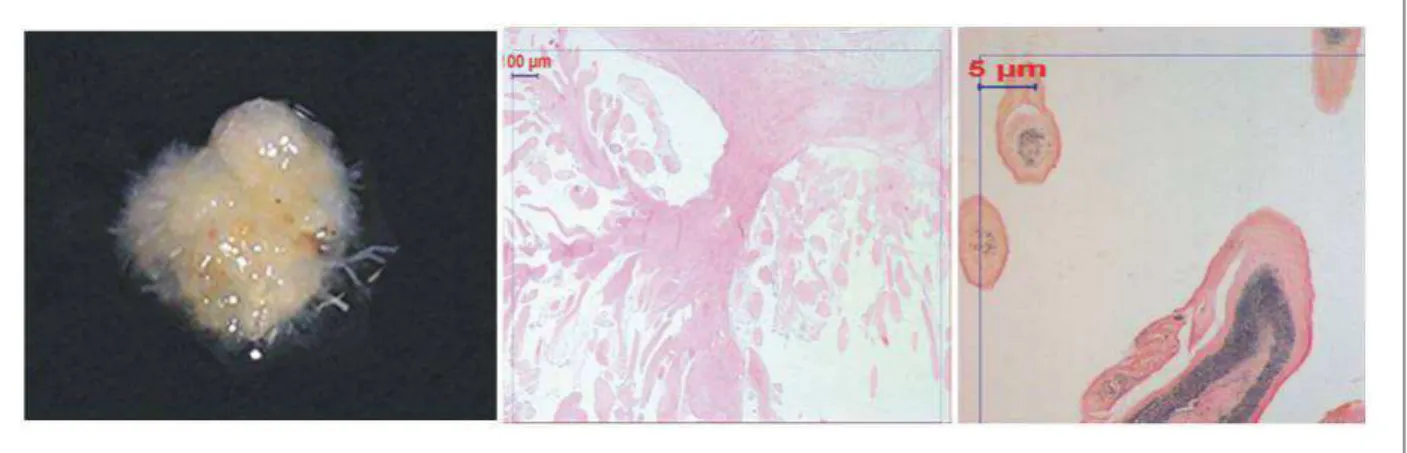
Figure 1 - Macroscopy: small papillae originating from one or more trunks, resulting in a spongy aspect (A). Microscopy (B) dendriform aspect, with branched papilla,
where elastic iber-rich thrombus ixation occurs (B.2).
Case Report
Mendes et al
Papillary fibroelastoma: Report of seven cases
Arq Bras Cardiol 2012;98(3):e59-e61
References
1. Gowda RM, Khan IA, Nair CK, Mehta NJ, Vasavada BC, Sacchi TJ. Cardiac papillary fibroelastoma: a comprehensive analysis of 725 cases. Am Heart J. 2003;146(3):404-10.
2. Law KB, Phillips KR, Cusimano RJ, Butany J. Multifocal ‘‘tapete’’ papillary fibroelastoma. J Clin Pathol. 2009;62(12):1066-70.
3. Mariscalco G, Bruno VD, Borsani P, Dominici C, Sala A. Papillary fibroelastoma: insight to a primary cardiac valve tumor. J Card Surg. 2010;25(2):198-205. 4. Biocic S, Puksic S, Vincelj J, Durasevic Z, Sutlic Z, Manojlovic S.
Pulmonary valve papillary fibroelastoma diagnosed by echocardiography: a case report. Eur J Echocardiogr. 2009;10(5):726-8.
5. Oliveira SF, Dias RR, Fernandes F, Stolf NA, Mady C, Oliveira SA. [Cardiac papillary fibroelastoma: experience of an institution]. Arq Bras Cardiol. 2005;85(3):205-7.
6. Chryssagis K, Liangos A, Westhof F, Batz G, Diegeler A. Transesophageal echocardiography for detection of a papillary fibroelastoma of the aortic valve. Hellenic J Cardiol. 2010;51(2):170-4.
7. Fabricius AM, Heidrich L, Gutz U, Mohr FW. Papillary fibroelastoma of the tricuspid valve chordae with a review of the literature. Cardiovasc JS Afr. 2002;13(3):122-4.
8. Vizzardi E, Faggiano P, Antonioli E, Zanini G, Chiari E, Nodari S, et al. Thrombus or tumor? a case of fibroelastoma as indicated during the submission process. Cases J. 2009;2(1):31.
9. Bicer M, Cikirikcioglu M, Pektok E, Muller H, Dettwiler S, Kalangos A. Papillary fibroelastoma of the left atrial wall: a case report. J Cardiothorac Surg. 2009;4:28.
Figure 2 - Magnetic Resonance: sessile mass in the atrial face of the tricuspid valve septal lealet (arrow).