PHYSICAL ACTIVITY AS A HEALTH FACTOR MODIFYING HEART RATE VARIABILITY (HRV) Nowosielska-Swadźba Danuta 1, Zwolińska Danuta 1
, Jendrysek Marek 1, Podstawski Robert 2. 1The Institute of Physical Culture, State Higher Vocational School in Racibórz, Poland 2Department of Physical Education and Sport, University of Warmia & Mazury in Olsztyn, Poland
Annotation:
Purpose: The aim of the research was the evaluation of the selected HRV factors of the training volleyball players in two training periods and non-training people. Materials and methods: The study involved 8 leading volleyball players aged 20-23 and 13 non-training persons aged 19-26. The study of the training players was conducted twice: in the pre-competition and in the pre-competition period. The study for the non-training persons was conducted once. The selected factors of the spectral analysis have been evaluated: TP [ms2], share of LF and HF power [n.u], LF/HF indicator and time analysis factors: RR [ms], HR [1/min], RMSSD [ms]. Results: Statistically significant differences appeared only in the selected time analysis factors (RR, HR), between the group of the training and non-training persons. Other differences in the evaluated parameters were not statistically significant. Conclusions: Physical activity influences on the HRV growth. HRV measurement may serve for the control of the changes taking place in the AUN under the influence of the physical activity.
Key words: Autonomic nervous system (AUN), heart rate variability (HRV), health, physical exercise adaptation.
Introduction1
Silvetti M. S. et al. [1] state that HRV is an important tool which can provide data about a modulatory influence of the autonomic nervous system (AUN) on the sinus node. The prognostic value of the heart rate variability (HRV) is used in the clinical medicine and sport [2]. As a result of the surveys, it has been stated that there is a correlation between disorders in the AUN and various diseases. HRV reduction has been noted in the cardiac disease e.g. chronic heart failure and non-cardiac e.g. diabetes [3, 4]. The factor influencing on HRV growth is physical activity.
Heart rate variability (HRV) is a normal, involuntary reaction to the changing environment conditions. The analysis of the heart rhythm variability gives the possibility to evaluate the activity of the sinus node [5]. HRV provides non-invasive information about AUN, evaluating the changes in the sympathetic and parasympathetic system at the sinus node level [6, 7, 2].
HRV is used in diagnosing many diseases, because it provides information about AUN and it is the factor of the changes in the cardio-vascular system [8]. HRV drop reflects the reduction of the vagus nerve tension and/or leads to electrical destabilization of the heart. Reduced HRV has been noted with people after a past heart attack, persons with hypertension, diabetic neuropathy, tetraplegia, depression [2, 9].The risk of the chronic diseases such as diabetes, hypertension, circulatory system diseases are frequently connected with obesity. Parasympathetic domination in the modulation of the heart rate appears with health people at rest, however, sympathetic domination predominates with people with heart diseases. HRV changes appear together with hypertension, AUN balance moves towards parasympathetic part. Analogical changes have been observed together with abdominal obesity evaluated on the base of waist circumference measurement and waist/hip indicator, on the other hand, such a dependence has not been noted relative to BMI factor [10]. According to Hottenrott K et al. [8], HRV factor can be used for the measurements of the endurance training effects. Furthermore, in the authors’ opinion, HRV can be a marker diagnosing overtraining and exhaustion [11]. Kiviniemi A.M. et al. [12] share similar opinion evaluating 26 young men divided into groups differentiated by the physical activity types. Additionally, the authors state that individually matched training may contribute to HRV growth. Quoted authors citing Hautal A.J. et al. [13] state that HRV growth in the high frequency band (HF: 0,15-0,4Hz), reflecting parasympathetic activity, is connected with growth of the VO2max factor. Bernardi L. et al. [14] state that changes in the levels of the physical activity are significant heart modulators. HRV higher values, extended intervals RR are connected with a higher activity of the vagus nerve. According to Buchheit M. et al. [15] moderate training raises vagal heart activity and contributes to reducing mortality caused by arrhythmia.
It is assumed that HRV is an effect of the different factors impact. The main factors affecting the sinoatrial node (sinus node pacemaker) are:
(a) tension oscillations between sympathetic and parasympathetic branch of the AUN, changes in the autonomic balance affect the amplitude spectrum proportions of low and high frequency,
(b) physical activity [2, 16].
Sport training, long-term physical activity trigger endobiotic changes which consolidate giving the image of the exercise adaptation [14, 16]. Numerous data from the written work prove that resting heart rate is lower with people doing sports than with not physically active ones [17, 18]. It is commonly believed that resting bradycardia appearing with athletes is caused by an increased tension of the vagus nerve. Such an observation may result from numerous surveys proving that hard (but not extreme), multi-month physical training of the athletes preparing for competitions leads to an increase in the activity of the parasympathetic component of the autonomic nervous system [19, 20].
© Nowosielska-Swadźba Danuta, Zwolińska Danuta, Jendrysek Marek, Podstawski Robert, 2015
O’Sullivan S.E. and Bell C. [21] state that duration of the exercise, and/or training type affects significantly the value of the HRV factors.
Materials and Methods Ethics
The study was conducted in accordance with the Helsinki Charter of Human Rights and it was approved by Ethics Committee of State Higher Vocational School in Raciborz. Each participant was willing to participate in the study voluntarily and confirmed that with a written agreement.
The study was conducted in the group of 8 contestants training volleyball. The study was carried out in two training periods: pre-competition and competition . The control group consisted of 13 physically inactive people.
The measurement of the tested persons was conducted with the use of Sport-Tester type Polar S 810i in the morning hours (7:00 – 10:00). The subjects were instructed to avoid physical activity, maintain a current food intake, with the exception of the consumption of alcohol and caffeine for 48 hours before testing. On a test day, the athletes were on empty stomach and the HRV measurement were made in the lying down position. The record of the systoles frequency lasted 15 minutes. Standard test conditions were kept for all of the contestants (temperature 20-22o C – thermo-neutral conditions) with keeping basic procedures compulsory in the sports metrology.
The record of the systoles frequency was transmitted to the computer memory and was processed in the statistics field with the use of the Polar Precision Performance 3 computer program, which is compatible with the HRV Analysis Software program (developed by The department of Applied Physics University of Kuopio in Finland). The methods of the spectral and time analysis were used for the development of the results. In the HRV Analysis program, the given incidence ranges were calculated: total power (TP – the range of the total power which is the sum of the ranges: ULF, VLF, LF, HF. The range between 0,01-0,5 Hz can be described as the sum of the ANS activity [22]), very Low Frequency (VLF – a band ranging within 0,0033 Hz – 0,05 Hz), LF (Low Frequency – a band ranging within 0,05 Hz - 0,15 Hz), HF (High Frequency – a band ranging within 0,15 Hz – 0.4 Hz) were calculated [23, 24].
From all the registered parameters, for further analysis the following indicators were chosen: TP [ms2], VLF [ms2, %], LF, HF [ms2, %, nu, the value LF/HF. In the time analysis were calculated: HR average [HR/min] systoles intensity. RR [ms] – the average time RR intervals between sinus stimulations, RMSSD [ms] – square root from the average sum of squares of differences between the next RR intervals [7, 25].
According to many authors, the component HF reflects mainly the influence of the parasympathetic system (vagal influence) [26]. They prove in many studies that LF factor shows the influences of both autonomic system branches i.e. sympathetic and parasympathetic parts [11]. The values of LF and HF can be given in the normalized units (nu). According to Malliani [27], the power of the LF and HF spectrum, especially when expressed in normalized units, reflects the balance between sympathetic and parasympathetic control. Moreover, sympathetic-parasympathetic balance is evaluated by the LF/HF indicator [28]. Hynen E. [29] states that the interpretation of the LF/HF indicator is doubtful, but it is applied as an indicator of the voltage state of an autonomic nervous system. The average RR interval time is a derivative of the systole HR incidence. The RMSSD indicator reflects the tension of the parasympathetic system. The factors of the spectral analysis: HF and time analysis: RMSSD, pNN50% correlate with each other, depend directly proportionally on the vagus nerve tension [2].
The results of the HRV analysis were juxtaposed in tables making standard statistical calculations according to the descriptive statistics module. Arithmetic mean, standard deviation and the significance of the differences between particular groups were calculated. In view of the small group size (8 and 13 people) and in case of some parameters in which the distribution was strongly agonic, to calculate statistically essential differences between the groups the non-parametric U Mann-Whitney test was used to compare two independent groups. For the level of significance, the value of p˂0,05 has been accepted. The calculations were made in the Statistica Pl v. 10 program [30].
Table 1 Somatic characteristic of 8 contestants training volleyball in the pre-competition and competition period and 13 young
men from the control group.
Parameters Pre-C period C period Control group
Mean ± standard deviation (min- max)
Age [yrs] 21,4 ± 1,14
(20 - 23)
22 ± 1,14 (20,6 - 23,6)
20,9 ±1,93 (19 - 26) Training experience
[yrs]
7,2 ± 2,28 (5 - 11)
7,8 ± 2,28 (5,6 - 11,6)
Body mass [kg] 86,35 ± 7,57
(76,9 - 97,2)
88,5 ± 6,52 (81 - 100)
82,4 ± 12,04 (65,7 - 107)
Body height [cm] 194,6 ± 4,96 (190 - 201)
194,5 ± 4,92 (190 - 201)
183,5 ± 4,85 (175 - 192)
BMI [kg/m2] 22,80 ± 1,9
(20,6 - 25,8)
23,37 ± 1,02 (22,2 - 24,8)
24,4 ± 2,76 (19,4 - 29)
Rohrer index [g/cm3] 1,17 ± 0,11 (1,03 - 1,36)
1,20 ± 0,06
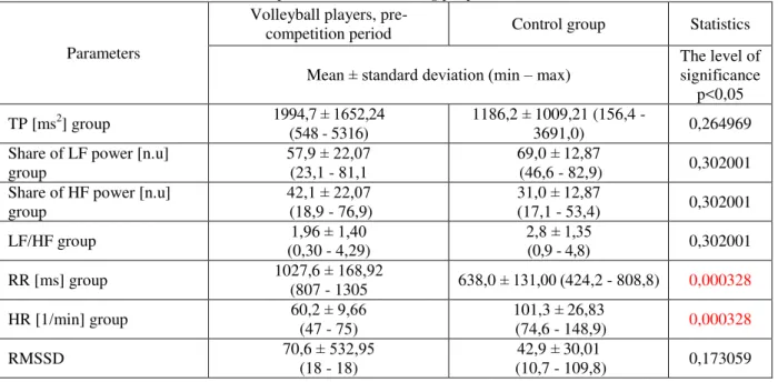
Table 2 Selected parameters of the spectral and time analysis in the tested volleyball players group in the pre-competition
period and non-training people.
Parameters
Volleyball players,
pre-competition period Control group Statistics
Mean ± standard deviation (min – max) The level of significance p<0,05
TP [ms2] group 1994,7 ± 1652,24
(548 - 5316)
1186,2 ± 1009,21 (156,4 -
3691,0) 0,264969
Share of LF power [n.u] group
57,9 ± 22,07 (23,1 - 81,1
69,0 ± 12,87
(46,6 - 82,9) 0,302001 Share of HF power [n.u]
group
42,1 ± 22,07 (18,9 - 76,9)
31,0 ± 12,87
(17,1 - 53,4) 0,302001
LF/HF group 1,96 ± 1,40
(0,30 - 4,29)
2,8 ± 1,35
(0,9 - 4,8) 0,302001
RR [ms] group 1027,6 ± 168,92
(807 - 1305 638,0 ± 131,00 (424,2 - 808,8) 0,000328
HR [1/min] group 60,2 ± 9,66
(47 - 75)
101,3 ± 26,83
(74,6 - 148,9) 0,000328
RMSSD 70,6 ± 532,95
(18 - 18)
42,9 ± 30,01
(10,7 - 109,8) 0,173059
Table 3 Selected parameters of the spectral and time analysis in the tested group of volleyball players in the competition period
and non-training people.
Parameters
Volleyball players,
competition period Control group Statistics
Mean ± standard deviation (min - max) significance The level of p<0,05
TP [ms2] group 2141,0 ± 2126,34 (856 - 7253)
1186,2 ± 1009,21 (156,4 -
3691,0) 0,148455
Share of LF power [n,u] group
64,2 ± 18,70 (30,2 - 83,3)
69,0 ± 12,87
(46,6 - 82,9) 0,649723
Share of HF power [n,u] group
35,8 ± 18,70 (16,7 - 69,8)
31,0 ± 12,87
(17,1 - 53,4) 0,649723
LF/HF group 2,4 ± 1,56
(0,43 - 4,98)
2,8 ±1,35
(0,9 - 4,8) 0,649723
RR [ms] group 944,5 ± 103,20
(753 - 1076)
638,0 ± 131,00
(424,2 - 808,8) 0,003754
HR [1/min] group 64,9 ± 7,60
(56 - 80)
101,3 ± 26,83
(74,6 - 148,9) 0,000611
RMSSD 61,8 ± 51,73
(30 - 187)
42,9 ± 30,01
(10,7 - 109,8) 0,173059
The evaluation by HRV of the changes taking place in AUN, affected by a long-term physical training volleyball players in the pre-competition and competition period and the control group showed that there were statistically significant changes in the field of the time analysis factors RR [ms] and HR [1/min]. In the physically inactive persons group there has been insignificant domination of the sympathetic part, in comparison to the physically active persons (LF n.u, LF/HF). In the volleyball players group, in the tested training periods, an insignificant growth in total power has been observed in comparison to the control group.
Discussion
in the sinus node and may cause a resting bradycardia observed with endurance athletes [33]. Stein R.M.C. et al. [34] state, in turn, that changes in the internal mechanisms of the sinus node, and not AUN, are responsible for a resting bradycardia. In our own research, there has been a decrease in heart rate with the volleyball players group, as a result of a long-term training. Both in the pre-competition and competition period, in comparison to the non-training group, decrease in heart rate (HR) and RR intervals showed a statistically significant difference. Baggish A.L. and Wood M.J. [35] state that heart rhythm disorders and changes in conduction, in trained athletes’ heart are well known. According to the cited authors, sinus bradycardia of the contestants is asymptomatic. It is caused by an increased activity of the parasympathetic system, and especially an increased activity of the vagus nerve. However, some data suggest that systematic, hard, but not extreme physical training can lead to internal chronotropic changes of the sinus node. Some of the changes may be treated as lesions [36, 37]. We cannot clearly determine where the heart’s adaptive changes finish in response to a long-term, regular physical effort. It may be assumed that cardiac changes with the people practicing sports professionally, do not need to be adaptive changes [38]. In our own researches, decreased heart rate in the group of people with an increased physical activity, has not been proved by the spectral analysis factors. In the obtain tests results, both in the contestants’ and control group, a sympathetic domination has been observed. However, in the contestants’ group, in the tested training periods, an insignificantly lower tension of the sympathetic part has been observed in comparison to the control group. In the competition period, a period of a high physical activity, in the volleyball players’ group, growth trends of the sympathetic part tension appeared in comparison to the pre-competition period. According to Iellamo F. et al. [20] growth of the sympathetic component, as a result of an intensified physical activity confirms good contestants’ preparation for starts and achieving high performances. Authors quoted above state that longer maintaining of sympathicotonia can contribute to contestants’ overtraining. Mourot L. et al. [39] confirm that too intensive trainings are connected with a balance shift in AUN towards sympathetic direction. The authors examined persons of a different physical activity (8 healthy men, 8 training, 7 with overtraining symptoms). On the base of the researches and the obtained results, it has been stated that the contestants with the overtraining symptoms showed an increase in sympathetic activity accompanied by the drop in parasympathetic activity and total power. Such a situation is connected with an overtraining of the Basedow type, which can appear with contestants [40]. Together with the training intensity reduction, the balance in AUN is restored. Nowosielska-Swadźba D [38]. examined boys aged 14-16 training athletics, volleyball and swimming. The results were compared to the physically inactive control group. A small domination of the sympathetic part has been marked in the control group. However, in the swimmers’ group, subjected to more physical load, there has been a trend reversal, from a characteristic vagotonia to sympathicotonia. In our own research, in the control group, with the sympathetic domination, a shortened RR intervals have been observed and a high frequency of the heart rate in comparison to volleyball players group. LF and LF/HF factors testify the balance shift towards sympathetic direction in the control group. In the control group, the average value of the heart rate was 101,3/min and it was 41,1 beats higher in comparison to the people training volleyball. In the control group, sympathetic predominance, low total power value may be the result of the reduced physical activity [2]. Karjalainen j. et al. [41] write that tachycardia is caused, among others, by an increased activity of the sympathetic system. Rajab M. et al. [42] state that the reduction of high-frequency component (HF) in the total heart rate variability may be a diabetes complication, a risk factor for a sudden death.
Many researchers believe that physical activity shifts the AUN balance towards parasympathetic direction, through which it reduces mortality among others in cardiac diseases [2]. Nowosielska-Swadźba D. [38] examining boys aged 10-12 with the increased physical activity and physically inactive stated that in the experimental groups, there has been an increase in the heart rate variability. At the same time, in the group with an increased physical activity, longer RR intervals and lower heart rate frequency have been marked. Sacknoff d. M. et al. [43] examining trained people and a control group state, that an endurance training raises vagal activity, which is connected with a reduction of a resting frequency of heart rate. Levy W. c. et al. [44] were examining two groups of people, a younger one, aged 24- 32 and older, aged 60-82. After a six-month sport training in the examined groups, they found a resting bradycardia through an increase in the vagal activity and sympathetic tension drop. A chronotropic activity drop has been observed. A resting HR drop and a growth of the resting HRV have been noticed in the examined group. Carter J.B. [45] came to similar observations examining mixed, recreational groups (6 men and 6 women) aged 19-21 and 40-45, in two periods before and after 12-month training. On the base of the heart rate variability record, he observed heart rate reduction and the total power growth with the examined persons. After the training period, the younger group showed a bigger growth of the total power. Melanson E.L. and Freedson P.S. [46] was examining men with the mean of age 36,6. After 16-month controlled, moderate power training, there has been a growth of the time and spectral analysis factors. However, many authors believe that HRV factors interpretation is often ambiguous. The surveys of the sportsmen showed that both HRV growth and drop can be connected with negative adaptation to exercise. Together with a positive adaptation of the circulatory system, a drop in the HRV value has been observed in the sportsmen group [47].
(BMI, Rohr factor) in the examined groups, it can be stated that both groups have a proper body weight and athletic type. However, in the group with a reduced physical activity, BMI factor is at the limit of the standard and overweight while Rohr factor is at the limit of the athletic and pyknic type (WHO Raport www.WHO.com) [50]. In the examined control group, with the lack of physical activity, BMI growth trends may occur.
Conclusions
1. HRV is an important tool which can provide data about the modulatory influence of the AUN on the sinus node.
2. Under the influence of a long-term sport training, there appear changes in the sinus node which is reflected in a resting bradycardia.
3. Physical activity affects total power growth which has a positive effect on health. 4. Sport training influences on a balance shift towards parasympathetic direction. 5. Too intensive physical exercise strengthens sympathetic domination.
References
1. Silvetti M.S., Drago F., Ragonese P. Heart rate variability in healthy children and adolescents is partially related to age and gender. Int J Cardiol, 2001, vol. 81(2-3), pp. 169-74.
2. Task Force. Task Force of the European Society of Cardiology and the North American Society of Pacing and Electrophysiology, Heart Rate Variability: standards of measurement physiological interpretation and clinical use. Eur. Heart. J., 1996, vol.17, pp. 354-381.
3. La Rovere M.T, Pinna G.D., Maestri R., Mortara A., Capomolla S., Febo O., Ferrari R., Franchini M., Gnemmi M., Opasich C., Riccardi P.G., Traversi E. & Cobelli F. Short-Term Heart Rate Variability Strongly Predicts Sudden Cardiac Death in Chronic Heart Failure Patients. Clinical Investigation and Reports.
Circulation, 2003, vol. 107, pp. 565-570.
4. Brunelli S., Traballesi M., Averna T., Porcacchia P., Polidori L. Di Carlo C., Di Giusto C., Marchetti M. Field tests for evaluating elite wheelchair basketball players. Fondazione Santa Lucia IRCCS, IWBF Europe, 2006, 200 p.
5. Jethon Z. Physical activity as a distress. Hygeia Public Health, 2013, vol. 48(2), pp. 156-161.
6. Sinnreich R., Kark J.D., Friedlander Y., Sapoznikov D., Luria M.H. Five minute recording of heart rate variability for population studies: repeatability and age-sex characteristics. Heart. 1998, vol. 80(2), pp. 156-162.
7. Cervantes Blásquez J. C., Rodas G., Capdevila Ortís L. Heart-rate variability and precompetitive anxiety in swimmers. Psicothema, 2009, vol. 21, (4), pp. 531-536.
8. Hottenrott K, Hoos O., Esperer H.D. Heart rate variability and physical exercise. Current status. Herz., 2006, Sep; vol. 31(6), pp. 544-52.
9. Lewis M.J., Short A.L. Exercise and cardiac regulation what can electrocardiographic time series tell us? Scand J Med. Sci Sports, 2010, vol. 20, pp. 794-804.
10. Farah B.Q., do Prado W.L., Tenório T.R., Ritti-Dias R.M. Heart rate variability and its relationship with central and general obesity in obese normotensive adolescents. Einstein, 2013, vol.11 (3), pp. 23-30.
11. Makivić B., Djordjević Nikić M., Willis M.S. Heart Rate Variability (HRV) as a Tool for Diagnostic and Monitoring Performance in Sport and Physical Activities. Journal of Exercise Physiologyonline, 2013, vol. 16 (3), pp.100-109.
12. Kiviniemi A.M., Hautala A.J., Kinnunen H., Tulpo M.P. Endurance training guided individually by daily heart rate variability measurements. Eur J Appl Physiol, 2007, http://orcid.org/10.1007/s00421-007-0552-2. 13. Hautala A.J., Mäkikallio T.H., Kiviniemi A., Laukkanen R.T., Nissilä S., Huikuri H.V., Tulppo M.P.
Cardiovascular autonomic function correlates with the response to aerobic training in health sedentary subjects. Am J Physiol Heart Circ Physiol, 2003, vol. 285(4), pp. 1747-1752.
14. Bernardi L., Valle a F., Coca a M., Calciati a A., Sleight P. Physical activity influences heart rate variability and very-low-frequency components in Holter electrocardiograms. Cardiovascular Research, 1996, vol. 32, pp. 234-237.
15. Buchheit M., Simon C., Piquard F., Ehrhart J., Brandenberger G. Effects of increased training load on vagal-related indexes of heart rate variability: a novel sleep approach. Am J Physiol Heart Circ Physiol, 2004, vol. 287(6), pp. 2813-2818.
16. Hautula A., Tulppo M.P., Mäkikallio T.H., Laukkanen R., Nissilä S., Huikuri H.V. Changes in cardiac auto-nomic regulation after prolonged maximal exercise. Clin.Physiol.,2001, vol. 21(2), pp. 238-245.
17. Shin K., Minamitani H., Onishi S., Yamazaki H., Lee M. Autonomic differences between athletes and non-athletes: spectral analysis approach. Med. Sci. Sports Exerc., 1997, vol. 29(11), pp. 1482-1490.
18. Goldsmidt R.L., Bigger T., Steinman R.C., Fleiss J.L. Comparison of 24-hours parasympathetic activity in endurance and untrained young men. J. Am. Coll. Cardiol., 1992, vol. 20, pp. 552-558.
20. Iellamo F., Legramante J.M., Pigozzi F., C Spataro A., Norbiato G., Lucini D., Pagani M. Conversion from vagal to sympathetic predominance with sternuous training in hight-performance world class athletes. Circulation, 2002, vol. 105, pp. 2719-2724.
21. O’Sullivan S.E., Bell C. The effects of exercise and training on human cardiovascular reflex control. J. Auton. Nerv. Syst., 2000, vol. 81(1-3), pp. 16-24.
22. Kazuma N., Otsuka K., Wakamatsu K., Shirase E., Matsuoka I. Heart rate variability in normotensive healthy children with aging. Clin Exp Hypertens, 2002, vol. 24, pp. 83-89.
23. Stein P.K., Bosner M.S., Kleiger R.E., Conger B.M. Heart rate variability: A measure of cardiac autonomic tone. Am. Heart. J., 1994, vol. 127(5), pp. 1376-1381.
24. Taylor J.A., Carr D.L., Myers C.W., Eckberg D.L. Mechanism underlying very-low-frequency R-R interval oscillations in humans. Circulation, 1998 vol. 6, pp. 547-555.
25. Lund V., Laine J., Laitio T., Kentala E., Jalonem J., Scheinin H. Instantaneous beat to beat variability reflects vagal tone during hyperbaric hyperoxia. UHM, 2003, vol. 30 (1), pp. 120-126.
26. Backers F., Seps B., Ramaekers D, Verheyden B., Aubert A.E. Parasympathetic heart rate modulation during parabolic flights. Eur J Appl Physiol, 2003, vol. 90, pp. 83-91.
27. Berntson G.G., Bigger J.T., J.R., Eckberg D.L., Grossman P., Kaufman P.G., Malik M., Nagaraja H.N., Porges S.W., Saul J.P., Stone P.H., van der Molen M.W. Heart rate variability: Origins, methods, and interpretive caveats. Psychophysiology, 1997, vol. 34, pp. 623-648.
28. Mandigout S., Melin A., Fauchier L., N'Guyen L. D., Courteix D., Obert P. Physical training increases heart rate variability in healthy prepubertal children. Eur J Clin Invest, 2002, vol. 32, pp. 479-487.
29. Hynynen E., Iglesias X., Feriche B., Calderon C., Abalos X., Vazquez J., Barrero A., Rodríguez L., Levine B. D, , Rodríguez F. A. Heart rate variability in orthostatic test during different training periods in elite swimmers. INEFC Barcelona Sport Sciences Research Group. Poster prezent AT the 59th Annual Meeting of the American College of Sports Medicine (ACSM), San Francisco, California, USA, 2012, pp. 145-154. 30. Stanisz A. Practical course in statistics with STATISTICA PL using examples from medicine. Cracow:
StatSoft Polska Press, 2001, 200 p.
31. Andrew M.E, Shengqiao L.I, Wacławski-Wende J. Adiposity, muscle, and physical activity: predictors of perturbations in heart rate variability. Am J Hum Biol, 2013, vol. 25, pp. 370-377.
32. Palil H.R., O’Keefe J.H. Cardiovascular damage resulting from chronic excessive endurance training. Mo Med., 2012, vol. 109, pp. 312-321.
33. Martinelli F.S., Chacon - Mikahil M.P.T., Martins L.E.B., Lima-Filho E.C., Golfetti R., Paschoal M.A., Gallo-Junior L. Heart rate variability In athletes and nonathletes at rest and during head-up tilt. Braz J Med. Biol Res, 2005, vol. 38(4), pp. 639-647.
34. Stein R.M.C., Rosito G.A., Zimerman L.I., Ribeiro J. Intrinsic sinus and atrioventricular node electrophysiologic adaptations in endurance athletes. Journal of the American College of Cardiology, 2002, vol. 39, pp. 1033-1038.
35. Baggish A.L., Wood M.J. Athlete's Heart and Cardiovascular Care of the Athlete Scientific and Clinical Update. Circulation., 2011, vol. 123, pp. 2723-2735.
36. Zeppilli P, Fenici R, Sassara M, Pirrami MM., Caselli G. Wenckebach second-degree A-V block in top-ranking athletes: an old problem revisited. Am Heart J. 1980, vol. 100, pp. 281-294.
37. Barold S., Padeletti L. Mobitz type II second-degree atrioventricular block in athletes: true or false? Br J Sports Med., 2008, September 18.
38. Nowosielska-Swadźba D. Heart rate variability (HRV)with boysin the age of 10-16 years old dependenting on sport discipline [Zmienność rytmu zatokowego (HRV) u chłopców w wieku 10-16 lat w zależności od uprawianej dyscypliny sportu]. Doctor's Thesis, University of Physical Education Wroclaw, 2008, 120 p. (in Polish)
39. Mourot L., Bouhaddi M., Perrey S., Cappelle S., Henriet M.T., Wolf J.P., Rouillon J.D., Regnard J. Decrease in heart rate variability with overtraining: assessment by the Poincare plot analysis. Clin Physiol Funct Imaging, 2004, vol. 24 (1), pp. 10-18.
40. Podstawski R., Boraczyński M., Nowosielska-Swadźba D., Zwolińska D. Heart rate variability during pre-competition and pre-competition periods in volleyball players. Biomedical Human Kinetics, 2014, vol. 6, pp. 19-26.
41. Karjalainen J, Kujala UM, Kaprio J, Sarna S, Viitasalo M: Lone atrial fibrillation in vigorously exercising middle aged men: case-control study. BMJ, 1998, vol. 316, pp. 1784-1785.
42. Rajab M., Jin H., Welzig Ch.M., Albano A., Aronovitz M., Zhang Y., Park H.J., Link M.S., Noujaim S.F., Galper J.B. Increased inducibility of ventricular tachycardia and decreased heart rate variability in a mouse model for type 1 diabetes: effect of pravastatin. American Journal of Physiology - Heart and Circulatory PhysiologyPublished, 2013, vol.305, no.15, pp. 1807-1816. http://orcid.org/10.1152/ajpheart.00979.2012 43. Sacknoff D.M., Glein G.W., Stachenfeld N., Coplan N.L. Effect of athletic training on heart rate variability.
44. Levy W.C., Cerqueira M.D., Harp G.D., Johannessen K.A., Abrass I.A., Schwartz R.S., Stratton J.R. Effect of Endurance Exercise Training on heart rate variability at rest In healthy young and older men. Am J Cardiol, 1998, vol. 82, pp. 1236-1241.
45. Carter J.B., Banister E.W., Blaber A.P. The effect of age and gender on heart rate variability after endurance training. Medicine and Science in Sports and Exercise, 2003, vol. 35(8), pp. 1333-1340.
46. Melanson E.L., Freedson P.S. The effect of endurance training on resting heart rate variability in sedentary adult males. European Journal of Applied Physiology, 2001vol.85, no.5, pp. 442-449.
47. Plews D.J., Laursen P.B., Stanley J., Kilding A.E., Buchheit M. Training adaptation and heart rate variability in elite endurance athletes: opening the door to effective monitoring. Sports Med., 2013, vol.43(9), pp. 773-81. 48. Wierzbicka - Damska I. Anthropological measurements in control of trainng`s efects. Physiological`s test in
assessment physical fitness [Pomiary antropologiczne w kontroli efektów treningu. [in]: Testy fizjologiczne w ocenie wydolności fizycznej]. PWN, Warsaw, 2010, pp 145-157. (in Polish)
49. Reilly J.J. Health consequences of obesity. Arch Dis Child., 2003, vol. 88, pp. 748-752.