Enhanced by the Cell Wall Hydrolases LytB and LytC of
Streptococcus pneumoniae
Elisa Ramos-Sevillano, Miriam Moscoso, Pedro Garcı´a, Ernesto Garcı´a, Jose Yuste*¤
Centro de Investigaciones Biolo´gicas, Consejo Superior de Investigaciones Cientı´ficas, and CIBER de Enfermedades Respiratorias (CIBERES), Madrid, Spain
Abstract
Background:Streptococcus pneumoniaeis a common colonizer of the human nasopharynx and one of the major pathogens
causing invasive disease worldwide. Dissection of the molecular pathways responsible for colonization, invasion, and evasion of the immune system will provide new targets for antimicrobial or vaccine therapies for this common pathogen.
Methodology/Principal Findings:We have constructed mutants lacking the pneumococcal cell wall hydrolases (CWHs) LytB
and LytC to investigate the role of these proteins in different phases of the pneumococcal pathogenesis. Our results show that LytB and LytC are involved in the attachment ofS. pneumoniaeto human nasopharyngeal cells both in vitro and in vivo. The interaction of both proteins with phagocytic cells demonstrated that LytB and LytC act in concert avoiding pneumococcal phagocytosis mediated by neutrophils and alveolar macrophages. Furthermore, C3b deposition was increased on thelytCmutant confirming that LytC is involved in complement evasion. As a result, thelytCmutant showed a reduced ability to successfully cause pneumococcal pneumonia and sepsis. Bacterial mutants lacking both LytB and LytC showed a dramatically impaired attachment to nasopharyngeal cells as well as a marked degree of attenuation in a mouse model of colonization. In addition, C3b deposition and phagocytosis was more efficient for the doublelytB lytCmutant and its virulence was greatly impaired in both systemic and pulmonary models of infection.
Conclusions/Significance:This study confirms that the CWHs LytB and LytC ofS. pneumoniaeare essential virulence factors involved in the colonization of the nasopharynx and in the progress of invasive disease by avoiding host immunity.
Citation:Ramos-Sevillano E, Moscoso M, Garcı´a P, Garcı´a E, Yuste J (2011) Nasopharyngeal Colonization and Invasive Disease Are Enhanced by the Cell Wall Hydrolases LytB and LytC ofStreptococcus pneumoniae. PLoS ONE 6(8): e23626. doi:10.1371/journal.pone.0023626
Editor:Lorenzo Aguilar, School of Medicine, Univ. Complutense, Spain
ReceivedJune 2, 2011;AcceptedJuly 21, 2011;PublishedAugust 23, 2011
Copyright:ß2011 Ramos-Sevillano et al. This is an open-access article distributed under the terms of the Creative Commons Attribution License, which permits unrestricted use, distribution, and reproduction in any medium, provided the original author and source are credited.
Funding:Partial support for these investigations was provided by grant SAF2009-10824 from Direccio´n General de Investigacio´n Cientı´fica y Te´cnica. Centro de Investigacio´n Biome´dica en Red de Enfermedades Respiratorias (CIBERES) is an initiative of the ISCIII. E.R-S. was supported by an FPU fellowship from the Ministerio de Ciencia e Innovacio´n. The funders had no role in study design, data collection and analysis, decision to publish, or preparation of the manuscript.
Competing Interests:The authors have declared that no competing interests exist.
* E-mail: jyuste@isciii.es
¤ Current address: Centro Nacional de Microbiologı´a, Instituto de Salud Carlos III, Madrid, Spain
Introduction
Streptococcus pneumoniae, the pneumococcus, is a major cause of bacterial sepsis and the most common etiologic agent in acute otitis media, community-acquired pneumonia as well as non-epidemic bacterial meningitis [1,2]. Pneumococcal disease is preceded by colonization, which is particularly common in children, with more than one serotype frequently colonizing the nasopharynx of the same individual at the same time [1]. Direct bacterial translocation from the nasopharynx to the bloodstream, generally known as occult bacteremia, is a well-recognized complication of pneumococcal carriage, particularly in early childhood [3]. Pneumococcal colonization involves binding of the bacterium to cell-surface carbohydrates such as N-acetyl-glycosamine on the respiratory epithelium and this process is mediated by cell-wall-associated surface proteins [1]. Cell wall hydrolases (CWHs) are surface enzymes that cleave specific covalent bonds of the cell wall and eventually, cause the lysis and death of the bacteria [4]. Among these proteins, LytB is a
interaction with the host surfaces or in the establishment of the pneumococcal pathogenesis has not been clearly defined. As a common colonizer of the upper respiratory tract,S. pneumoniaehas developed an arsenal of components that are of great importance for biofilm formation and efficient colonization of the nasopharynx which is the first step of pneumococcal virulence [12]. Inactivation of LytB and LytC has been shown to hinder biofilm formation whereas simultaneous disruption of both CWHs markedly reduced biofilm establishment, which may suggest that both enzymes are important pneumococcal components with additive or synergistic effects upon bacterial adhesion [13]. In this sense, using singlelytB
andlytCmutants on an unencapsulated type 4 strain, it has been shown that loss of LytC but not LytB showed a moderate impaired attachment to nasopharyngeal cells at 30uC but not a 37uC whereas no attenuation was found in pneumococcal sepsis for both mutations [14]. Besides, as the most common cause of community acquired pneumonia, the ability of S. pneumoniae to invade the pulmonary compartment and escape from the immune system is a key event in the pathogenesis of pneumococcal pneumonia and invasive disease. Therefore, the characterization of virulence factors involved in pathogenesis is necessary to understand the molecular basis of pneumococcal disease and allow identification of new targets for the prevention and treatment of S. pneumoniae
infection.
In this study, we have constructed isogenic mutants lacking LytB and/or LytC to analyze their role in the engagement and persistence ofS. pneumoniaein the upper respiratory tract. We have also investigated the role of these proteins in evasion of complement immunity and phagocytosis. Finally, we have explored the implication of LytB and LytC in the establishment of pneumococcal pneumonia and bacterial dissemination through-out the systemic circulation.
Materials and Methods
Ethics Statement
The part of the study including serum sampling from healthy volunteer controls as a source of complement was approved by the Centro de Investigaciones Biolo´gicas (CIB) Research Ethics Committee and was obtained following institutional guidelines. Animals were bred at our Institution’s animal facilities whereas the scientific procedures were performed at Fundacio´n Jime´nez Dı´az (FJD) following institutional guidelines for animal use and care. Infection experiments conformed to the Spanish government legislation (RD 1201/2005) and European Community regulations (86/609/EEC). The Animal Care and Use Committees of both Institutions approved all the experiments involving animals in this study (Approval Reference: CIB-FJD 06010017).
Bacterial strains and growth conditions
TheS. pneumoniaeclinical isolates used in the present study were TIGR4 (serotype 4) [15] and D39 (serotype 2) [16]. IsogeniclytB
orlytC mutants of TIGR4 and D39 strains were constructed by transformation with DNA prepared from mutants previously characterized in our laboratory [5,13]. Kanamycin (250mg ml21)
and tetracycline (0.5mg ml21) were added to blood agar plates
when required. ThelytB lytCdouble mutants were constructed by transforminglytBmutants with DNA obtained from alytCstrain. The accuracy of the mutations was confirmed by PCR using specific primers.S. pneumoniaestrains were incubated at 37uC on blood agar plates (with or without antibiotics) in a 5% CO2
atmosphere, or in Todd-Hewitt broth supplemented with 0.5% yeast extract, to an optical density at 550 nm of 0.5, corresponding
toca. 108CFU ml21
, and stored at –70uC in 10% glycerol as single-use aliquots.
In vitro studies
a) Attachment to nasopharyngeal cells. Monolayers of human nasopharyngeal Detroit-562 (D562) cells (CCL138; ATCC) were cultured to 90–95% confluence in tissue culture flasks or in plates containing RPMI 1640 supplemented with 10% heat-inactivated fetal calf serum, 1% glutamine, and 1 mM sodium pyruvate [17]. Tissue culture 24-well plates containing 105cells per well were infected in triplicate with the pneumococcal
isolates at a ratio of 10 bacteria:1 cell and incubated at 30uC or 37uC for 1 h. Afterwards, infected plates were washed three times with phosphate-buffered saline (PBS; pH 7.3) and adherent bacteria were lifted off by treatment firstly with 200ml per well of a solution containing 0.25% trypsin-1 mM EDTA and then with 200ml per well of 0.025% Triton X-100 (in PBS) as previously described [17]. To determine the proportion of bacteria recovered from infected cells, serial dilutions were plated and counted. Experiments were repeated three times and results were expressed as the proportion of bacteria recovered from D562 cells infected with the different mutants in comparison to the control groups of cells infected with the D39 and TIGR4 wild-type strains.
b) C3b binding toS. pneumoniae. Serum samples from five healthy male volunteer controls (median age of 40 years) were obtained according to institutional guidelines and stored as single-use aliquots at –70uC to use as a source of complement. C3b deposition on theS. pneumoniaesurface was analyzed using a flow cytometry assay [18,19]. Briefly, C3b deposition was investigated by incubating 107CFU of S. pneumoniae with 10ml of pooled human serum (diluted to 20% in PBS) for 20 minutes at 30uC or 37uC. After two washes in PBS-Tween 20 (0.01%), C3b bound to the different strains was labeled with 50ml of a 1/500 dilution of
fluorescein isothiocyanate-conjugated polyclonal goat anti-human C3b antibody (ICN). The detection of C3b binding was performed using a FACS Calibur flow cytometer (BD Biosciences) with gating based on the analysis of at least 25,000 bacteria [18]. Experiments were repeated three times and the results were expressed as the proportion of C3b deposition on the surface of the different mutants compared to the C3b deposition on the D39 wild-type strain. Bacteria incubated with PBS instead of serum were included as a negative control [18,19]. Additional experiments of C3b deposition were performed in the presence or absence of 2mg of the purified proteins LytB and/or LytC to assess restoration of C3b levels.
c) Phagocytosis of S. pneumoniae. c1- By mice alveolar macrophages. Experiments testing murine alveolar macrophage phagocytosis were performed as previously described [20]. Briefly, monolayers of MH-S cells (CRL-2019; ATCC) were grown in RPMI 1640 tissue culture medium supplemented with 10% heat-inactivated fetal calf serum and HEPES (10 mM). Cells seeded in 24-well plates containing 76105cells per well were infected in triplicate with 50ml of a suspension of the pneumococcal isolates
at a ratio of 50 bacteria:1 cell and incubated at 37uC. For adhesion assays, cells were infected for 1 h, washed five times with PBS and lysed with 300ml of a solution containing 0.025% saponin-PBS for 10 minutes at room temperature. Viable bacteria recovered from infected cells were obtained by plating serial dilutions on blood agar plates. For phagocytosis assays, cells previously infected with the different strains for 1 h were washed five times with PBS and incubated for an additional hour in tissue culture medium containing penicillin (10mg/ml) and gentamicin (200mg ml21)
Experiments were repeated three times and results were expressed as CFU/ml of bacteria recovered from MH-S cells infected with the different mutants in comparison to cells infected with the D39 wild-type strain as a control.
c2- By human neutrophils. Experiments investigating human neutrophil phagocytosis were performed using a flow cytometry opsonophagocytic assay includingS. pneumoniae labeled with 5, 6-carboxyfluorescein succinimidyl ester (Molecular Probes) and HL-60 cells (CCL-240; ATCC) differentiated to granulocytes [21,22]. This study was performed using the D39 wild type strain and isogenic singlelytB, lytC and the doublelytB lytCmutant strains. Differentiation into granulocytes was confirmed before the assays using a monoclonal antibody to CD11b (kindly supplied by Prof. C. Bernabeu, CIB-CSIC) which is a marker of granulocytic differentiation [23]. A minimum of 6,000 cells were analyzed using a Cytomics FC500 Beckman Coulter flow cytometer equipped with a 488 nm Ar-ion laser. Experiments were repeated three times and bacteria incubated in PBS instead of pooled human serum (20% diluted in PBS) were used as a negative control. Results were expressed as a fluorescence index defined as the proportion of positive cells for fluorescent bacteria multiplied by the geometric mean of fluorescence intensity which correlates with the amount of bacteria phagocytosed per cell [21,24]. Additional experiments of phagocytosis by human neutrophils were per-formed in the presence or absence of 2mg of the purified proteins LytB and/or LytC to assess prevention of phagocytosis by the CWHs LytB and LytC ofS. pneumoniae.
In vivo studies
a) Colonization mice model. To investigate the ability of the wild-type strains to induce a prolonged colonization of mice, groups of five C57BL/6 mice (8 to 16 weeks old) under anesthesia with isofluorane were inoculated by the intranasal route with 10ml
of a suspension containing 109CFU/ml of each of the wild-type strains (TIGR4 and D39). Animals were sacrificed daily during a period of 7 days and bacterial counts were obtained from the nasopharyngeal lavage fluid. For the studies analyzing nasopharyngeal colonization of the wild-type strains (used as controls) vs. the single and double mutant strains, groups of at least five C57BL/6 mice under anesthesia with isofluorane were inoculated intranasally with 109CFU/ml (in a volume of 10ml
containing 107CFU) of each strain in separate groups of mice [25,26]. At 24 h and 120 h after challenge, a lethal dose of pentobarbital was administered. Nasopharyngeal lavage fluid was collected, diluted and plated for determination of viable bacteria. Experiments were repeated twice using 5 mice in each group. Results were expressed as Log CFU/ml of bacteria recovered from the nasopharyngeal lavage fluid.
b) Pneumonia and sepsis mice model. For experiments investigating the role of the different CWHs in the establishment of pneumococcal pneumonia and sepsis, inoculation and sample collection were performed in groups of 5 CD-1 mice (8 to 16 weeks old) as previously described [18]. Mixed infection experiments in a 1:1 ratio, were used to calculate the competitive index (CI) defined as the ratio of the test strain (single or double mutant) compared to the control strain (wild-type or a single mutant strain, respectively) recovered from mice divided by the ratio of the test strain to the control strain in the inoculum [18,27]. A CI of,1 indicates that the test strain is attenuated in virulence compared to the control strain. The lower the CI the more attenuated the strain. For the sepsis model of infection, mice were inoculated by the intraperitoneal route with a total of 56105CFU/ml (in a
challenge suspension of 200ml containing 56104CFU of each
strain as a mixed infection in a 1:1 ratio) and the CI was calculated
for bacteria recovered from the spleen after 24 h. For the pneumonia model, mice under anesthesia with isofluorane were infected by the intranasal route with a total of 46108CFU/ml (in a volume of 50ml containing 16107CFU of each strain as a mixed infection in a 1:1 ratio) and the CIs were calculated from bacteria recovered from bronchoalveolar lavage fluid (BALF), lung and blood after 24 h. CI experiments were repeated twice using 5 mice in each group and were performed using D39 wild-type strain and isogenic lytB, lytC and the double lytB lytC mutant strains.
Statistical analyses
Data are representative of results obtained from repeated independent experiments, and each data point represents the mean and standard deviations (SD) for 3 to 5 replicates. Statistical analysis was performed by using two-tailed Student’sttest (for two groups), whereas analysis of variance (ANOVA) was chosen for multiple comparisons. GraphPad InStat version 3.0 (GraphPad Software, San Diego, CA) was used for statistical analysis.
Results
LytB and LytC are important surface proteins involved in the attachment ofS. pneumoniae to human
nasopharyngeal cells
The ability oflytBandlytCmutants to adhere to nasopharyngeal cells at 37uC or 30uC was investigated using two different strains (TIGR4 and D39). Loss of either LytB or LytC in both serotypes caused a significant reduction in the capacityof S. pneumoniae to attach to the D562 cells at either temperature (Fig. 1) demon-strating that both CWHs play an important role in nasopharyn-geal colonization. The proportion of bacterial attachment appeared to be slightly lower (P ,0.05) for D39 lytC (48.3%68 SD) than for D39lytBmutants (65%612 SD) when incubated at 30uC which is the optimal temperature of the LytC lysozyme activity. To investigate whether both LytB and LytC hydrolases collaborate in promoting efficient attachment to nasopharyngeal cells, double mutants were also evaluated. The proportion oflytB lytCTIGR4 and D39 derivatives attached to the D562 cells was greatly reduced at both temperatures compared to the wild-type and the single mutant strains, with a very significant impairment at 30uC for both serotypes (Fig. 1).
LytB and LytC are key cell wall hydrolases in nasopharyngeal colonization
surfaces lining the nasopharynx (Fig. 2B–2C). Moreover, thelytB lytCdouble mutants were even less capable of colonizing the mice than the corresponding single mutants, which confirm that the combination of LytB and LytC is highly effective for the establishment of the carrier state.
C3b deposition is enhanced on the surface oflytCand
lytB lytCstrains
To analyze the interaction of the different CWHs with the complement system, a flow cytometry assay was performed. C3b deposition was measured on the bacterial surface of the different mutants and the wild-type strain on a D39 background as this strain has been commonly used worldwide to investigate the interaction of different proteins of S. pneumoniae with the complement system [18,28,29]. Binding of the key complement component C3b to the lytB strain was similar to the wild-type strain at both 30uC and 37uC showing that LytB is not required for binding of the C3b component (Fig. 3). However, C3b deposition was increased on the lytC background at both temperatures suggesting that LytC avoids complement immunity by targeting C3b (Fig. 3). The double mutant was also found to bind C3b at higher level than wild-type or single mutants, confirming that the combination of both LytB and LytC is highly
effective in the inhibition of the complement activity. Addition of exogenous purified proteins restored C3b levels to those found for the wild-type strain demonstrating that the increased C3b deposition on the lytC and lytB lytC mutants was due to the absence of the proteins (Fig. 3E).
LytB and LytC hinderS. pneumoniaerecognition and engulfment by murine alveolar macrophages
The lung contains alveolar macrophages which are both sentinels and the first line of defense against infection and clearance ofS. pneumoniaefrom lungs and blood depends on the efficiency of host phagocytes to recognize and destroy the pathogen. Phagocytosis experiments were performed using the murine MH-S cell line to investigate the ability of alveolar macrophages to phagocytose the mutants lacking different CWHs compared to the D39 wild-type strain. Adhesion to the alveolar macrophage cell line was slightly, but significantly increased for thelytBstrain compared to the wild-type strain while no difference was detected between the wild-type and thelytCmutant (Fig. 4A). However, phagocytosis was significantly increased for both lytB
andlytCmutants suggesting that each of the CWHs are involved in evasion of phagocytosis by alveolar macrophages (Fig. 4B). Furthermore, loss of both CWHs had a marked increased on
Figure 1. Proportion of bacteria recovered from D562 cells infected with wild-type or defective strains in CWHs.D562 cells were incubated at 37uC (solid bars) or 30uC (hatched bar). Error bars represent the SDs, and asterisks mark results that are statistically significant compared with wild-type strains (two-tailed Student’sttest; *,P,0.05; **,P,0.01; ***,P,0.001). (A) TIGR4 strain and isogenic mutants.P,0.01 (lytBvs.lytB lytC) andP= 0.09 (lytCvs.lytB lytC) (Student’s unpairedttest, 2-tailed).P,0.001 for the overall comparison (one-way ANOVA with a post hoc Dunnett test). (B) D39 strain and isogenic mutants. For thelytB lytCdouble mutant vs.lytBorlytCsingle mutants:P,0.001 (Student’s unpairedttest, two-tailed). P,0.001 for the overall comparison (one-way ANOVA with a post hoc Dunnett test).
pneumococcal adhesion and phagocytosis compared to the wild-type and the single mutant strains, indicating that the presence of LytB and LytC is of great importance for resistance to phagocytosis mediated by alveolar macrophages (Fig. 4C).
LytB and LytC enhance the resistance to neutrophil phagocytosis
The complement system is a key component of the immune system involved in phagocytosis of invading pathogens in the systemic circulation and indeed, complement receptors for C3b / iC3b are one of the primary receptors on human neutrophils which mediate opsonophagocytosis of pneumococci and other encapsulated bacteria. The presence of complement receptors on HL-60 granulocytes has been previously documented [23] and therefore expression of CD11b (iC3b receptor and CR3a-chain),
a marker of granulocytic differentiation, was measured prior to phagocytic assays to confirm the presence of the receptor (data not shown). Phagocytosis of lytB and lytC single mutants was significantly increased indicating that both CWHs are involved in the evasion of uptake by human neutrophils (Figs. 5A and B). Moreover, the phagocytosis of thelytB lytCdouble mutant strain was markedly increased compared to the wild-type and the single mutants, demonstrating that LytB and LytC together, are highly effective in diverting neutrophil-mediated phagocytosis.
It is well documented that choline-binding proteins of S. pneumoniae, such as the CWHs studied here, bind rapidly and specifically to the choline residues located at the pneumococcal surface when added to a bacterial culture [4,5]. To investigate further whether LytB and LytC in combination are more effective at preventing phagocytosis of S. pneumoniae than each protein alone, phagocytosis assays were repeated in the presence of the corresponding purified proteins. These experiments confirmed that the enhanced phagocytosis of the mutant strains was mainly due to the absence of the CWHs providing further evidence that the proteins in combination had a pronounced activity on pneumococcal avoidance of phagocytosis (Fig. 5C).
LytB and LytC contribute to pneumococcal sepsis
To investigate the effect oflytBandlytCmutations on the virulence ofS. pneumoniae, CIs were determined in a mouse sepsis model. The CI for thelytBmutant strain was close to 1 indicating that LytB by itself does not have a significant role in the establishment of pneumococcal sepsis (Fig. 6A). However, the CI was reduced for the
lytCmutant compared to the wild-type strain, showing a significant attenuation in virulence in this model of infection (Fig. 6A). In addition, reduced CIs were also obtained for the doublelytB lytC
mutant strain compared to the D39 parent strain, demonstrating that loss of both LytB and LytC impairs virulence significantly compared
Figure 2. Nasopharyngeal colonization by clinicalS. pneumoniaeisolates and CWHs mutants in a mouse model.(A) Colonization curve of mice inoculated intranasally withS. pneumoniaeTIGR4 (triangles) and D39 (circles). Number of pneumococci recovered from the nasopharynx of the infected mice is expressed as Log10CFU ml21. (B) Results of the nasopharyngeal colonization levels at 24 h from mice infected with TIGR4 (open
to the single mutants and the wild-type strain (Fig. 6A). To confirm that loss of both LytB and LytC results in a cooperative reduction in virulence, mixed infections of thelytB lytCmutant vs. thelytCstrain were performed. As thelytCmutation is present in both strains, the CI (lytCvs.lytB lytC) should be similar to the corresponding CI for thelytB
vs. wild-type strain. However, if the CI is lower, the results would indicate an additive or synergistic phenotype of mutations affecting independent functions involved in systemic infection [18,27]. Mixed infections of thelytCvs.lytB lytCwere performed in a sepsis model of
infection showing a significantly lower CI than the CI found for the
lytBstrain vs. the wild-type D39 strain (Fig. 6B), confirming that loss of both LytB and LytC has additive effects on virulence.
Lack of both LytB and LytC is associated with an impaired pneumococcal pneumonia
To further analyze the role of LytB and LytC on pneumococcal pneumonia, strains lacking each CWH and the wild-type strain were inoculated as mixed infections and their role in virulence
Figure 3. C3b deposition onlytB,lytCandlytB lytCD39 strains using a flow cytometry assay.(A) Proportion of C3b deposition at 30uC. (B) Example of a flow cytometry histogram for C3b deposition at 30uC. (C) Proportion of C3b deposition at 37uC. (D) Example of a flow cytometry histogram for C3b deposition at 37uC. Error bars represent the SDs and asterisks indicate statistical significance compared to the wild-type strain (two-tailed Student’sttest; *,P,0.05; **,P,0.01; ***,P,0.001).P,0.001 (at 30uC and 37uC) for the comparison of the results forlytB lytCversus the single mutants. For the results for all defective strains compared to wild-type strain at both temperatures,P,0.001 (one-way ANOVA with Dunnett’s post hoc test). (E) Restoration of the C3b levels onlytB,lytC,andlytB lytCmutants in the presence of 2mg of the indicated purified CWHs. Open bars
indicate C3b deposition without the addition of exogenous proteins (–), whereas blackened bars represent C3b levels in the presence of LytB, LytC or a mixture of both proteins (+).
were calculated as a CI for bacteria recovered from BALF, lung, and blood. Loss of LytB did not show impaired virulence at any of the target sites analyzed confirming that the single mutation of
LytB does not have any effect on bacterial virulence within the lung airway compartment (Figs. 7A to C). The CI oflytC vs. the wild-type strain was significantly reduced in BALF and lung
Figure 4. Phagocytosis mediated by murine alveolar macro-phages.(A) Adhesion of D39 wild-type strain andlytB,lytCandlytB lytC strains to alveolar macrophages. (B and C) Phagocytosis of the wild-type D39 and CWHs defective strains by alveolar macrophages. Error bars represent the SDs and asterisks indicate statistical significance compared to the wild-type strain (two-tailed Student’s t test; *, P,0.05; **, P,0.01; ***, P,0.001). P,0.05 and P,0.01 for the comparison of the results in adhesion and phagocytosis forlytB lytCvs. lytB or lytC respectively. P,0.001 for the overall comparison in phagocytosis (one-way ANOVA with a post hoc Dunnett test). doi:10.1371/journal.pone.0023626.g004
Figure 5. Opsonophagocytosis mediated by human neutro-phils using a flow cytometry assay.(A) Phagocytosis of D39 strain andlytB,lytCandlytB lytCstrains incubated in 20% human serum and expressed as percent fluorescent indices relative to the results for the wild-type D39 strain. (B) Example of a flow cytometry histogram for phagocytosis. (C) Restoration of the levels of phagocytosis oflytB,lytC andlytB lytCmutants in the presence of 2m]g of the indicated purified
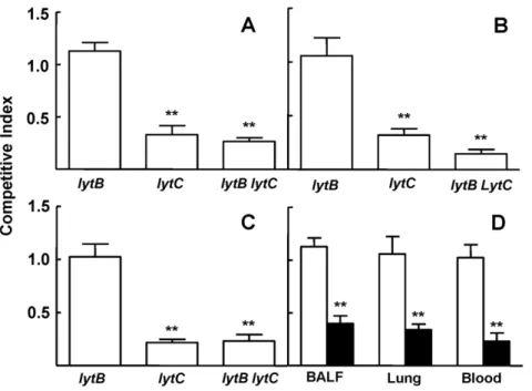
confirming that LytC is important for the establishment of pneumococcal pneumonia in the respiratory tract (Fig. 7A and B). Moreover, the bacteremia caused by thelytCmutant strain was reduced after intranasal inoculation compared to the wild-type strain, suggesting that LytC plays a significant role in the spread of the bacteria from the lung to the systemic circulation (Fig. 7C). The virulence of the double mutantlytB lytCstrain was explored using competitive indices and showed that loss of both CWHs had a consistent impaired effect on virulence suggesting that the combination of LytB and LytC is of great importance for full virulence during pneumonia and invasive dissemination.
To explore whether loss of both LytB and LytC might have a cooperative role on pneumococcal pneumonia, mixed infections were performed by the intranasal route to compare the CIs of the doublelytB lytCstrain vs.lytCwith those obtained for the mixed infection oflytBmutant vs. the wild-type strain. Our results show that the CI of thelytB lytCvs.lytCwas between 2–3 times lower in the lung compartment (1.13 vs. 0.39 in BALF and 1.06 vs. 0.34 in the lung;P,0.01) and nearly five times lower in blood (1.02 vs. 0.23;P,0.01) than the CI of thelytBvs. wild-type strain (Fig. 7D) confirming that, although LytB may not have a direct role itself in virulence, this CWH improves the pathogenesis mediated by LytC in the establishment of pneumococcal pneumonia and enhances the spread of the bacteria throughout the systemic circulation.
Discussion
S. pneumoniae is one of the most important human pathogens responsible for serious diseases associated to high morbidity and mortality rates worldwide [30]. Although the pneumococcal capsule is essential for full virulence by preventing complement immunity and phagocytosis [22,31], there are strong evidences suggesting that structural components of the bacteria are also important at different steps of the pathogenesis process [32]. Identification and functional analysis of gene products involved in colonization, inflammation, and invasion is a key tool to understand the host-pathogen interaction of S. pneumoniae and provides essential knowledge that can be used to fight against this pathogen with the discovery of new therapeutic targets or vaccine-based strategies. Peptidoglycan is a major component of the bacterial cell envelope that can be modified by the activity of bacterial CWHs [33]. CWHs are a group of proteins whose role in pathogenesis is not fully understood, although it is thought to be important [4]. As peptidoglycan is one of the main pathogen-associated molecular patterns targeted by the host innate immune system, modifications of this essential and unique cell wall component may be used by bacterial pathogens to subvert host innate immunity [34,35]. Peptidoglycan hydrolases are required for initial attachment to hydrophobic surfaces and contribute to bacterial pathogenesis and full virulence by increasing the survival within the systemic circulation, therefore enhancing the spread of the bacteria throughout the host [36,37,38,39]. In this study we have constructed single and double mutants in LytB and LytC to investigate the impact of both proteins on pneumococcal pathogenesis by exploring their role in the establishment of the carrier state, invasive disease, and evasion of several host defence mechanisms. Our findings demonstrate that both LytB and LytC are important pneumococcal surface proteins involved in the attachment to human nasopharyngeal cells, with a more marked effect of LytC at 30uC, which is the physiological temperature found in the upper respiratory tract and the optimal temperature for this enzyme [10]. Previous results by Gosink and coworkers [14] have shown that loss of function of LytC only moderately reduced (30%) the adherence of S. pneumoniae to D562 cells at 30uC, whereas no effect was observed at 37uC. It should be noted, however, that those authors employed a fluorescein isothiocya-nate-labeled, nonencapsulated derivative of TIGR4 strain and a very different methodology, i.e., pneumococcal cells were fixed with glutaraldehyde before counting, and adherent bacteria were quantified visually. These experimental conditions may have introduced a significant bias on the results reported by those authors. Using doublelytB lytCdefective TIGR4 and D39 strains we evaluated the role of both CWHs in adhesion to nasopharyn-geal cells. Our results demonstrate for the first time that the attachment of S. pneumoniae to human nasopharyngeal cells was dramatically impaired in the absence of both LytB and LytC proteins with a maximum reduction at 30uC. The role of both CWHs was also investigated in a mouse model of nasopharyngeal colonization using a high bacterial challenge (107CFU). This inoculum results in colonization as well as invasion of the adjacent mucosal sites without bacteremia [40]. Bacterial levels in the nasopharynx were maintained between 104and 105CFU ml21 throughout the 7 days of the study. These results confirmed that this model of nasopharyngeal colonization was self-limited and similar to pneumococcal carriage in humans [41]. Our results demonstrated that LytB and LytC are important CWHs involved in the initial attachment to the nasopharynx although other pneumococcal proteins also participate at this early stage as there was some degree of colonization in the absence of both CWHs and
Figure 6. Role in virulence of the different strains using a sepsis model of infection.(A) Virulence represented as a competitive index (CI) oflytB,lytCandlytB lytCstrains compared to their parental D39 wild-type strain. Error bars represent SDs of the means. For the comparisons of the results of the CI of thelytCstrain vs.lytB**,P,0.01 (two-tailed Student’sttest). For the comparisons of the doublelytB lytC strain vs. the single lytB or lytC strains ***,P,0.001 and *, P,0.05 respectively (two-tailed Student’sttest). (B) CIs oflytBstrain compared to strain D39 and CIs oflytB lytCmutants compared tolytCstrain to assess the combined effect of LytB and LytC in sepsis. **,P,0.01 for the comparison of both CIs (two-tailed Student’sttest).
confirmed that the combination of LytB and LytC is required for effective colonization of the respiratory tract over time. Targeting mechanisms of preventing bacterial factors involved in the colonization process during pneumococcal pathogenesis might be an attractive strategy to prevent the pneumococcal carrier state. Following adherence and asymptomatic colonization, S. pneu-moniaemust overcome host defense mechanisms implicated in the recognition and clearance of the microorganism to gain access to the alveolar space and/or the systemic circulation, producing invasive disease. The complement system is an important component of host immunity participating in the recognition and phagocytosis of invading pathogens [42]. Evidence suggesting that different proteins ofS. pneumoniaeare involved in the inhibition of complement immunity and phagocytosis has been previously published [18,29,43]. Our results demonstrate that loss of LytC, but not LytB, increased C3b deposition at both 30uC and 37uC demonstrating that LytC diverts C3b deposition on the bacterial surface. This is the first evidence showing complement evasion mediated by the pneumococcal lytic lysozyme. In addition, loss of both LytB and LytC was associated with a markedly increased C3b deposition on the bacterial surface confirming that the combination of both proteins is very efficient in the diversion of complement immunity.
The interaction with phagocytic cells was also investigated using two different cell lines that are representatives of pneumococcal phagocytosis [44,45]. lytB or lytC single mutants displayed an increased uptake of S. pneumoniae by alveolar macrophages and neutrophils, whereas the phagocytosis of the double mutantlytB lytCwas greatly enhanced compared to the single mutants. These differences do not seem to be attributable to variations in cell separation among the strains as it has been reported that the average number of pneumococci per phagocyte is the same between pneumococcal long-chain and short-chain variants [46].
Our results indicate that both CWHs are important pneumococcal proteins involved in resistance to phagocytosis and demonstrate that the presence of both LytB and LytC is highly effective in evasion of pneumococcal phagocytosis. A clear correlation exists between resistance to phagocytosis and carriage, where strains more resistant to neutrophil clearance have an advantage to persist in the nasopharynx [47]. The results of our study support this hypothesis and suggest that one of the main virulence mechanisms of LytB and LytC might be to avoid the recognition by professional phagocytes within the upper respiratory tract allowing the bacterium to efficiently colonize the nasopharynx. Targeting these proteins by the use of a vaccine or a chemical drug might help to reduce the carrier state by increasing the ability of the host immune defense system to efficiently recognizeS. pneumoniaefrom the human nasopharynx. The addition of exogenous LytB and LytC proteins to the single and double mutant strains restored C3b levels to those found on the wild-type strain and increased the resistance to phagocytosis to a similar degree than in the wild-type isolate strongly suggesting that the phenotypes observed were mainly due to the lack of LytB and LytC rather than other unexpected genetic differences among the strains such as insertions, duplications or second site mutations. Moreover, the addition of choline binding proteins such as LytB and LytC to a bacterial culture has been shown to recognize specifically the choline residues located at the bacterial surface. Addition of GFP-LytB to alytB mutant strain binds the specific target (the polar ends) to which it remains bound for a long time exhibiting a dispersing effect typical of LytB, whereas exogenous addition of the LytC lysozyme to a lytC deficient strain showed that this enzyme was kept until regulatory control by the ‘‘cured’’ cells [4,5]. These observations suggest that the addition of purified hydrolases to the defective strains had the ability to compensate the phenotype due to the lack of these enzymes.
Figure 7. Impact of mutations in the genes encoding LytB and LytC on pneumococcal pneumonia.(A to C) CIs oflytB,lytCand the doublelytB lytCstrains compared to their parental D39 wild-type strain in BALF (A), lungs (B), and blood (C) after intranasal inoculation of a mixed culture of the corresponding mutant with the wild-type strain. Error bars represent SDs of the mean and asterisks indicate results that are statistically significant compared to those for thelytBstrain which was outcompeted (two-tailed Student’sttest; *,P,0.05; **,P,0.01; ***,P,0.001). (D) CIs of lytBvs. D39 (open bars) andlytB lytCvs.lytC(blackened bars) in BALF, lungs, and blood after intranasal infection. **,P,0.01 for the comparison of both CIs at the corresponding sites of infection (two-tailed Student’sttest).
There is evidence supporting the idea that the factors needed for pneumococcal infection may also be important in nasopharyngeal colonization, as the majority of the genes required for invasive disease were found to be required for bacterial carriage [48]. The role of LytB and LytC in pneumococcal pneumonia and sepsis was investigated using a mouse model of infection. Loss of LytB did not influence virulence compared to the wild-type strain indicating that LytB by itself does not participate in the establishment of pneumonia or in systemic dissemination. These results are in agreement with those found by other authors using a sepsis model in which the virulence of a D39lytBstrain was very similar to the corresponding wild-type strain [49]. However, the virulence of the
lytCmutant strain was attenuated in both models of systemic and pulmonary infection with an impaired ability to spread from the lung to the bloodstream confirming that LytC is an important virulence factor ofS. pneumoniae. In addition, the doublelytB lytC
mutant strain was even more attenuated in virulence than the corresponding single mutants or the wild-type strain. This confirms that the presence of both LytB and LytC permits highly effective establishment of pneumococcal pneumonia and sepsis and indicates that both proteins enhance the dissemination of the bacteria from the lung compartment to the systemic circulation. This is in agreement with other authors confirming that different virulence factors ofS. pneumoniaeact in concert increasing bacterial
virulence and reinforces the idea that several bacterial components ofS. pneumoniaeare needed to efficiently produce invasive disease [18,29,43,50].
In summary, we have shown that LytB and LytC are surface exposed proteins that play a significant role in essential phases of the pneumococcal pathogenesis such as nasopharyngeal coloniza-tion, pneumonia, and sepsis. Our data suggest that pneumococcal cell wall hydrolases are important proteins ofS. pneumoniaeinvolved in the attachment of the bacteria to the nasopharynx and in the progress of the pneumococcal pneumonia and sepsis by avoiding complement immunity and phagocytosis.
Acknowledgments
The authors wish to thank Carlos Castilla from Fundacio´n Jime´nez Dı´az, and E. Cano for skilful technical assistance. We also thank Rachel Exley for critical reading of the manuscript.
Author Contributions
Conceived and designed the experiments: ER-S MM PG EG JY. Performed the experiments: ER-S MM JY. Analyzed the data: ER-S EG JY. Wrote the paper: EG JY. Reviewed and approved the manuscript: ER-S MM PG EG JY.
References
1. Bogaert D, de Groot R, Hermans PWM (2004) Streptococcus pneumoniae colonisation: the key to pneumococcal disease. Lancet Infect Dis 4: 144–154. 2. Wardlaw T, Salama P, Johansson EW, Mason E (2006) Pneumonia: the leading
killer of children. Lancet 368: 1048–1050.
3. Weiser JN (2010) The pneumococcus: why a commensal misbehaves. J Mol Med 88: 7–102.
4. Lo´pez R, Garcı´a E (2004) Recent trends on the molecular biology of pneumococcal capsules, lytic enzymes, and bacteriophage. FEMS Microbiol Rev 28: 553–580.
5. De las Rivas B, Garcı´a JL, Lo´pez R, Garcı´a P (2002) Purification and polar localization of pneumococcal LytB, a putative endo-b-N-acetylglucosaminidase: the chain-dispersing murein hydrolase. J Bacteriol 184: 4988–5000. 6. Garcı´a P, Gonza´lez MP, Garcı´a E, Lo´pez R, Garcı´a JL (1999) LytB, a novel
pneumococcal murein hydrolase essential for cell separation. Mol Microbiol 31: 1275–1277.
7. Moscoso M, Obrego´n V, Lo´pez R, Garcı´a JL, Garcı´a E (2005) Allelic variation of the polymorphic locus lytB, encoding a choline-binding protein, from streptococci of the mitis group. Appl Environ Microbiol 71: 8706–8713. 8. Wizemann TM, Heinrichs JH, Adamou JE, Erwin AL, Kunsch C, et al. (2001)
Use of a whole genome approach to identify vaccine molecules affording protection againstStreptococcus pneumoniaeinfection. Infect Immun 69: 1593–1598. 9. Pe´rez-Dorado I, Gonza´lez A, Morales M, Sanles R, Striker W, et al. (2010) Insights into pneumococcal fratricide from the crystal structures of the modular killing factor LytC. Nat Struct Mol Biol 17: 576–581.
10. Garcı´a P, Gonza´lez MP, Garcı´a E, Garcı´a JL, Lo´pez R (1999) The molecular characterization of the first autolytic lysozyme ofStreptococcus pneumoniaereveals evolutionary mobile domains. Mol Microbiol 33: 128–138.
11. Eldholm V, Johnsborg O, Haugen K, Ohnstad HS, Ha˚varstein LS (2009) Fratricide in Streptococcus pneumoniae: contributions and role of the cell wall hydrolases CbpD, LytA and LytC. Microbiology 155: 2223–2234.
12. Moscoso M, Garcı´a E, Lo´pez R (2009) Pneumococcal biofilms. Int Microbiol 12: 77–85.
13. Moscoso M, Garcı´a E, Lo´pez R (2006) Biofilm formation by Streptococcus pneumoniae: role of choline, extracellular DNA, and capsular polysaccharide in microbial accretion. J Bacteriol 188: 7785–7795.
14. Gosink KK, Mann ER, Guglielmo C, Tuomanen EI, Masure HR (2000) Role of novel choline binding proteins in virulence ofStreptococcus pneumoniae. Infect Immun 68: 5690–5695.
15. Tettelin H, Nelson KE, Paulsen IT, Eisen JA, Read TD, et al. (2001) Complete genome sequence of a virulent isolate ofStreptococcus pneumoniae. Science 293: 498–506.
16. Lanie JA, Ng W-L, Kazmierczak KM, Andrzejewski TM, Davidsen TM, et al. (2007) Genome sequence of Avery’s virulent serotype 2 strain D39 ofStreptococcus pneumoniaeand comparison with that of unencapsulated laboratory strain R6. J Bacteriol 189: 38–51.
17. Hendriksen WT, Kloosterman TG, Bootsma HJ, Estevao S, de Groot R, et al. (2008) Site-specific contributions of glutamine-dependent regulator GlnR and GlnR-regulated genes to virulence ofStreptococcus pneumoniae. Infect Immun 76: 1230–1238.
18. Yuste J, Botto M, Paton JC, Holden DW, Brown JS (2005) Additive inhibition of complement deposition by pneumolysin and PspA facilitates Streptococcus pneumoniaesepticemia. J Immunol 175: 1813–1819.
19. Yuste J, Khandavilli S, Ansari N, Muttardi K, Ismail L, et al. (2010) The effects of PspC on complement-mediated immunity toStreptococcus pneumoniaevary with strain background and capsular serotype. Infect Immun 78: 283–292. 20. Marti-Lliteras P, Regueiro V, Morey P, Hood DW, Saus C, et al. (2009)
Nontypeable Haemophilus influenzae clearance by alveolar macrophages is impaired by exposure to cigarette smoke. Infect Immun 77: 4232–4242. 21. Yuste J, Sen A, Truedsson L, Jo¨nsson G, Tay L-S, et al. (2008) Impaired
opsonization with C3b and phagocytosis ofStreptococcus pneumoniaein sera from subjects with defects in the classical complement pathway. Infect Immun 76: 3761–3770.
22. Hyams C, Yuste J, Bax K, Camberlein E, Weiser JN, et al. (2010)Streptococcus pneumoniaeresistance to complement-mediated immunity is dependent on the capsular serotype. Infect Immun 78: 716–725.
23. Fleck RA, Romero-Steiner S, Nahm MH (2005) Use of HL-60 cell line to measure opsonic capacity of pneumococcal antibodies. Clin Diagn Lab Immunol 12: 19–27.
24. Yuste J, Sen A, Truedsson L, Jo¨nsson G, Hyams C, et al. (2010) Impaired opsonization with complement and phagocytosis ofStreptococcus pyogenesin sera from subjects with inherited C2 deficiency. Microbes Infect 12: 626–634. 25. McAllister LJ, Tseng HJ, Ogunniyi AD, Jennings MP, McEwan AG, et al.
(2004) Molecular analysis of the psa permease complex ofStreptococcus pneumoniae. Mol Microbiol 53: 889–901.
26. Quin LR, Onwubiko C, Moore QC, Mills MF, McDaniel LS, et al. (2007) Factor H binding to PspC of Streptococcus pneumoniaeincreases adherence to human cell lines in vitro and enhances invasion of mouse lungs in vivo. Infect Immun 75: 4082–4087.
27. Beuzo´n CR, Holden DW (2001) Use of mixed infections withSalmonellastrains to study virulence genes and their interactions in vivo. Microbes Infect 3: 1345–1352.
28. Abeyta M, Hardy GG, Yother J (2003) Genetic alteration of capsule type but not PspA type affects accessibility of surface-bound complement and surface antigens ofStreptococcus pneumoniae. Infect Immun 71: 218–225.
29. Quin LR, Moore QC, III, McDaniel LS (2007) Pneumolysin, PspA, and PspC contribute to pneumococcal evasion of early innate immune responses during bacteremia in mice. Infect Immun 75: 2067–2070.
30. O’Brien KL, Wolfson LJ, Watt JP, Henkle E, Deloria-Knoll M, et al. (2009) Burden of disease caused byStreptococcus pneumoniaein children younger than 5 years: global estimates. Lancet 374: 893–902.
31. Hyams C, Camberlein E, Cohen JM, Bax K, Brown JS (2010) TheStreptococcus pneumoniaecapsule inhibits complement activity and neutrophil phagocytosis by multiple mechanisms. Infect Immun 78: 704–715.
32. Kadioglu A, Weiser J, Paton JC, Andrew PW (2008) The role ofStreptococcus pneumoniaevirulence factors in host respiratory colonization and disease. Nat Rev Microbiol 6: 288–301.
34. Dziarski R, Gupta D (2005) Peptidoglycan recognition in innate immunity. J Endotoxin Res 11: 304–310.
35. Mengin-Lecreulx D, Lemaitre B (2005) Structure and metabolism of peptidoglycan and molecular requirements allowing its detection by the Drosophilainnate immune system. J Endotoxin Res 11: 105–111.
36. Heilmann C, Hussain M, Peters G, Gotz F (1997) Evidence for autolysin-mediated primary attachment ofStaphylococcus epidermidisto a polystyrene surface. Mol Microbiol 24: 1013–1024.
37. Cabanes D, Dussurget O, Dehoux P, Cossart P (2004) Auto, a surface associated autolysin of Listeria monocytogenes required for entry into eukaryotic cells and virulence. Mol Microbiol 51: 1601–1614.
38. Wang L, Lin M (2008) A novel cell wall-anchored peptidoglycan hydrolase (autolysin), IspC, essential for Listeria monocytogenes virulence: genetic and proteomic analysis. Microbiology 154: 1900–1913.
39. Jung CJ, Zheng QH, Shieh YH, Lin CS, Chia JS (2009)Streptococcus mutans autolysin AtlA is a fibronectin-binding protein and contributes to bacterial survival in the bloodstream and virulence for infective endocarditis. Mol Microbiol 74: 888–902.
40. van Ginkel FW, McGhee JR, Watt JM, Campos-Torres A, Parish LA, et al. (2003) Pneumococcal carriage results in ganglioside-mediated olfactory tissue infection. Proc Natl Acad Sci USA 100: 14363–14367.
41. McCool TL, Weiser JN (2004) Limited role of antibody in clearance of Streptococcus pneumoniaein a murine model of colonization. Infect Immun 72: 5807–5813.
42. Walport MJ (2001) Complement. First of two parts. N Engl J Med 344: 1058–1066.
43. Dalia AB, Standish AJ, Weiser JN (2010) Three surface exoglycosidases from Streptococcus pneumoniae, NanA, BgaA, and StrH, promote resistance to opsonophagocytic killing by human neutrophils. Infect Immun 78: 2108–2116. 44. Romero-Steiner S, Libutti D, Pais LB, Dykes J, Anderson P, et al. (1997) Standardization of an opsonophagocytic assay for the measurement of functional antibody activity againstStreptococcus pneumoniaeusing differentiated HL-60 cells. Clin Diagn Lab Immunol 4: 415–422.
45. Stegenga ME, Florquin S, de Vos AF, van der Poll T (2009) The thiazolidinedione ciglitazone reduces bacterial outgrowth and early inflamma-tion duringStreptococcus pneumoniae pneumonia in mice. Crit Care Med 37: 614–618.
46. Austrian R (1953) Morphologic variation in pneumococcus. I. An analysis of the bases for morphologic variation in pneumococcus and description of a hitherto undefined morphologic variant. J Exp Med 98: 21–34.
47. Weinberger DM, Trzcin´ski K, Lu Y-J, Bogaert D, Brandes A, et al. (2009) Pneumococcal capsular polysaccharide structure predicts serotype prevalence. PLoS Pathog 5: e1000476.
48. Hava DL, Camilli A (2002) Large-scale identification of serotype 4Streptococcus pneumoniaevirulence factors. Mol Microbiol 45: 1389–1406.
49. Kharat AS, Tomasz A (2006) Drastic reduction in the virulence ofStreptococcus pneumoniaeexpressing type 2 capsular polysaccharide but lacking choline residues in the cell wall. Mol Microbiol 60: 93–107.