BGD
8, 10697–10724, 2011
Indications for a ubiquitous dissolved
pigment
R. R ¨ottgers and B. P. Koch
Title Page
Abstract Introduction
Conclusions References
Tables Figures
◭ ◮
◭ ◮
Back Close
Full Screen / Esc
Printer-friendly Version Interactive Discussion
Discussion
P
a
per
|
Dis
cussion
P
a
per
|
Discussion
P
a
per
|
Discussio
n
P
a
per
|
Biogeosciences Discuss., 8, 10697–10724, 2011 www.biogeosciences-discuss.net/8/10697/2011/ doi:10.5194/bgd-8-10697-2011
© Author(s) 2011. CC Attribution 3.0 License.
Biogeosciences Discussions
This discussion paper is/has been under review for the journal Biogeosciences (BG). Please refer to the corresponding final paper in BG if available.
Indications for a ubiquitous dissolved
pigment degradation product in
subsurface waters of the global ocean
R. R ¨ottgers1and B. P. Koch2,3
1
Helmholtz-Zentrum Geesthacht, Center for Materials and Coastal Research, Institute for Coastal Research, Max Planck Str. 1, 21502 Geesthacht, Germany
2
Alfred Wegener Institute for Polar and Marine Research, Am Handelshafen 12, 27570 Bremerhaven, Germany
3
University of Applied Sciences, An der Karlstadt 8, 27568 Bremerhaven, Germany
Received: 12 October 2011 – Accepted: 17 October 2011 – Published: 31 October 2011
Correspondence to: R. R ¨ottgers (rroettgers@hzg.de)
BGD
8, 10697–10724, 2011
Indications for a ubiquitous dissolved
pigment
R. R ¨ottgers and B. P. Koch
Title Page
Abstract Introduction
Conclusions References
Tables Figures
◭ ◮
◭ ◮
Back Close
Full Screen / Esc
Printer-friendly Version Interactive Discussion
Discussion
P
a
per
|
Dis
cussion
P
a
per
|
Discussion
P
a
per
|
Discussio
n
P
a
per
|
Abstract
Measurements of light absorption by chromophoric dissolved organic matter (CDOM) from sub-surface waters of the tropical Atlantic and Pacific showed a distinct absorp-tion shoulder at 410–415 nm, indicating an underlying absorpabsorp-tion of a pigment. A sim-ilar absorption maximum at ∼410 nm was also found in the particulate fraction and 5
is usually attributed to absorption by respiratory pigments of heterotrophic unicellu-lar organisms. The CDOM absorption shoulder was described earlier in the Indian Ocean at 600 m depth and was related to a “deep red fluorescence” found in the same depth, i.e. in the oxygen minimum zone (Breves et al., 2003; Broenkow et al., 1983). In our study, fluorescence measurements of pre-concentrated DOM samples
10
confirmed that the absorption at ∼410 nm was related to a specific fluorescence at
650 nm. The absorption characteristic of this specific fluorophor was examined by flu-orescence emission/excitation analysis and this showed a clear excitation maximum at 415 nm (in methanol) that can explain the absorption shoulder in the CDOM spectra. The spectral characteristics of the substance found in the dissolved and particulate
15
fraction did not match with those of chlorophylla degradation products (as found in a sample from the sea surface) but can be explained by the occurrence of respiratory pig-ments from heterotrophs. Combining the observations of the “deep red fluorescence” and the 410 nm-absorption shoulder suggests that there are high concentrations of a pigment degradation product (cytochrome c) in DOM of all major oceans. Most
pro-20
nouncedly we found this signal in the deep chlorophyll maximum and the oxygen min-imum zone of tropical regions. The origin, chemical nature, turn-over rate, and fate of this molecule is so far unknown.
1 Introduction
Dissolved organic matter (DOM) in the ocean forms the largest reservoir of reduced
or-25
BGD
8, 10697–10724, 2011
Indications for a ubiquitous dissolved
pigment
R. R ¨ottgers and B. P. Koch
Title Page
Abstract Introduction
Conclusions References
Tables Figures
◭ ◮
◭ ◮
Back Close
Full Screen / Esc
Printer-friendly Version Interactive Discussion
Discussion
P
a
per
|
Dis
cussion
P
a
per
|
Discussion
P
a
per
|
Discussio
n
P
a
per
|
a large proportion of which is refractory. The overall distribution of DOM in the world ocean is determined by autochtonous and only partly allothonous sources, by pho-todegradation near the sea surface, and particulate matter re-mineralization in deeper waters. Measurement of DOM light absorption is one method to describe the biogeo-chemistry and hydrography of different oceanic regions and water masses. In
combi-5
nation with fluorescence characterization it is suitable to get an insight into the chem-ical nature and origin of DOM (Stedmon et al., 2003). Chromophoric and fluorescent moieties of the DOM (CDOM and FDOM, respectively), represent only a small part of the DOM pool, but can easily be accessed by optical methods. The absorption of CDOM in the aquatic environment has a characteristic spectral shape, with a strong
10
absorption in the UV to visible range, which decreases exponentially towards higher wavelengths. The slope of this decrease is used e.g. to characterize CDOM sources (e.g. Vodacek et al., 1997) and to describe its molecular composition (e.g. Helms et al., 2008). Because of the generally low CDOM absorption in oligotrophic regions, the measurements are mostly performed at UV rather than at visible wavelengths.
Com-15
monly, samples are measured near sea surface where dissolved organic carbon (DOC) concentration is highest and CDOM absorption can be retrieved by satellite remote sensing. Therefore most scientific studies addressed CDOM absorption features at the sea surface. However, due to photo-degradation at the surface, CDOM absorption is higher below the surface mixing zone, and often reflects the hydrographic origin of
20
a water mass, as a major proportion of DOM is refractory and degrades only slowly. A pronounced shoulder in the CDOM absorption spectrum can usually be found only in the UV (∼270 nm) due to high concentrations of aromatic structures in DOM. Also
its fluorescence emission originates from chromophors absorbing in the UV (<400 nm, Coble 2007), fluorescing in the UV to shorter VIS wavelengths. A previous studies
25
BGD
8, 10697–10724, 2011
Indications for a ubiquitous dissolved
pigment
R. R ¨ottgers and B. P. Koch
Title Page
Abstract Introduction
Conclusions References
Tables Figures
◭ ◮
◭ ◮
Back Close
Full Screen / Esc
Printer-friendly Version Interactive Discussion
Discussion
P
a
per
|
Dis
cussion
P
a
per
|
Discussion
P
a
per
|
Discussio
n
P
a
per
|
Breves et al., 2003). A red fluorescence in subsurface waters was also reported earlier from the Pacific (Broenkow et al., 1983, 1985, 1992; Lewitus and Broenkow, 1985). The authors concluded that this “deep red fluorescence” is a global feature found in all major oceans and at all latitudes, and that its maximum is associated with the oceanic oxygen minimum zone (OMZ). Chlorophyll or some of its degradation products in
phy-5
toplanktonic particles are thought to be a possible molecular precursor. To our knowl-edge it is still unresolved whether the red fluorescence occurs in the particulate and/or dissolved fraction. Moreover, the exact molecular nature of this phenomenon is also unknown. Breves et al. (2003) concluded from sample filtration and a specific fluo-rescence emission at∼650 nm, which appeared in samples from mesopelagic depth 10
when excited with light of 420 nm, that it is most likely in the dissolved phase. This contradicts earlier results (Broenkow et al., 1992) which often found a close correlation of the “deep red fluorescence” with light attenuation (mainly scattering by particles). It is possible that the fluorescence at 650 nm emits from the chromophor that absorbs at 410 nm, and that this fluorescence is collected by common in situ chlorophyll
flu-15
orometers. Hence, the observation of this absorption shoulder in the visible spectral range and the “deep red fluorescence” in the subsurface waters are most probably linked. If this is true, the observed “deep red fluorescence” provides evidence that this specific chromophor/fluorophor occurs globally and throughout the water column, its optical characteristic provides evidence that it is of pigment origin and unique among
20
other chromophoric/fluorescent DOM molecules in the subsurface ocean. The aims of the work presented here are (1) to give evidence that the absorption at 410 nm and the fluorescence at 650 nm are originating from the same molecule, (2) to clarify whether it is found in either the dissolved or the particulate fraction, and (3) to investigate the distribution of this chromophor/fluorophor in the water column and on a latitudinal
tran-25
BGD
8, 10697–10724, 2011
Indications for a ubiquitous dissolved
pigment
R. R ¨ottgers and B. P. Koch
Title Page
Abstract Introduction
Conclusions References
Tables Figures
◭ ◮
◭ ◮
Back Close
Full Screen / Esc
Printer-friendly Version Interactive Discussion
Discussion
P
a
per
|
Dis
cussion
P
a
per
|
Discussion
P
a
per
|
Discussio
n
P
a
per
|
2 Materials and methods
2.1 Sampling
Several oceanographic cruises were conducted to collect samples for measurements of particulate and CDOM absorption, and for DOM extracts. The first cruise (ANT XXIII/1, RV “Polarstern”, October/November 2005) covered a transect from Germany
5
to South Africa crossing the Atlantic Ocean along the African continent. During this cruise CDOM samples were mainly taken from the surface and 200 m depth. On 5 additional stations the entire water column was sampled. DOM extracts were collected from the surface and once from 200 m depth. On a second transect cruise (ANT XXV/1, RV “Polarstern”, November 2008) which covered the same region, DOM was extracted
10
throughout the water column across the Atlantic. Two other cruises were used to collect single samples for CDOM and particulate absorption measurements from the surface and from 80–200 m depths: (1) in the West-Pacific offshore of New Caledonia (VALHY-BIO, RV “L’Alis”, March/April 2008), and (2) in the Santa Barbara Channel (California, North-East Pacific, RaDyO, RV “Kilo Moana”, September 2008).
15
2.2 Light absorption measurements
The light absorption of dissolved (CDOM) and particulate matter,acdomandap,
respec-tively, was measured using a point-source integrating cavity absorption meter (PSI-CAM) as described in R ¨ottgers et al. (2007) and R ¨ottgers and Doerffer (2007). For this, the sample absorption was measured before and after a filtration through 0.2 µm,
20
and the absorption of the filtered fraction was subtracted from the sample absorption to obtain particulate absorption. During the RaDyO cruise the absorption by CDOM was measured additionally using a 2 m path length liquid core waveguide capillary cell (LWCC), as e.g. described in Miller et al. (2002). All PSICAM and LWCC measure-ments were done shortly after sampling (<2 h) onboard of the ship, using ultrapure
25
BGD
8, 10697–10724, 2011
Indications for a ubiquitous dissolved
pigment
R. R ¨ottgers and B. P. Koch
Title Page
Abstract Introduction
Conclusions References
Tables Figures
◭ ◮
◭ ◮
Back Close
Full Screen / Esc
Printer-friendly Version Interactive Discussion
Discussion
P
a
per
|
Dis
cussion
P
a
per
|
Discussion
P
a
per
|
Discussio
n
P
a
per
|
Measurements were done at least in triplicates and averaged, and the absorption co-efficient, a, was calculated as a=−ln T/L[m−1], where T is the transmission and L the corrected optical path length of the capillary cell, as provided by the manufac-turer (WPI Inc.). CDOM filtration was done using cleaned glass filtration units. After rinsing with 50–100 mLs of the sample, a first filtration was performed through
pre-5
combusted GF/F filters (Whatman) under low vacuum (<100 mbar) to avoid breakage of phytoplankton cells. A second filtration through 0.2 µm (washed GWSP filters, Mil-lipore) was done under vacuum of 200–300 mbar. Prior to absorption measurements, filtered samples were adjusted to the same temperature as the reference purified water to minimize temperature effects. However, slight temperature difference as well as the
10
difference induced by the difference in salinity between sample and reference water were individually corrected, using instrument-specific correction coefficients obtained from measurements of concentrated NaCl solutions. Light absorption (350–700 nm, 2 nm resolution and slit width) of the methanol DOM extracts were measured back in the lab against pure methanol as a reference in a Lambda 850 spectrophotometer
15
(Perkin Elmer) using 1 cm path length quartz glass cuvette.
2.3 DOM extraction
DOM was extracted from 5 l filtered (GF/F) sea-water samples using solid phase ad-sorption (1 g PPL cartridges, Varian) according to Dittmar et al. (2008). Solid phase extractable DOC (SPE-DOC) amounted to 42±6 % of the total DOC (n=138) and 20
22±11 % of the DON was solid phase extractable (n=128). For an in-depth
descrip-tion of the extracdescrip-tion method refer to Flerus et al. (2011).
2.4 Fluorescence measurements
Fluorescence emission and excitation spectra from the methanolic DOM extracts were recorded with an Aminco-Bowman Serie 2 Luminescence Spectrometer in a 1 cm path
25
BGD
8, 10697–10724, 2011
Indications for a ubiquitous dissolved
pigment
R. R ¨ottgers and B. P. Koch
Title Page
Abstract Introduction
Conclusions References
Tables Figures
◭ ◮
◭ ◮
Back Close
Full Screen / Esc
Printer-friendly Version Interactive Discussion
Discussion
P
a
per
|
Dis
cussion
P
a
per
|
Discussion
P
a
per
|
Discussio
n
P
a
per
|
(EEM) were recorded for selected samples only using different wavelength ranges and signal amplification in order to amplify low fluorescence features at >500 nm. EEM measurements were done with a resolution of 2 nm, band widths of 4 nm for exci-tation and emission, and scan speed of 5–10 nm s−1 for (1) Ex: 250–500 nm, Em: 270−500 nm, (2) Ex: 380–680 nm, Em: 400–700 nm, (3) Ex: 380–480 nm, Em: 550– 5
700 nm. Single emission spectra (270–700 nm) were recorded for several samples for excitation between 250 and 500 nm. Single excitation spectra (>350 nm) were recorded for emissions at 490, 650, 670, and 720 nm. For these single scans the signal to noise level was reduced by a slower scan speed (1–2 nm s−1), multiple scans (n=5−10) that were averaged, and a band width of 8–6 nm for the fixed monochroma-10
tor (i.e. for emission during excitation scans, and excitation during emission scans). For all measurements pure methanol was used with the same scan speed, band width, and amplification settings for a subsequent subtraction of Raman and Rayleigh scattering. Internally recorded light intensity of the excitation was used to correct for spectral varia-tion in the excitavaria-tion light spectrum. Further correcvaria-tions for fluorescence re-absorpvaria-tion
15
or other quenching were not applied. When possible the fluorescence was normalized to the measured absorption at 415 nm.
3 Results
3.1 CDOM absorption
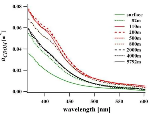
CDOM absorption spectra from the sea surface showed a typical spectral distribution
20
in the UVA to VIS range (380–700 nm), i.e. an exponential decrease with increasing wavelength without pronounced structures. Absolute absorption varied due to water mass differences along the latitudinal transect from north to south. Over a depth pro-file CDOM absorption was lowest at the sea surface and typically highest in depths of the oxygen minimum zone. Figure 1 shows one example of CDOM absorption
spec-25
BGD
8, 10697–10724, 2011
Indications for a ubiquitous dissolved
pigment
R. R ¨ottgers and B. P. Koch
Title Page
Abstract Introduction
Conclusions References
Tables Figures
◭ ◮
◭ ◮
Back Close
Full Screen / Esc
Printer-friendly Version Interactive Discussion
Discussion
P
a
per
|
Dis
cussion
P
a
per
|
Discussion
P
a
per
|
Discussio
n
P
a
per
|
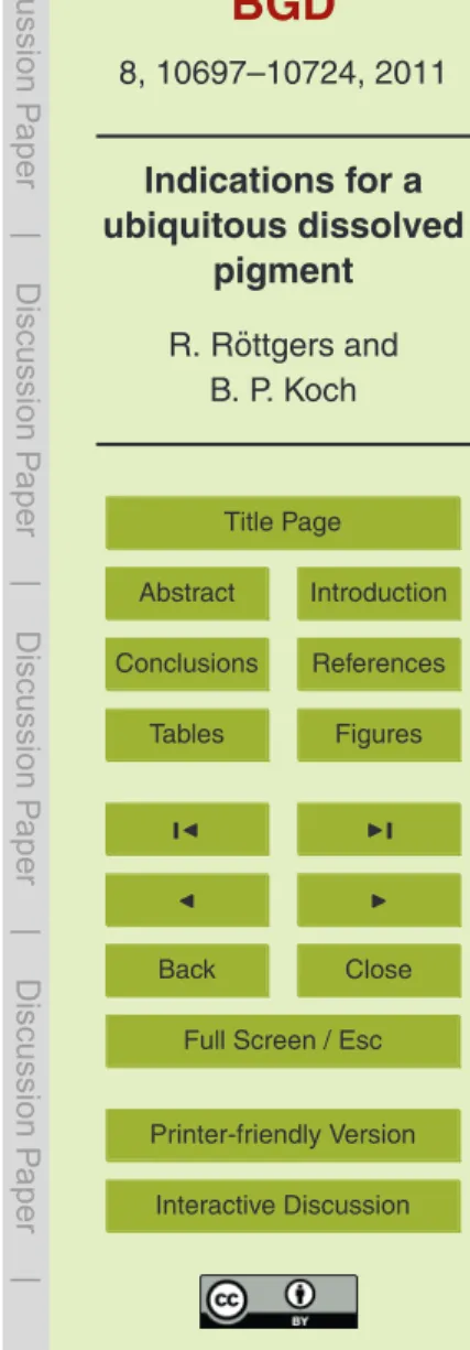
in sub-surface water is higher, there is also a pronounced absorption shoulder at ca. 410 nm for intermediate depth of 110–800 m. Even at higher depths this shoulder is still observable by a linear deflection of the exponential curve. This “typical” exponen-tial decrease in the CDOM absorption spectra was only found in samples from the sea surface whereas the shoulder was found in nearly all samples from depths below the
5
surface mixing zone (40–100 m, Fig. 2), as well as in depths of the deep chlorophyll maximum (DCM) just below the mixing layer (e.g. 82 m, Fig. 1). It was most pronounced in and below the DCM, up to a depth of 800 m. This corresponds to the typical extent of the oxygen minimum zone in this region. The maximum of the shoulder was found be-tween 110 and 500 m (Fig. 1) where the oxygen minimum occurred. However, sample
10
resolution was too low to determine the exact maximum appearance of the shoulder for each profile. Regular sampling was carried out at 200 m water depth. The highest extent of the shoulder in 200 m was found in samples from the tropics (25◦N–25◦S), the lowest in samples from higher latitudes (ca. 40◦N, data not shown).
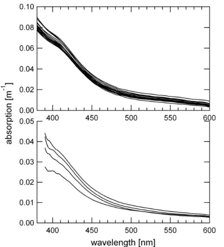
During two additional cruises, samples for CDOM absorption from depths up to
15
200 m were taken to determine the occurrence of this absorption shoulder in other oceanographic regions. A few samples were taken from the shallow Santa Barbara Channel (SBC, Californian coast, West Pacific, bottom depth ca. 200 m) and offshore of New Caledonia (South East Pacific, bottom depth>2000 m). During the SBC cruise, CDOM absorption was measured using a liquid waveguide system in addition to the
20
PSICAM to test two independent methods. In both regions (and with both methods in case of the SBC) a shoulder at 410 nm was found in depths below the mixed layer of the sea surface (Fig. 3), whereas the surface samples showed typical surface CDOM spectra without a visible shoulder (data not shown).
3.2 Particulate absorption
25
BGD
8, 10697–10724, 2011
Indications for a ubiquitous dissolved
pigment
R. R ¨ottgers and B. P. Koch
Title Page
Abstract Introduction
Conclusions References
Tables Figures
◭ ◮
◭ ◮
Back Close
Full Screen / Esc
Printer-friendly Version Interactive Discussion
Discussion
P
a
per
|
Dis
cussion
P
a
per
|
Discussion
P
a
per
|
Discussio
n
P
a
per
|
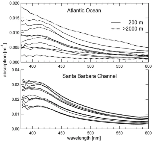
fraction (>0.2 µm) as well. At the surface the particulate absorption in the ocean is usually dominated by phytoplankton pigment absorption and in the deep sea by that of heterotrophs (mainly bacteria) and detritus. Surface samples were ignored as the phy-toplankton pigment maxima and shoulders would mask the distinct shoulder at 410 nm. On the other hand particulate absorption in the deep sea is very low and, thus, difficult
5
to measure. The PSICAM provides a sensitive method to determine particulate ab-sorption; nevertheless the absorption was often below the detection limit. Significant measurements could only be made for the samples from the SBC and a few sam-ples from the Atlantic Ocean (Fig. 4). The spectra showed an absorption maximum at 410 nm in all samples from 90–200 m, and no pronounced maxima in samples taken
10
from depth>2000 m in the Atlantic Ocean. The absorption spectra from these higher depths showed a typical detritus absorption, i.e. an exponential decrease with increas-ing wavelength with a slope lower than the slope for CDOM absorption. The absolute particulate absorption at 410 nm was lower than the respective CDOM absorption due to the high concentration of DOM compared to POM (particulate organic matter).
How-15
ever, if the absorption of the shoulder is regarded as an absorption maximum on top of the normal CDOM absorption, the absorption of the underlying pigment in the CDOM fraction is in the same range than that of the similar particulate absorption, at least for samples which possessed higher particulate absorption.
3.3 DOM fluorescence and absorption from ANTXXV/1
20
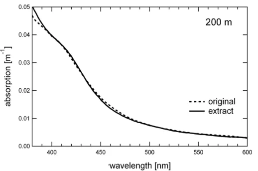
During ANT XXIII/1 a DOM extract was collected from a 200 m sample to verify the occurrence of the shoulder in concentrated DOM samples and to further analyze a possible fluorescence associated with the 410 nm-absorption shoulder. This DOM methanolic extract showed the same absorption shoulder at 410 nm than the original water sample (Fig. 5), proving that DOM extraction was a good method to concentrate
25
BGD
8, 10697–10724, 2011
Indications for a ubiquitous dissolved
pigment
R. R ¨ottgers and B. P. Koch
Title Page
Abstract Introduction
Conclusions References
Tables Figures
◭ ◮
◭ ◮
Back Close
Full Screen / Esc
Printer-friendly Version Interactive Discussion
Discussion
P
a
per
|
Dis
cussion
P
a
per
|
Discussion
P
a
per
|
Discussio
n
P
a
per
|
and fluorescence characteristics for all samples from 200 m and for a few samples from the surface and from higher depths were determined. First, emission-excitation matrices were measured for a few samples but for a large wavelength range (Ex: 250– 600 nm, Em: 270–700 nm) as fluorescence measurements of the single ANTXXIII/1 extract had shown a specific fluorescence emission at>600 nm. As the absolute
flu-5
orescence is highest in the UV to short-VIS, the matrices were repeated at longer wavelengths with higher detector signal amplification. Figure 6 shows the composite of three matrices of one sample from the sea surface and one from 200 m. There is a strong maximum at 370 nm/420 nm (Ex/Em) in both samples and some minor shoulders in the UV and short-VIS. The surface sample showed a low but distinct
max-10
imum at 410 nm/670 nm, whereas the maximum in the 200 m sample was located at 415 nm/650 nm. The relevant emission spectra are explicitly shown in Fig. 7a. The pos-sible absorption related to these emissions were examined by single excitation spectra (Ex: 370–630/−650/−690 nm, for Em: 650/670/720 nm, respectively). The emission
wavelength of 720 nm was used to examine absorption features around the emission
15
maxima for each sample. Figure 7b shows the excitation spectra for these two sam-ples (surface and 200 m). The surface sample showed a strong excitation maximum at 410 nm and smaller, rather narrow maxima at ca. 509, 538, 608 and 665 nm. The sample from 200 m depth showed a distinct maximum at 415 nm and smaller, broad maxima at 512 and 552 nm, and no significant features at >600 nm. The excitation
20
maximum at 415 nm most likely corresponds to the CDOM absorption shoulder found at the same wavelength in the extract; as slight shift in wavelength compared to the absorption of the water sample (ca. 410 nm) can be explained by the difference in sol-vent (seawater vs. methanol). To examine the geographic distribution of the obtained excitation maxima at 415 nm, excitation spectra of each sample were recorded with the
25
BGD
8, 10697–10724, 2011
Indications for a ubiquitous dissolved
pigment
R. R ¨ottgers and B. P. Koch
Title Page
Abstract Introduction
Conclusions References
Tables Figures
◭ ◮
◭ ◮
Back Close
Full Screen / Esc
Printer-friendly Version Interactive Discussion
Discussion
P
a
per
|
Dis
cussion
P
a
per
|
Discussion
P
a
per
|
Discussio
n
P
a
per
|
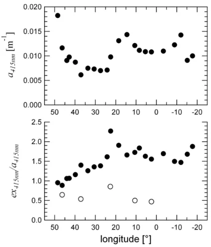
north to south is shown in Fig. 9 (samples from>20◦S were not available). The CDOM
absorption at 200 m showed high values in the highest latitudes (Gulf of Biscay), low values in the subtropics (40–23◦N), and high values in the tropics with distinct max-ima north and south of the equator. The relative 415 nm excitation maximum in 200 m depth was low in higher latitudes, steadily increased throughout the subtropics, and
5
was highest in the tropics. Distinct maxima were found on the onset of the CDOM in-crease/decrease north and south of the equator at ca. 23◦N and 20◦S. Samples from
the surface showed only small excitation maxima, with higher values at ca. 23◦N only, however the number of samples was limited.
4 Discussion
10
4.1 CDOM and particulate absorption of subsurface waters
The CDOM absorption spectra from subsurface water samples in the Eastern Atlantic Ocean showed a clear absorption shoulder at ca. 410 nm that, in its maximum extent and wavelength position (see Figs. 1 and 2), is similar to the shoulder found previously in the Arabian Sea in 600 m (Breves and Reuter, 2000; Breves, 2001; Breves et al.,
15
2003). The shoulder was absent in samples from the sea surface, most pronounced in depths below the surface mixing layer, i.e. in and below the DCM, and extended into the OMZ. Samples from the deep ocean showed no pronounced shoulder but a clear deviation from the typical exponential decrease of surface samples that can still be an indication of a possible underlying absorption maximum at 410 nm. The shoulder
20
is found as well in sub-surface coastal waters of the Santa Barbara Channel and in offshore waters near New Caledonia, both in the Pacific Ocean. Hence, it is possible that the 410 nm-absorption shoulder is a typical feature of the subsurface CDOM in all major oceans, although our samples are taken mainly in the subtropics and tropics.
When the particulate absorption exceeded the detection limit, spectra from
sub-25
BGD
8, 10697–10724, 2011
Indications for a ubiquitous dissolved
pigment
R. R ¨ottgers and B. P. Koch
Title Page
Abstract Introduction
Conclusions References
Tables Figures
◭ ◮
◭ ◮
Back Close
Full Screen / Esc
Printer-friendly Version Interactive Discussion
Discussion
P
a
per
|
Dis
cussion
P
a
per
|
Discussion
P
a
per
|
Discussio
n
P
a
per
|
at 410 nm (Fig. 4). The extent of this maximum is in the same range than that of an absorption maximum underlying the shoulder in the CDOM spectra. If the same chro-mophor in the particulate fraction also occurs in the dissolved fraction, it is likely that particles are the source for the dissolved chromophor. It should be noted that in most cases the particulate absorption of deeper water was below the detecting limit. The
5
particulate fraction in deeper water consists mainly of detrital matter and bacteria, and not many measurements exist for samples from these depths. Surface detritus showed a similar exponentially decreasing absorption as CDOM; this can be assumed for open ocean deep waters as well. Absorption by non-colored marine bacteria (or heterotrophic flagellates, ciliates) usually show a strong maximum of the respiratory
10
enzyme (e.g. cytochromec) which absorbs strongest around 410 to 415 nm (Yentsch, 1962; Morel and Ahn, 1990, 1991; Stramski and Kiefer, 1990). The presented data showed that there is a possible particulate source for the observed chromophor in these depths.
4.2 CDOM extracts
15
The same CDOM absorption shoulder that was found in seawater samples was found in methanolic DOM extracts (Fig. 5) that were taken for later fluorescence analyses. The extraction provided a simple way to concentrate the DOM. The fluorescence analy-sis was stimulated by the observation of a specific fluorescence at 660 nm (Ex: 420 nm) in the OMZ of the Arabian Sea (Breves, 2000; Breves et al., 2003), which was related
20
to the 410 nm-CDOM absorption shoulder. The fluorescence EEM analysis showed Ex/Em maxima typical for oceanic DOM (e.g. Coble, 1996; Coble et al., 1998) with the strongest one being 340/425 nm. In addition, there are distinct maxima with emission at longer wavelengths, which are not measured in most DOM-EEM studies due to the very low fluorescence signal at these wavelengths (exception: Coble et al. 1998).
Sam-25
BGD
8, 10697–10724, 2011
Indications for a ubiquitous dissolved
pigment
R. R ¨ottgers and B. P. Koch
Title Page
Abstract Introduction
Conclusions References
Tables Figures
◭ ◮
◭ ◮
Back Close
Full Screen / Esc
Printer-friendly Version Interactive Discussion
Discussion
P
a
per
|
Dis
cussion
P
a
per
|
Discussion
P
a
per
|
Discussio
n
P
a
per
|
in Coble et al., 1998). The small wavelength shift in the emission and excitation max-imum compared to that found by Coble et al. (1998) could be explained by the diff er-ent solver-ent (methanol vs. water). The peak at 415 nm/650 nm was to our knowledge not described earlier, neither for surface nor for sub-surface DOM samples. Only a 660 nm-emission (for 420 nm excitation) was observed in situ (Breves, 2000; Breves
5
et al., 2003). Considering the small solvent-dependent wavelength shift and the asso-ciated 415 nm-excitation maximum, it is most likely that this 660 nm emission peak is derived from the same common fluorophor.
The excitation spectrum can be used to get information on the related absorption efficiency for each emission maximum. The fluorescence at 650 nm is linked to an
10
absorption maximum at 415 nm, whereas the emission at 670 nm related to absorption at 410 nm. The absorption characteristics of each fluorophor were further analyzed by more precise excitation spectra (Fig. 7). The underlying chromophor for the 670 nm fluorescence at the sea surface showed the characteristic absorption of a chlorophylla
degradation product, e.g. pheophytina, pheophorbidea, protopheophorbidea (which
15
all have the same absorption characteristics, e.g. Jeffrey et al., 2007). The absorption characteristics of the chromophor in the subsurface water were clearly different from these chlorophyll a derivatives. First, there was no strong absorption maximum in the red region (650–670 nm), and second the strong Soret maximum was shifted to 415 nm, with the minor maxima being broader and at longer wavelengths (Fig. 7). In
20
conclusion, the specific chromophor/fluorophor of the sub-surface samples was not directly related to that found in surface waters; the surface signal was clearly derived from a chlorophylladegradation product. Hence, the 415 nm-chromophor was not an early degradation product from chlorophylla, as the typical red absorption maxima was missing and the fluorescence was shifted to shorter wavelengths.
25
4.3 Geographic distribution
BGD
8, 10697–10724, 2011
Indications for a ubiquitous dissolved
pigment
R. R ¨ottgers and B. P. Koch
Title Page
Abstract Introduction
Conclusions References
Tables Figures
◭ ◮
◭ ◮
Back Close
Full Screen / Esc
Printer-friendly Version Interactive Discussion
Discussion
P
a
per
|
Dis
cussion
P
a
per
|
Discussion
P
a
per
|
Discussio
n
P
a
per
|
and normalized to the absorption at 415 nm. The normalized excitation at 415 nm was used as an indication for the relative chromophor concentration over the latitudinal tran-sect in the Atlantic Ocean between 50◦N and 20◦S. In a few surface samples this value was relatively low throughout the transect (Figs. 8 and 9) and the visible maximum re-lated to fluorescence of the chlorophylladegradation product that is still measurable at
5
650 nm. In samples from the deep sea this excitation maximum was generally low. The strongest 415 nm-excitation were found in the samples from 200 m depth (only samples from the surface, 200 m, and the deep sea were analyzed, so, the 200 m sample might not be representing the maximum of the shoulder in the water column). The absorption at 415 nm showed higher values at 200 m throughout the tropics and lower values in the
10
subtropics. The chromophor concentration was related to this high CDOM absorption, showing the highest concentration in the tropics around the equator.
The results presented here provide strong evidence that the 415 nm-absorption shoulder is related to the fluorescence at 650 nm. This fluorescence most probably can be detected by regular chlorophylla fluorometers, and, as there was no
chlorophyll-15
like fluorescence detectable in subsurface water, the same fluorescence was likely detected earlier as “deep red fluorescence” (Broenkow et al., 1983, 1985, 1992; Le-witus and Broenkow, 1985; their “third” fluorescence maximum), and by Breves and co-workers (Breves and Reuter, 2000; Breves, 2001; Breves et al., 2003). Breves and co-workers reported this fluorescence from the Arabian Sea in the tropical Indian
20
Ocean, and Broenkow et al., (1983, 1985, 1992) from the eastern Pacific Ocean rang-ing from tropical to temperate regions. Broenkow et al. were one of the first measurrang-ing red fluorescence in deeper waters, as those measurements were mainly conducted in surface waters considering phytoplankton in the euphotic zone as the main source. Already Lewitus and Broenkow (1985) speculated from their observations that the
max-25
BGD
8, 10697–10724, 2011
Indications for a ubiquitous dissolved
pigment
R. R ¨ottgers and B. P. Koch
Title Page
Abstract Introduction
Conclusions References
Tables Figures
◭ ◮
◭ ◮
Back Close
Full Screen / Esc
Printer-friendly Version Interactive Discussion
Discussion
P
a
per
|
Dis
cussion
P
a
per
|
Discussion
P
a
per
|
Discussio
n
P
a
per
|
the Pacific Ocean. Concentrations in the temperate to subtropical Atlantic were rela-tively low, but still detectable. The “third” fluorescence maximum (sensu Broenkow et al., 1983) was found in depths of the OMZ, and the extent of the 410 nm-absorption shoulder was high in the OMZ, however, the absorption shoulder was also high in depths of the DCM below the vertical mixing zone in the tropical Atlantic. Any red
fluo-5
rescence by this specific fluorophor might be covered by chlorophyll fluorescence from phytoplankton in the DCM. Broenkow et al., (1983, 1985, 1992), however, observed a clear fluorescence minimum between the surface maximum and the “third” maximum, which could not be observed yet for the absorption shoulder due to the low vertical resolution. More information is needed to resolve the relationship between OMZ and
10
this fluorophor, but from the absorption results it is clear that it does not exclusively occur in the OMZ, e.g. the absorption shoulder was found in about 100–200 m of the Santa Barbara Channel, a coastal environment. More work is needed to quantitatively determine the concentration of this chromophor in the global ocean and to unravel its molecular origin.
15
4.4 Possible origin of the chromophor/fluorophor
The data presented here show that a specific absorption is observed in the dissolved and in the particulate fraction at around 410–415 nm. In most cases the particulate absorption was too low to be measured accurately, but the absorption in the dissolved fraction (CDOM) was always high enough and, hence, often higher than that in the
20
particulate absorption. Only when the particulate absorption was highest in deeper waters the extent of this absorption was the same in the particulate as in the dissolved fraction. It can be stated that this specific chromophor was generally found in the dis-solved phase. However, it probably originates from the particulate fraction, either from bacteria or from detrital particles sinking down from the euphotic zone. Earlier
ob-25
BGD
8, 10697–10724, 2011
Indications for a ubiquitous dissolved
pigment
R. R ¨ottgers and B. P. Koch
Title Page
Abstract Introduction
Conclusions References
Tables Figures
◭ ◮
◭ ◮
Back Close
Full Screen / Esc
Printer-friendly Version Interactive Discussion
Discussion
P
a
per
|
Dis
cussion
P
a
per
|
Discussion
P
a
per
|
Discussio
n
P
a
per
|
originated from the particulate fraction, whereas Breves et al. (2003) found it in the dissolved fraction. Here it is shown clearly that the specific absorption is detectable in the particles (Fig. 4), but that the main signal is from the dissolved fraction. No samples were taken to test whether the particulate absorption at 410 nm is giving a similar fluorescence signal than that in the CDOM, but due to the spectral similarity
5
it is most likely that the particles serve as the source of the dissolved chromophor. As the absorption takes place in the visible spectral region this chromophor by defini-tion is part of a pigment, and, hence, this observadefini-tion shows the occurrence of high concentration of a pigment degradation product in the dissolved phase that is accu-mulated in sub-surface waters by either a high production or a low remineralization
10
rate. The particulate absorption found in sub-surface waters is similar to earlier ob-servations of bio-detritus (e.g. Kishino et al., 1985; Bricaud and Stramski, 1990). The absorption maxima at ca. 410 nm can be explained by absorption of heterotrophs, like bacteria, ciliates, flagellates (e.g. Stramski and Kiefer, 1990; Morel and Ahn, 1990, 1991; Stramski and Kiefer, 1998), and is due to absorption of the respiration pigments
15
(e.g. cytochromec). The similarity to the 410 nm absorption shoulder in CDOM gives evidence that this chromophor originates from respiration pigments in the particles, hence, from heterotrophs.
Thus, the optical characteristics of the chromophor/fluorophor did not support that the molecule is a chlorophylladegradation product, but fits in its absorption and
fluo-20
rescence to that of a cytochromec. The particulate absorption of heterotrophic bac-terial cultures showed maxima at 409 nm (Stramksi and Kiefer, 1998) or 412–415 nm (Morel and Ahn, 1990), and specific weak absorption maxima at 525 and 555 nm (Morel and Ahn, 1991). Methanolic extract of the heterotrophic pigment showed an absorp-tion maximum around 415 nm. These absorpabsorp-tion features correspond exactly to the
25
BGD
8, 10697–10724, 2011
Indications for a ubiquitous dissolved
pigment
R. R ¨ottgers and B. P. Koch
Title Page
Abstract Introduction
Conclusions References
Tables Figures
◭ ◮
◭ ◮
Back Close
Full Screen / Esc
Printer-friendly Version Interactive Discussion
Discussion
P
a
per
|
Dis
cussion
P
a
per
|
Discussion
P
a
per
|
Discussio
n
P
a
per
|
regions, whereProchlorococcussp. andSynecchococcussp. often dominate the phy-toplankton communities, the high chromophor concentrations found in CDOM from the DCM suggest both organism classes to be a potential source.
5 Conclusions
The detected fluorescence and absorption characteristics of an unknown chromophor
5
in sub-surface waters of the sub-tropical to tropical ocean links observations of an ab-sorption maxima at ca. 410 nm with earlier observations of a “deep red fluorescence” in these waters. This chromophor is not a chlorophyll-degradation product as speculated earlier but might originate from bacterial respiration pigments. The specific fluores-cence at 650 nm and absorption at 410 nm are features of a single, relatively high
con-10
centrated pigment molecule (or set a similar molecules) in DOM, which makes it unique among all other molecules in the DOM pool of sub-surface waters. If the biogeochem-istry (rates of production, remineralization, degradation etc.) of this chromophor is known, the easy optical detection of this chromophor could make it suitable as a tracer for e.g. bacterial production, or POM and DOM remineralization. Its molecular identity,
15
origin, global occurrence, and concentration currently remain unknown.
Acknowledgements. We thank captains and crews of RV “Polarstern”, RV “L’Alis”, and RV “Kilo Moana” for their help and support. The DOM extracts were kindly taken by Walter Geibert, Oliver Lechtenfeld, and Ruth Flerus. The participation of R. R ¨ottgers on the RV “L′Alis” cruise was supported by the “VALHYBIO/PNTS” project.
20
References
Breves, W.: Bio-Optik im Arabischen Meer: Datenanalyse und Modellierung, Dissertation, Uni Oldenburg – VII, 172 Bl., Oldenburg, 2001.
Breves, W. and Reuter, R.: Bio-optical properties of gelbstoffin the Arabian Sea at the onset of the southwest monsoon, Proc. Ind. Acad. Sci. EPS, 109, 415–425, 2000.
BGD
8, 10697–10724, 2011
Indications for a ubiquitous dissolved
pigment
R. R ¨ottgers and B. P. Koch
Title Page
Abstract Introduction
Conclusions References
Tables Figures
◭ ◮
◭ ◮
Back Close
Full Screen / Esc
Printer-friendly Version Interactive Discussion
Discussion
P
a
per
|
Dis
cussion
P
a
per
|
Discussion
P
a
per
|
Discussio
n
P
a
per
|
Breves, W., Heuermann, R., and Reuter, R.: Enhanced red fluorescence emission in the oxy-gen minimum zone of the Arabian Sea, Ocean Dynam., 53, 86–97, doi:10.1007/s10236-003-0026-y, 2003.
Bricaud, A. and Stramski, D.: Spectral absorption coefficients of living phytoplankton and non-algal biogenous matter: A comparison between the Peru upwelling area and the Sargasso 5
Sea, Limnol. Oceanogr., 35, 562–582, 1990.
Broenkow, W. W., Lewitus, A. J., Yarbrough, M. A., and Krenz, R. T.: Particle fluorescence and bioluminescence distributions in the eastern tropical Pacific, Nature, 302, 329–331, 1983. Broenkow, W. W., Lewitus, A. J., and Yarbrough, M. A.: Spectral observations of pigment
fluorescence in intermediate depth waters of the North Pacific, J. Mar. Res., 43, 875–891, 10
1985.
Broenkow, W. W., Yuen, M. A., and Yarbrough, M. A.: VERTEX: biological implications of total attenuation and chlorophyll and phycoerythrin fluorescence distributions along a 2000 m deep section in the Gulf of Alaska, Deep-Sea Res., 39, 417–437, 1992.
Coble, P. G.: Characterization of marine and terrestrial DOM in seawater using excitation-15
emission matrix spectroscopy, Mar. Chem. 51, 325–346, 1996.
Coble, P. G.: Marine optical biogeochemistry: the chemistry of ocean color, Chem. Rev., 107, 402–418, doi:10.1021/cr050350, 2007.
Coble, P. G., Carlos E. Del Castillo, C. E., and Avril, B.: Distribution and optical properties of CDOM in the Arabian Sea during the 1995 Southwest Monsoon, Deep-Sea Res. Part II, 45, 20
2195–2223, doi:10.1016/S0967-0645(98)00068-x, 1998.
Dittmar, T., Koch, B., Hertkorn, N., and Kattner, G.: A simple and efficient method for the solid-phase extraction of dissolved organic matter (SPE-DOM) from seawater, Limnol. Oceanogr. Methods, 6, 230–235, 2008.
Flerus, R., Koch, B. P., Lechtenfeld, O. J., McCallister, S. L., Schmitt-Kopplin, P., Benner, R., 25
Kaiser, K., and Kattner G.: Ageing of marine dissolved organic matter: a molecular perspec-tive, to be submitted to Biogeosciences, 2011.
Helms, J. R., Stubbins, A., Ritchie, J. D., Minor, E. C., Kieber, D. J., and Mopper, K.: Absorption spectral slopes and slope ratios as indicators of molecular weight, source, and photobleach-ing of chromophoric dissolved organic matter, Limnol. Oceangr., 53, 955–969, 2008. 30
Jeffrey, S. W., Mantoura, R. F. C., and Wright, S. W.: Phytoplankton Pigments in Oceanography: Guidelines to ModernMethods, UNESCO, Paris, 1997, p. 207, Chapter 7, 2007.
BGD
8, 10697–10724, 2011
Indications for a ubiquitous dissolved
pigment
R. R ¨ottgers and B. P. Koch
Title Page
Abstract Introduction
Conclusions References
Tables Figures
◭ ◮
◭ ◮
Back Close
Full Screen / Esc
Printer-friendly Version Interactive Discussion
Discussion
P
a
per
|
Dis
cussion
P
a
per
|
Discussion
P
a
per
|
Discussio
n
P
a
per
|
coefficients of phytoplankton in the sea, Bull. Mar. Sci., 37, 634–642, 1985
Lewitus, A. J., Broenkow, W. W.: Intermediate depth pigment maxima in oxygen minimum zones, Deep-Sea Res. 32, 1101–1115, 1985.
Miller, R. L., Belz, M., Del Castillo, C., and Trzaska, R.: Determining CDOM absorption spectra in diverse coastal environments using a multiple pathlength, liquid core waveguide system, 5
Cont. Shelf Res., 22, 1301–1310, 2002.
Morel, A. and Ahn, Y.-H.: Optical efficiency factors of free-living marine bacteria: Influence of bacterioplankton upon the optical properties and particulate organic carbon in oceanic waters, J. Mar. Res., 48, 145–175, 1990.
Morel, A. and Ahn Y.-H.: Optics of heterotrophic nanoflagellates and ciliates: a tentative as-10
sessment of their scattering role in oceanic waters compared to those of bacteria and algal cells, J. Mar. Res., 49, 177–202, 1991.
R ¨ottgers, R. and Doerffer, R.: Measurements of optical absorption by chromophoric dissolved organic matter using a point-source integrating-cavity absorption meter, Limnol. Oceanogr. Methods, 5, 126–135, 2007.
15
R ¨ottgers, R., H ¨ase, C., and Doerffer, R.: Determination of the particulate absorption of microal-gae using a point-source integrating-cavity absorption meter: verification with a photometric technique, improvements for pigment bleaching, and correction for chlorophyll fluorescence, Limnol. Oceanogr. Methods, 5, 1–12, 2007.
Stedmon, C. A., Markager, S., and Bro, R.: Tracing dissolved organic matter in aquatic envi-20
ronments using a new approach to fluorescence spectroscopy, Mar. Chem., 82, 239–254, doi:10.1016/S0304-4203(03)00072-0, 2003.
Stramski, D. and Kiefer, D. A.: Optical properties of marine bacteria, Proc. SPIE 1302, 250, doi:10.1117/12.21450, 1990.
Stramski, D. and Kiefer, D. A.: Can heterotrophic bacteria be important to marine light absorp-25
tion?, J. Plankton Res., 20, 1489–1500, doi:10.1093/plankt/20.8.1489, 1998.
Vodacek, A., Blough, N. V., DeGranpre, M. D., Peltzer, E. T., and Nelson, R. K.: Seasonal vari-ation of CDOM and DOC in the Middle Atlantic Bight: terrestrial inputs and photooxidvari-ation, Limnol. Oceanogr., 42, 674–686, 1997.
Yentsch, C. S.: Measurement of visible light absorption by paniculate matter in the ocean, 30
BGD
8, 10697–10724, 2011
Indications for a ubiquitous dissolved
pigment
R. R ¨ottgers and B. P. Koch
Title Page
Abstract Introduction
Conclusions References
Tables Figures
◭ ◮
◭ ◮
Back Close
Full Screen / Esc
Printer-friendly Version Interactive Discussion
Discussion
P
a
per
|
Dis
cussion
P
a
per
|
Discussion
P
a
per
|
Discussio
n
P
a
per
|
Fig. 2. Normalized CDOM absorption spectra from a transect in the Western Atlantic Ocean (ANT XXIII/1), showing absorption for samples from (a) the surface and from depths of (b)
BGD
8, 10697–10724, 2011
Indications for a ubiquitous dissolved
pigment
R. R ¨ottgers and B. P. Koch
Title Page
Abstract Introduction
Conclusions References
Tables Figures
◭ ◮
◭ ◮
Back Close
Full Screen / Esc
Printer-friendly Version Interactive Discussion
Discussion
P
a
per
|
Dis
cussion
P
a
per
|
Discussion
P
a
per
|
Discussio
n
P
a
per
|