Aquaporin 9 (AQP9) Localization in the
Adult Dog Testis Excurrent Ducts
by Immunohistochemistry
RAQUEL FANTIN DOMENICONI,1,2*ANTONIO MARCOS ORSI,2LUIS ANTONIO JUSTULIN JR,1CE´ LIA CRISTINA LEME BEU,4 ANDSE´ RGIO LUIS FELISBINO3
1Department of Cell Biology, Institute of Biology, UNICAMP, Campinas, SP, Brazil 2Department of Anatomy, Institute of Biosciences, UNESP, Botucatu, SP, Brazil 3
Medical and Pharmaceutical Sciences Center, UNIOESTE, Cascavel, PR, Brazil
4
Department of Morphology, Institute of Biosciences, UNESP, Botucatu, SP, Brazil
ABSTRACT
Aquaporins (AQPs) are small, intrinsic membrane proteins that are present in many cell types involved in fluid transport. AQP9 is a major api-cal water channel that is expressed throughout the efferent ducts, epididy-mis, and vas deferens, as well as in other regions of the human and rodent male reproductive tract. The target of this study was to examine the expression of AQP9 in epithelial cells in the adult dog efferent ducts, epidi-dymis, and vas deferens. Samples of dog male reproductive tract compris-ing fragments of the testis; initial segment, caput, corpus, and cauda of the epididymis; and vas deferens were obtained from eight adult mongrel dogs. Immunohistochemistry and Western blotting procedures were used to show AQP9 localization and distribution. AQP9 expression was not detected either in dog seminiferous tubules or rete testis. However, apical labeling for AQP9 was detected in the different regions of epididymis and vas deferens, with the reaction being less intense in the caput epididymis. Thus, AQP9 is abundantly expressed in dog male reproductive tract, in which it is an important apical pathway for transmembrane flow of water and neutral solutes. Anat Rec, 290:1519–1525, 2007. Ó2007 Wiley-Liss, Inc.
Key words: efferent ducts; epididymis; vas deferens; aqua-porin 9; immunohistochemistry; water channel; epith elial transport
The composition of the luminal fluid is modified dur-ing its passage throughout the efferent ducts, epididy-mis, and vas deferens (Robaire and Viger, 1995). Signifi-cant fluid reabsorption occurs in the efferent duct and epididymis (Wong et al., 1978; Clulow et al., 1994). Fluid secretion and absorption are vital processes in the physi-ology of male reproduction and alterations in fluid home-ostasis are related to infertility (Russell et al., 1989). Fluid reabsorption by epididymal epithelium causes high spermatozoa concentrations forward to the cauda epididymis (Turner, 1991; Robaire and Viger, 1995).
Water can slowly permeate the lipid bilayer by simple diffusion. However, some specialized cell membranes show higher water permeability, suggesting the existence of additional pathways for water moving through the membranes (Agre, 2004; Matsuzaki et al., 2002). Some
hypotheses have been suggested until the discovery, by Preston et al. (1992), of a set of proteins involved in water transport in erythrocytes, the aquaporins (AQPs).
*Correspondence to: Raquel Fantin Domeniconi, Fellowship from the Department of Anatomy, Institute of Biosciences, Sao Paulo State University (UNESP), PO-Box 510, 18618-000, Botu-catu, Sao Paulo, Brazil. Fax: 55-14-3811-6361.
E-mail: rdomeniconi@ibb.unesp.br
Grant sponsor: FAPESP; Grant numbers: 04/05578-1 and 04/ 05579-8.
Received 3 July 2007; Accepted 9 August 2007 DOI 10.1002/ar.20611
Published online 24 October 2007 in Wiley InterScience (www. interscience.wiley.com).
Ó2007 WILEY-LISS, INC.
AQPs are small, intrinsic membrane proteins that are present in many cell types involved in fluid transport (Verkman and Mitra, 2000). Thirteen isoforms of AQPs, AQP0 to AQP12, have been identified in mammalian cells and different genetic codes in each isoform. The AQPs are composed of a single chain of approximately 270 amino acids, which spans the membrane six times and the amino- and carboxyl-terminal ends are both in the cytoplasm (Agre et al., 1995; Da Silva et al., 2006).
The AQPs make the membrane 10- to 100-fold more permeable to water than membranes lacking such chan-nels (Agre, 2004). The movement of water across cell membranes by AQPs is accomplished by bulk flow driven by an osmotic gradient through the membrane. Given the relevance of the AQPs for fluid transport, they have been described in many other tissues, such as leukocytes, liver, kidney, brain, lung, small and large intestines, skin, and in the male reproductive tract (Cho et al., 2003; and for review, see Da Silva et al., 2006).
Until now, many AQPs have been detected in the male reproductive tract, being AQP1, AQP2, AQP7, AQP8, and AQP9 the most investigated (Brown et al., 1993; Nelson et al., 1998; Elkjaer et al., 2000; Stevens et al., 2000; Badran and Hermo, 2002). Among the AQPs detected in male reproductive tract, AQP9 represents an important apical pathway for transmembrane move-ments of water and others solutes—such as carbimides, polyols, purines and pirimidines—which are sometimes referred as aquaglyceroporins (Tsukaguchi et al., 1998; Matsuzaki et al., 2002).
The rat and the human AQP9 genes were cloned in 1998 (Ishibashi et al., 1998). AQP9 mRNA has been found in liver, testes, and brain. The cellular localization in these tissues was shown by immunostaining and also by in situ hybridization to be in hepatocytes, in seminif-erous tubules and Leydig cells (Tsukaguchi et al., 1998). In the efferent ducts, the reaction was noted over the apex of the nonciliated cells corresponding to the
stain-Figure 1.
Figure 2.
ing of their microvilli. In the epididymis, reactivity to anti-AQP9 was noted over the microvilli of the principal cells, although the intensity of the AQP9 expression was cell- and region-specific (Badran and Hermo, 2002).
In addition to being expressed in epididymis efferent ducts, AQP9 was present along the entire length of the rat vas deferens (Pastor-Soler et al., 2001). Thus, AQP9 repre-sents a major apical water channel that is expressed throughout the efferent ducts, epididymis, and vas deferens, as well as in other regions of the male reproductive tract.
So, the aim of this study was to examine the expres-sion of AQP9 in epithelial cells in the efferent ducts, epi-didymis, and vas deferens from adult dogs. It is known that the dog is a biomedical key species, being a model for study of many human diseases, some of them sponta-neous and others experimentally induced. Moreover, the canine epididymal proteins are similarly organized to
the human epididymis, also having a similar tissue dis-tribution (Kirchhoff, 2002). A previous work also has shown that the dog is an excellent model for compara-tive reproduccompara-tive studies about development of sperm cryopreservation protocols (Anderson et al., 2001).
MATERIALS AND METHODS Animal and Tissues
Samples of dog male reproductive tract were obtained from eight adult mongrel dogs (Canis familiaris), during castration surgery realized in the Clinical Hospital of the Veterinary Medical School of UNESP at Botucatu. Fragments of testis; initial segment, caput, corpus and cauda of the epididymis, and vas deferens were col-lected. A previous histological epididymal zonation of the dog was accomplished according to Schimming and
Figure 4.
Figs. 1–4. Aquaporin 9 (AQP9) immunolocalization in dog excurrent ducts. The reaction was observed at the apical pole of the cells. AQP9 immunoreaction was absent in rete testis (Fig. 1A,B), but an intense immunostaining was observed in the apical brush border at
testicular efferent ducts (Fig. 2A,B). In the epididymal efferent ducts, only nonciliated cells (arrow) showed a positive immunostaining for AQP9 (Fig. 3A,B). AQP9 was located in the long apical stereocilia at the initial segment epididymidis (Fig. 4A,B). Scale bar520mm. Figure 3.
Vicentini (1997). The biological samples were immedi-ately immersed in fixative, and some alternate samples were randomly assigned and snap frozen.
Immunohistochemistry
The tissues were fixed in 4% paraformaldehyde dis-solved in phosphate buffered saline (PBS) for 24 hr. Fixed samples were washed in PBS for 24 hr, dehy-drated in graded ethanol series, clarified in xylene, and embedded in ParaplastTM(Sigma, St. Louis, MO). Para-plast sections (5mm thick) were stained with hematoxy-lin–eosin (H&E) for general morphological view or pre-treated with a citric acid monohydrate antigen retrieval method, and afterward, immunostained with rabbit poly-clonal antibody against AQP9 (Chemicon Temecula, CA) at 1:100 dilution. The secondary biotinylated antibody goat anti-rabbit IgG (Santa Cruz Biotechnology, Santa Cruz, CA) was diluted 1:70. The reaction was visualized with diaminobenzidine tetrachloride as a chromagen, and sections were counterstained with hematoxylin.
Negative controls were obtained from reactions per-formed without the primary antibody incubation step. The sections were analyzed in an Olympus BX 411
Microscope connected to a Olympus DP12 camera, and the images digitalized using image analyzer Olympus Image - ProExpress WindowsTM.
Western Blotting
Frozen samples from the different epididymal regions focused on and the proximal part of the vas deferens were homogenized in 50 mM Tris buffer (pH 7.5) plus 0.25% Triton X-100 by Polytron for 30 sec at 48C and centrifuged; the protein fraction was extracted on super-natant and quantified as per Bradford (1976). A protein sample (70 mg) was loaded into 8% sodium dodecyl sul-fate-polyacrylamide gel electrophoresis under reducing conditions. After electrophoresis, the proteins were transblotted onto a nitrocellulose membrane (Sigma). The blot was blocked with 10% nonfat dry milk in TBST (10 mM Tris-HCl, pH 7.5; 150 mM NaCl; 0.1%
Tween-Figure 6. Figure 5.
20) for 1 hr. The blot was then incubated overnight at 48C with 3% bovine serum albumin containing 1–1,000 dilution of the AQP9 (Chemicon, USA) orb-actin (Santa Cruz Biotechnology) primary antibodies. The blot mem-brane was then washed for 20 min three times in TBST and incubated for 1 hr at room temperature with peroxi-dase-conjugated goat anti-rabbit IgG antibody. The blot was again washed for 20 min three times in TBST. Pro-teins were detected using the Chemiluminescent Peroxi-dase Substrate (Sigma).
RESULTS
Strong apical labeling for AQP9 was detected by immu-nohistochemistry in many regions of the excurrent ducts in the adult dog. However, AQP9 expression was not detected in dog seminiferous tubules (not shown) or rete
testis (Fig. 1). In testicular efferent ducts, the staining was expressed in the entire apical brush border (Fig. 2) and in epididymal efferent ducts, the staining was restricted to the apical brush border of nonciliated cells (Fig. 3).
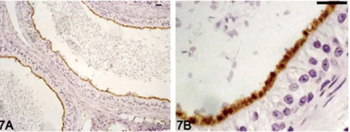
In the initial segment of the epididymis, a strong labeling was observed in the long apical stereocilia from principal cells and little intracellular staining was de-tectable (Fig. 4). The reaction was less intense in the caput epididymis (Fig. 5) than in the corpus (Fig. 6) and cauda epididymidis (Fig. 7). AQP9 staining was abun-dantly expressed on apical stereocilia of principal cells in the initial portion of the vas deferens (Fig. 8). Baso-lateral staining or intracellular staining for AQP9 was not detectable in the epithelial cells of vas deferens.
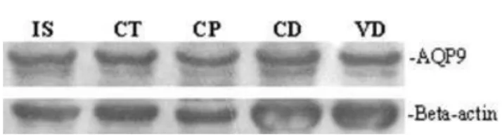
The Western blotting procedure for AQP9 in the extracts from different portions of the epididymis and vas deferens of dog detected one main band
approxi-Figure 7.
Figure 8.
Figs. 5–8. Aquaporin 9 (AQP9) immunolocalization in dog excurrent ducts. The apical region of caput epididymidis showed a weak to moderate intensity of AQP9 immunostaining (Fig. 5A,B). Immunostain-ing intensity of AQP9 in the cellular stereocilia increases in the corpus
(Fig. 6A,B) and cauda (Fig. 7A,B). AQP9-positive immunoreaction was present on the cellular stereocilia in the proximal region of the vas de-ferens (Fig. 8A,B). Scale bar520mm.
mately 30 kDa (Fig. 9), confirming the antibody specific-ity. The antibody also detected other higher bands that represent differentially glycosylated AQP9 forms con-firming the findings of Pastor-Soler et al. (2002). The beta-actin protein, used as an internal control of the reaction, showed an equal amount of protein loaded in each lane.
DISCUSSION
AQPs mediate the efficient movement of water across the cell membranes in different tissues. However, much has been elucidated concerning from molecular structure to cellular distribution in AQPs, hitherto the real knowl-edge of AQPs is not sufficient to obtain a comprehensive view of their role as channel proteins acting on the overall physiology of cell membranes (Matsuzaki et al., 2002).
Especially the AQP9 is a water channel that allows the passage not only of water but also of neutral solutes (Pastor-Soler et al., 2001). Here, AQP9 was detected in efferent ducts, in different parts of the epididymis and vas deferens epithelium in the adult dog by immunohis-tochemistry and by Western blotting methods. Our pres-ent data show that the AQP9 is abundantly expressed in excurrent ducts of the testis in adult dog, where it could represent an important apical pathway for transepithe-lial water flow (Pastor-Soler et al., 2001).
AQP9 in testicular efferent ducts was detected in the entire apical brush border. In epididymal efferent ducts, the staining was restricted to the apical brush border of nonciliated cells in adult dog. These findings are in agree-ment with those reported for rodents, in which AQP9 has been identified in the apical membrane of nonciliated cells of the efferent ducts (Fisher et al., 1998; Pastor-Soler et al., 2001; Badran and Hermo, 2002).
Although AQP1 plays the major role in the fluid resorption of the seminiferous tubules and in the effer-ent ducts (Clulow et al., 1994), it is possible that the presence of AQP9 in the efferent ducts takes part in resorption. So AQP9 perhaps might compensate, at least partially, for the loss of AQP1 from those tissues in the AQP1 knockout mice aiming to preserve their fertility (Da Silva et al., 2006). Furthermore, AQP9, to facilitate the rapid movement of water across the epithelia, could also be involved in other functions, such as the passage of glycerol, which has been proposed as a source of meta-bolic substrate for sperm (Cooper and Brooks, 1981; Da Silva et al., 2006).
In the testis, the interstitial cells have been demon-strated to express AQP9 (Elkjaer et al., 2000; Badran and Hermo, 2002). However, in the present study, as
shown for human testis by Tsukaguchi et al. (1999), AQP9 was not detected in dog testis. On the other hand, AQP9 is an abundant apical membrane protein in all regions of the dog epididymis. Its reactivity was less intense in the caput epididymidis than in the corpus and cauda epididymidis, showing a pattern similar to that described for rats (Pastor-Soler et al., 2001) and humans (Tsukaguchi et al., 1999). In these tissues, AQP9 appears to be a constitutive epithelial membrane protein that may be responsible for apical membrane permeability of water and solutes (Pastor-Soler et al., 2001).
AQP9 was abundantly expressed in the dog vas defer-ens at the apical membrane of principal cells along the proximal region of this duct. In the rat, AQP9 staining was also found throughout the entirety of the vas deferens while intracellular or basolateral staining was absent (Pastor-Soler et al., 2001). In addition to AQP9, both AQP1 and AQP2 are also present in the rat vas deferens, suggest-ing that the composition of the luminal compartment, in which spermatozoa terminate their maturation and are stored (Robaire and Hermo, 1988), involves a complex reg-ulation of transepithelial water and solute transport (Pastor-Soler et al., 2001; Da Silva et al., 2006).
For some years, the vas deferens was considered to be simply a tubular organ whereby sperm exit the epididy-mis at the time of ejaculation. Although, presently there are a large number of investigations concerning to the structure and functions of the vas deferens epithelium cells, which regulate the vas deferens role on the sperm emission and storage throughout its luminal compart-ment (Robaire and Hermo, 1988; Hermo et al., 1994).
Moreover, the vas deferens presents regional differen-ces in its morphology and functions, as well as in the tis-sular distribution and cellular-specific location of the three AQPs (AQP1, AQP2, AQP9) cited, in accordance to Stevens et al. (2000). Nevertheless, the mechanism for transepithelial fluid that occurs in the vas deferens still remains to be elucidated. Perhaps, the vas deferens must play a role to provide in its microenvironment the functional conditions necessary for continuous spermato-zoa the maturation, as well viability and protection of sperm during their passage and storage into the duct, as proposed by Hinton et al. (1996).
Our data also showed that AQP9 distribution along the male reproductive tract in dog is very similar to that verified in humans, allowing one to conclude that the dog apparently could be a good model for comparative and experimental reproductive biology studies, targeting the human andrology. In conclusion, here we described by immunohistochemistry and Western blotting that AQP9 is abundantly expressed along the male reproduc-tive tract of the dog, being an important apical pathway for transmembrane water and neutral solutes flow.
ACKNOWLEDGMENTS
Special thanks are due to Dr. Ste´lio Pacca Loureiro Luna Associate Professor of Veterinary School of UNESP at Botucatu. Also thanks to Dr. Patrı´cia F.F. Pinheiro and Dr. Se´rgio Pereira from our Department for collabo-ration and helpful discussions. This study is part of the PhD Thesis presented by R.F.D. to the State University of Campinas - UNICAMP, Brazil. R.F.D. was funded by FAPESP.
Fig. 9. Western blot analysis of aquaporin 9 (AQP9) in the initial segment (IS), caput (Ct), corpus (CP), cauda (CD) of epididymis and vas deferens (VD) protein extracts from dog. Each line represents 70
mg of protein from different tissues. The beta-actin protein was used as an internal control of the reaction. The antibody recognized a main band of AQP9 of approximately 30 kDa.
LITERATURE CITED
Agre P. 2004. Aquaporin water channels (Nobel Lecture). Angew Chem Int Ed Engl 43:4278–4290.
Agre P, Brown D, Nielsen S. 1995. Aquaporin water channels: unanswered questions and unresolved controversies. Curr Opin Cell Biol 7:472–483.
Anderson AC, Amodeo DV, Westman I, Olson MA, Durrante BS. 2001. Analysis of characteristics and cryosurvival of sperm from the caput, corpus and cauda epididymis of the domestic dog. Biol Reprod Suppl 64:222–223.
Badran HH, Hermo LS. 2002. Expression and regulation of Aquapor-ins 1, 8 and 9 in the testis, efferent ducts, and epididymis of adult rats and during postnatal development. J Androl 23:358–373. Bradford MM. 1976. A rapid and sensitive method for the
quantita-tion of microgram quantities of protein utilizing the principle of protein-dye binding. Anal Biochem 72:248–254.
Brown D, Verbavatz JM, Valenti G, Lui B, Sabolic I. 1993. Localiza-tion of the CHIP28 water channel in reabsorptive segments of the rat male reproductive tract. Eur J Cell Biol 61:264–273.
Cho YS, Svelto M, Calamita G. 2003. Possible functional implica-tions of aquaporin water channels in reproductive physiology and medically assisted procreation. Cell Mol Biol 49:515–519. Clulow J, Jones RC, Hansen LA. 1994. Micropuncture and
cannula-tion studies of the fluid composicannula-tion and transport in the ductuli efferentes testis of the rat: comparisons with the homologous metanephric proximal tubule. Exp Physiol 79:915–928.
Cooper TG, Brooks DE. 1981. Entry of glycerol into the rat epididy-mis and its utilization by epididymal spermatozoa. J Reprod Fer-til 61:163–169.
Da Silva N, Silberstein C, Beaulieu V, Pietrement C, Hoek ANV, Brown D, Breton S. 2006. Postnatal expression of aquaporins in epithelial cells of the rat epididymis. Biol Reprod 74:427–438. Elkjaer M, Vadja Z, Nejsum LN, Kwon T, Jensen UB,
Amiry-Mog-haddam M, Frokiaer J, Nielsen S. 2000. Immunolocalization of AQP9 in liver, epididymis, testis, spleen, and brain. Biochem Bio-phys Res Commun 276:1118–1128.
Fisher JS, Turner KJ, Fraser HM, Saunders PTK, Brown D, Sharpe M. 1998. Immunoexpression of aquaporin-1 in the efferent ducts of the rat and marmoset monkey during developmente, its modu-lation by estrogens, and its possible role in fluid resorption. Endo-crinology 139:3935–3945.
Hermo L, Oko R, Morales C. 1994. Secretion and endocytosis in the male reproductive tract: a role in sperm maturation. Int Rev Cytol 154:106–189.
Hinton BT, Palladino MA, Rudolph D, Lan ZJ, Labus JC. 1996. The role of the epididymis in the protection of spermatozoa. Curr Top Dev Biol 33:61–102.
Ishibashi K, Kuwahara M, Gu Y, Tanaka Y, Marumo F, Sasaki S. 1998. Cloning and functional expression of a new aquaporin (AQP9) abundantly expressed in the peripheral leukocytes perme-able to water and urea, but not to glycerol. Biochem Biophys Res Commun 244:268–274.
Kirchhoff C. 2002. The dog as a model to study human epidi-dymal function at a molecular level. Mol Hum Reprod 8:695– 701.
Matsuzaki T, Tajiki Y, Tserentsoodol N, Suzuki T, Aoki T, Hagiwara H, Takata K. 2002. Aquaporins: a water channel family. Anat Sci Int 77:85–93.
Nelson RD, Stricklett P, Gustafson C, Stevens A, Ausiello D, Brown D, Kohan DE. 1998. Expression of an AQP2 Cre recombinase transgene in kidney and male reproductive system of transgenic mice. Am J Physiol Cell Physiol 275:C216–C226.
Pastor-Soler N, Bagnis C, Sabolic I, Tyszkowski R, Mckee M, Hoek AV, Breton S, Brown D. 2001. Aquaporin 9 expression along the male reproductive tract. Biol Reprod 65:384–393.
Pastor-Soler N, Isnard-Bagnis C, Herak-Kramberger C, Sabolic I, Hoek AV, Brown D, Breton S. 2002. Expression of aquaporin 9 in the adult rat epididymal epithelium is modulated by androgens. Biol Reprod 66:1716–1722.
Preston GM, Carroll TP, Guggino WB, Agre P. 1992. Appearance of water channels in Xenopus oocytes expressing red cell CHIP28 protein. Science 256:385–387.
Robaire B, Hermo L. 1988. Efferent ducts, epididymis, and vas de-ferens: structure, functions, and their regulation. In: Knobil E, Neill JD editors. The physiology of reproduction. New York: Raven Press. p 999–1080.
Robaire B, Viger RS. 1995. Regulation of epidydimal epithelial cell functions. Biol Reprod 52:226–236.
Russell LD, Bartke A, Goh JC. 1989. Postnatal development of the Sertoli cell barrier, tubular lumen, and cytoskeleton of Sertoli and myoid cells in the rat, and their relationship to tubular fluid secretion and flow. Am J Anat 184:179–189.
Schimming BC, Vicentini CA. 1997. Ultrastructural features in the epididymis of the dog (Canis familiaris, L.). Anat Histol Embryol 30:327–332.
Stevens AL, Breton S, Gustafson CE, Bouley R, Nelson RD, Kohan DE, Brown D. 2000. Aquaporin-2 is a vasopressin-independent, constitutive apical membrane protein in rat vas deferens. Am J Physiol Cell Physiol 278:C791–C802.
Tsukaguchi H, Shayakul C, Berger UV, Mackenzie B, Devidas S, Guggino WB, Van Hoek AN, Hediger MA. 1998. Molecular char-acterization of a broad selectivity neutral solute channel. J Biol Chem 273:24737–24743.
Tsukaguchi H, Weremowicz S, Morton CC, Hediger MA. 1999. Functional and molecular characterization of the human neutral solute channel aquaporin-9. Am J Physiol 277:F685– F696.
Turner TT. 1991. Spermatozoa are exposed to a complex microenvir-onment as they traverse the epididymis. Ann N Y Acad Sci 637:364–383.
Verkman AS, Mitra AK. 2000. Structure and function of aquaporin water channels. Am J Physiol Renal Physiol 278:F13–F28. Wong YC, Wong PY, Yeung CH. 1978. Ultrastructural correlation of
water reabsorption in isolated rat cauda epididymidis. Experien-tia 34:485–487.