Volume 2012, Article ID 340787,6pages doi:10.1155/2012/340787
Research Article
Antifungal Activity of Maytenin and Pristimerin
Fernanda P. Gullo,
1Janaina C. O. Sardi,
1Vˆania A. F. F. M. Santos,
2Fernanda Sangalli-Leite,
1Nayla S. Pitangui,
1Su´elen A. Rossi,
1Ana C. A. de Paula e Silva,
1Luciana A. Soares,
1Julhiany F. Silva,
1Haroldo C. Oliveira,
1Maysa Furlan,
2Dulce H. S. Silva,
2Vanderlan S. Bolzani,
2Maria Jos´e S. Mendes-Giannini,
1and Ana Marisa Fusco-Almeida
11Laboratory of Clinical Mycology, Department of Clinical Analysis, Faculty of Pharmaceutical Sciences, UNESP,
Rua Expedicion´arios do Brasil 1621, 14801-902 Araraquara, SP, Brazil
2Institute of Chemistry, UNESP, Rua Professor Francisco Degni 55, 14800-900 Araraquara, SP, Brazil
Correspondence should be addressed to Ana Marisa Fusco-Almeida,[email protected]
Received 5 January 2012; Revised 17 February 2012; Accepted 23 March 2012
Academic Editor: Guillermo Schmeda-Hirschmann
Copyright © 2012 Fernanda P. Gullo et al. This is an open access article distributed under the Creative Commons Attribution License, which permits unrestricted use, distribution, and reproduction in any medium, provided the original work is properly cited.
Fungal infections in humans have increased alarmingly in recent years, particularly in immunocompromised individuals. Among the infections systemic candidiasis, aspergillosis, cryptococcosis, paracoccidioidomycosis, and histoplasmosis mortality are more prevalent and more severe in humans. The current high incidence of dermatophytosis is in humans, especially as the main etiologic agentsTrichophyton rubrumandTrichophyton mentagrophytes. Molecules pristimerin and maytenin obtained from the plantMaytenus ilicifolia(Celastraceae) are known to show various pharmacological activities. This study aimed to evaluate the spectrum of antifungal activity of maytenin and pristimerin and their cytotoxicity in human keratinocytes (NOK cells of the oral mucosa). It was concluded that the best spectrum of antifungal activity has been shown to maytenin with MIC varying from 0.12 to 125 mg/L, although it is also active with pristimerin MIC ranging between 0.12 and 250 mg/L. Regarding the toxicity, both showed to have high IC50. The SI showed high pristimerin against some species of fungi, but SI maytenin was above 1.0 for all fungi tested,
showing a selective action of fungi. However, when comparing the two substances, maytenin also showed better results. The two molecules can be a possible prototype with a broad spectrum of action for the development of new antifungal agents.
1. Introduction
Fungal infections are mainly caused by opportunistic fungi and are usually associated with immunosuppression [1]. Over the past two decades, invasive fungal infections have emerged as a major threat to immunocompromised patients,
since species of Aspergillus, Candida, Cryptococcus, and
Histoplasma emerging pathogens such as Fusarium and
Trichophytoncan cause infection when defenses of host are
broken [1]. Paracoccidioidomycosis is a fungal infection that
is very important, which affects a large percentage of the rural
population of Latin America [2].
There is now a great interest in the discovery of new molecules of natural origin for the treatment of various diseases [3]. Natural products have provided a wide variety of drugs and have become an alternative to large demand
for new antifungal drugs [4]. In Brazil, the use of medicinal plants in traditional medicine has increased considerably in recent years. The wide distribution of natural resources in Brazilian ecosystems and natural diversity of chemical com-ponents and provides the country with potential bioactive materials [5].
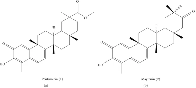
Maytenus ilicifolia (Celastraceae), popularly known as
“espinheira santa,” has been used in traditional medicine since the mid-1920s [6]. The secondary metabolites, maytenin and pristimerin (Figure 1), are classified as quinonemethide triterpenoids and mainly isolated from
the bark of the roots of mature M. ilicifolia plants [7].
Several studies have shown that maytenin exhibits strong antimicrobial activity against positive and Gram-negative organisms, but there are no studies detailing the
O
O
O
HO
Pristimerin (1)
(a)
O
O
HO
Maytenin (2)
(b)
Figure1: Structures of the isolated quinonemethide triterpenes fromM. ilicifolia, pristimerin (1) and maytenin (2).
The need to discover new antifungal molecules and natural products is of great importance for the development of new therapeutic tools. This paper proposes the study of the potential antifungal potential of maytenin and pristimerin against fungi, an agent of important mycoses. The potential broad spectrum of these molecules was evaluated, and the cytotoxicity of these substances in cell lines was evaluated, suggesting possible prototypes of a broad spectrum of components for the treatment of mycoses.
2. Methods
2.1. Microorganisms. ATCC strains and clinical isolates
belong to the mycology collection of the Laboratory of Clinical Mycology, Department of Clinical Analysis, Faculty of Pharmaceutical Sciences, UNESP, Araraquara, were used
in the current study. The strains used were:Candida albicans
ATCC 90028, Candida krusei ATCC 6258,Candida
parap-silosisATCC 22019,Candida glabrataATCC 90030,Candida
tropicalis ATCC 750, Cryptococcus neoformans var. grubii
ATCC 90012, Histoplasma capsulatum(G217B),Aspergillus
niger ATCC 16404, Aspergillus fumigatus ATCC 7100,
Tri-chophyton interdigitalis ATCC 40131.Clinical isolates from
Paracoccidioides brasiliensis (18), Trichophyton rubrum and
Trichophyton mentagrophytes(102),Cryptococcus neoformans
var.grubiiresistant to fluconazole (R30), two clinical isolates
fromCryptococcus neoformansvar.grubiisusceptible to
flu-conazole (S26 and S27), andCryptococcus gattii(118) isolate
from animals, resistant to fluconazole, and Histoplasma
capsulatum(M238P) (lung bat) were also used.
2.2. Minimum Inhibitory Concentration (MIC). All
microor-ganisms were tested for susceptibility to specific commercial drugs for the treatment of each gender. The test for yeast was carried out in accordance with the microdilution method described according to the M27-S3 of the CLSI (Clinical and Laboratory Standards Institute) (2008), with
modifications. The filamentous fungi susceptibility testing was performed according to the M38-A2 of the CLSI (2008) [9] with modifications to determine the minimum inhibitory concentration (MIC).
Two pure substances extracted from Maytenus
ilicifo-lia, maytenin and pristimerin, were prepared as described byScorzoni et al. 2007 [10]. The antifungal drugs were diluted according to CLSI M27-S3 [9]. Inoculums were prepared in RPMI-1640 (Sigma-Aldrich, St. Louis, MO, USA) with L-glutamine, without sodium bicarbonate,
sup-plemented with 2% glucose, and buffered to a pH of 7.0
using 0.165 M MOPS, (Sigma-Aldrich, St. Louis, MO, USA). The yeast suspension was adjusted to a final concentration
of 1.0 ×10⁴CFU/mL in RPMI-1640 and for filamentous
fungi, a suspension of microconidia was adjusted to 2.5 to
5.0×10³. In the 96-well plates, substances were added in
serial dilutions, starting from a concentration of 250 mg/L to 0.48 mg/L. The plates were incubated in a shaker at
37◦C/150 rpm to a specific time determined for each
microorganism. The reading of MIC was performed by spectrophotometry at 490 nm and confirmed using Alamar Blue (Sigma-Aldrich, St. Louis, MO, USA).
2.3. Minimum Fungicide Concentration (MFC). A qualitative
analysis of fungal viability was performed, by transferring a portion of the wells to a plate with Sabouraud (Sigma-Aldrich, St. Louis, MO, USA) medium and incubated at
37◦C during the time determined for each fungal agent. The
MFC was determined as the lowest concentration of the extract that did not allow the growth of any fungal colony on the solid medium after the incubation period [11]. A visual reading was performed to confirm the death or growth inhibition provided by the antifungal substances, maytenin and pristimerin.
2.4. Cell Cytotoxicity Assay. The cytotoxicity of maytenin
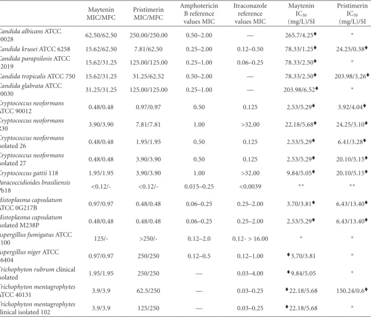
Table1: MIC values and quantitative analysis of fungal cellular viability of pure substances maytenin and pristimerin front of the yeasts and
filamentous pathogenic strains causing the most mycoses and evaluation of cytotoxic activity in NOK cells and selectivity index from the ratio of the IC50and MFC.
Maytenin MIC/MFC
Pristimerin MIC/MFC
Amphotericin B reference values MIC
Itraconazole reference values MIC
Maytenin IC50
(mg/L)/SI
Pristimerin IC50
(mg/L)/SI
Candida albicansATCC
90028 62.50/62.50 250.00/250.00 0.50–2.00 — 265.7/4.25
∗
Candida kruseiATCC 6258 15.62/62.50 7.81/62.50 0.25–2.00 0.12–0.50 78.33/1.25 24.25/0.38
Candida parapsilosisATCC
22019 15.62/31.25 125.00/125.00 0.25–1.00 0.06–0.25 78.33/2.50
∗
Candida tropicalisATCC 750 15.62/31.25 31.25/62.52 0.50–2.00 — 78.33/2.50 203.98/3.26
Candida glabrataATCC
90030 31.25/31.25 125.00/125.00 0.25–1.00 — 203.98/6.52
∗
Cryptococcus neoformans
ATCC 90012 0.48/0.48 0.97/0.97 0.50 0.125 2.53/5.29
3.92/4.04
Cryptococcus neoformans
R30 3.90/3.90 7.81/7.81 1.00 >32.00 22.18/5.68
24.25/3.10
Cryptococcus neoformans
isolated 26 0.48/0.48 1.95/1.95 0.50 0.125 2.53/5.29
6.41/3.28
Cryptococcus neoformans
isolated 27 0.48/0.48 3.90/3.90 0.50 0.125 2.53/5.29
20.10/5.15
Cryptococcus gattii118 1.95/1.95 3.90/3.90 1.00 >32.00 9.84/5.05 20.10/5.15
Paracoccidioides brasiliensis
Pb18 <0.12/- <0.12/- 0.015–0.25 <0.0039 ∗∗ ∗∗
Histoplasma capsulatum
ATCC 0G217B 0.97/0.97 0.48/0.48 0.06–0.25 0.25–2.00 3.70/3.81
6.43/13.40
Histoplasma capsulatum
isolated M238P 0.48/0.48 0.48/0.48 0.06–0.25 0.25–2.00 2.53/5.29
6.43/13.40
Aspergillus fumigatusATCC
7100 125/- >250/- 0.12–2.0 0.12->16.00 ∗ ∗
Aspergillus nigerATCC
16404 0.97/0.97 250/250 0.12–0.5 0.12–1.00
3.70/3.81 ∗
Trichophyton rubrumclinical
isolated 1.95/1.95 250/250 — 0.03–4.00
9.84/5.05 ∗
Trichophyton mentagrophytes
ATCC 40131 3.9/3.9 62.5/250 — 0.03–0.25
22.18/5.68 150.24/0.6
Trichophyton mentagrophytes
clinical isolated 102 3.9/3.9 125/250 — 0.03–0.25
22.18/5.68 ∗
MIC: minimum inhibitory concentration; MFC: minimum fungicide concentration. IC
50/IS values in NOK cells.
∗IC
50/IS not apply because the MIC and MFC values are above 62.5 mg/L.
∗∗IC50/IS not apply because the MIC and MFC values are below 0.12 mg/L.
IC50: 50% inhibitory concentration. SI: selectivity index obtained from the relationship between the IC50(NOK) by MFC for each fungi.
(keratinocyte oral mucosa), obtained from the American Type Culture Collection (Manassas, VA, USA). The cells were grown in own culture medium NOK (Keratinocytes—SFM,
GIBCO), and maintained at 36.5◦C. A concentration ranging
from 2.5 to 5.0×10⁴ cells/mL was used for the formation
of monolayer cells. The concentrations of pure substances were kept in contact with the cells for 24 hours. After the incubation period, the cells treated with the MTT reagent has an additional 5 mg/mL added, (Sigma-Aldrich, St. Louis, MO, USA) and the cells were incubated again for another
4 h. After the formation of formazan crystals, 100µL of
isopropanol was added to solubilize the precipitate and allow the reading of the result by changing the color of the medium
[12]. The absorbance of formazan was quantified using an ELISA reader (enzyme-linked immunosorbent assay) set at 560 nm. As a positive control test, hydrogen peroxide was used.
3. Results
3.1. Minimum Inhibitory Concentration (MIC). Table 1 present the results of the antifungal activity of maytenin
and pristimerin, isolated fromMaytenus ilicifolia. Maytenin
between 0.12 and 62.5 mg/L. Pristimerin previously showed a significant variation in the action potential. For yeasts, the
pristimerin showed potent activity, with the exception ofC.
glabrataATCC 90030 which showed moderate activity andC.
albicansATCC 90028, which presented low antifungal
activ-ity. For the filamentous fungi, pristimerin showed moderate
activity, with the exception ofTrichophyton mentagrophytes
ATCC 40131, which showed a MIC of 62.5 mg/L and was classified as having potent activity.
3.2. Minimum Fungicide Concentration (MFC). Minimum
fungicide concentration was performed to confirm cell death in MIC through observation of no colony growth in a rich medium. The maytenin showed fungicide activity for most
yeasts. No growth ofC. albicanscolonies was observed, and
the death of yeast occurred in a higher dilution concentration (Table 1). For the filamentous fungi, MFC was confirmed by testing cell viability, which occurs in MIC fungal cell death (Table 1).
Pristimerin presented a greater difference between the
MIC and MFC. Moreover, the difference between the MIC
and the value of qualitative analysis of fungal viability was
greater than the difference presented by maytenin (Table 1).
For the fungusParacoccidioides brasiliensisand the
fila-mentous fungiAspergillus nigerandfumigatus, the qualitative
test of fungal viability was flawed, since the growth of colonies cannot be observed on a solid medium.
3.3. Cell Cytotoxicity Assay. The cytotoxicity assay for the
NOK cell line showed more than 80% of cell viability in MIC maytenin concentrations. The substance pristimerin showed cell viability above 80% in normal cells (Table 1).
4. Discussion
Plants have been used in medicine for a long period of time, since they are easy to obtain and apply various diseases
[4, 13]. Regarding the search for new antifungal agents,
the ideal must have a broad spectrum of fungicidal activity without causing toxicity to the host [14]. The treatment of
fungal infections is not always effective because of resistance
to drugs in addition to presenting high toxicity for human cells. For this reason, there is a continuing search for new drugs which are more potent antifungal, but safer, than existing drugs [15].
The present study showed that pristimerin and maytenin had potente action on the fungi studied (Table 1), but maytenin showed the best results. The exception was
Histoplasma capsulatumisolated M238P andParacoccidioides
brasiliensiswhich showed MIC equal for both substances.
Alan´ıs-Garza et al., 2007 [16] studied the
anti-Histoplasmaactivity of extracts obtained from various plants
and showed MICs between 16 and 125 mg/L. This study
showed an MIC for ATCC strains ofH. capsulatum0.97 and
less than 0.48 mg/L pristimerin and maytenin respectively, while for clinical isolate, the MIC for the two substances was less than a 0.48 mg/L. Compared with few literary data of natural products active against this fungal, pristimerin and
maytenin can be considered prototypes that are excellent anti-Histoplasma.
Few studies have searched for new drugs from natural
products for P. brasiliensis [17, 18]. There are reports of
the activity of anti-Paracoccidioides of Piper regnellii and
“Baccharis dracunulifolia” 30 mg/L [17, 19]. The results
expressed in this study were excellent forP. brasiliensis, MIC
of 0.12 mg/L for both molecules at a concentration 65 times lower than the results of the studies mentioned above.
The systemic mycosis caused by yeastsCryptococcusspp.
has increased because of AIDS [20]. In the current results indicate that the molecules had an excellent MIC for the
yeasts this genus,ranging from 0.48 to 3.9 mg/L for maytenin
and 0.97 to 7.8 for pristimerin. Another study revealed the
antifungal activity of extracts ofMaytenus undataagainstC.
neoformans, which showed MIC of 0.09 mg/L after 24 h, and
0.18 mg/L after 48 h [21]. The speciesCandidaare classified
as the fourth most common pathogen in hospitals and are associated with increased mortality of bloodstream infec-tions due to these fungi having high resistance to existing antifungal [22]. For this yeast, the pristimerin showed potent
activity, with the exception of C. glabrata ATCC 90030,
which showed moderate activity, and C. albicans ATCC
90028, which obtained low antifungal activity. Maytenin showed moderate antifungal activity for this specie. For C. albicans, maytenin and pristimerin showed fungicide
activity; however, the Candida non-albicans the result was
fungistatic, except forC. glabratain contact pristimerin.
Although dermatophyte infections are restricted to cer-tain areas of the epidermis, they can be invasive and cause serious injury [23]. Due to the high incidence there is a great need to find new drugs which act on the dermatophytes. Maytenin showed potent activity with MIC ranging from 1.95 to 3.9 mg/L. For filamentous fungi, the pristimerin showed moderate activity, with the exception
ofTrichophyton mentagrophytesATCC 44131, which showed
a MIC of 62.5 mg/L. Many studies have demonstrated the plant antifungal potential against these dermatophytes [23– 25], as the study of Lau et al. 2010 [24], that evaluated
extractsEucalypti FoliumandFructus Psoraleae Globuli. Both
the pure compounds effectively inhibit the growth of T.
mentagrophytesandT. rubrum[25].
Invasive aspergillosis is an important cause of mortality in transplant patients [26]. Maytenin had better results
against Aspergillus niger with an MIC value of 0.97 mg/L,
whereas the MIC for A. fumigatus was 125 mg/L and
pristimerin was similar for both 250 mg/L. Maytenin showed good fungicide activity for most filamentous fungi, while pristimirin showed high MIC (Table 1).
On the other hand, the cytotoxicity tests performed with NOK (keratinocytes oral mucosa) showed that the exper-imental substances are not cytotoxic to this cell examined in experiment (Table 1). Although most of the antifungal agents available on the market are of synthetic origin, natural products of the study received the attention of researchers, mainly, due to the occurrence of unwanted factors, such as the resistance of some strains the conventional antifungal agents—especially in immunocompromised individuals—
found antimalarial activity of various substances, including the pristimerin but the cytotoxicity was only for the 17-(methoxycarbonyl)-28-nor-isoiguesterin in adenocarcinoma cell line HT-29 [27].
With respect to selectivity index (SI), it is known that the higher the SI of a substance, the greater is its security. In our study we found that the maytenin substance had SI above 1.0 for all species tested, so we can demonstrate the safe use of this. Likewise, the pristimerin presented high SI
against some fungal species, as described forH. capsulatum
(SI 13.40). However, when comparing the two substances, maytenin still showed better results (Table 1).
5. Conclusion
The results of this study indicate the potential use of maytenin and pristimerin for the treatment of fungal infections, which showed a potent antifungal activity against the fungi studied. Therefore, the data obtained are promis-ing. Although the medicinal plant “Maytenus ilicifolia” is consolidated in the treatment of gastritis and ulcers more pharmacological studies will be necessary to evaluate these molecules as antifungal prototypes.
Acknowledgments
The authors are grateful to the Fundac¸˜ao de Amparo Pesquisa do Estado de S˜ao Paulo (FAPESP) and the Conselho Nacional de Desenvolvimento Cient´ıfico and Tecnol ´ogico (CNPq) for their financial support. V. Santos thanks CAPES and is also grateful to Pr ´o-Reitoria de Pesquisa (PROPe/UNESP) for providing a research fellowship.
References
[1] S. Shoham and S. M. Levitz, “The immune response to fungal infections,”British Journal of Haematology, vol. 129, no. 5, pp. 569–582, 2005.
[2] M. A. Shikanai-Yasuda, F. D. Q. Telles Filho, R. P. Mendes et al., “Guideliness in paracoccidioidomycosis,”Revista da Sociedade
Brasileira de Medicina Tropical, vol. 39, no. 3, pp. 297–310,
2006.
[3] H. Hostettman,Strategy of the biological and chemical
evalua-tion of plant extracts, vol. 70, IUPAC, 1998.
[4] J. C. O. Sardi, A. M. F. Almeida, and M. J. S. Mendes Giannini, “New antimicrobial therapies used against fungi present in subgingival sites—a brief review,”Archives of Oral Biology, vol. 56, no. 10, pp. 951–959, 2011.
[5] M. C. T. Duarte, G. M. Figueira, A. Sartoratto, V. L. G. Rehder, and C. Delarmelina, “Anti-Candida activity of Brazilian medicinal plants,”Journal of Ethnopharmacology, vol. 97, no. 2, pp. 305–311, 2005.
[6] R. Santos-Oliveira, S. Coulaud-Cunha, and W. Colac¸o, “Review of Maytenus ilicifolia Mart. ex Reissek, Celastraceae. Contribution to the studies of pharmacological properties,”
Brazilian Journal of Pharmacognosy, vol. 19, no. 2B, pp. 650–
659, 2009.
[7] V. A. De Freitas Formenton Macedo Dos Santos, D. P. Dos Santos, I. Castro-Gamboa, M. V. B. Zanoni, and M. Furlan,
“Evaluation of antioxidant capacity and synergistic associa-tions of quinonemethide triterpenes and phenolic substances from maytenus ilicifolia (celastraceae),”Molecules, vol. 15, no. 10, pp. 6956–6973, 2010.
[8] L. de Le ´on, M. R. L ´opez, and L. Moujir, “Antibacterial prop-erties of zeylasterone, a triterpenoid isolated from Maytenus blepharodes, against Staphylococcus aureus,”Microbiological
Research, vol. 165, no. 8, pp. 617–626, 2010.
[9] Clinical Laboratory Standard Institute (CLSI), Document M38-A2. Reference method for broth dilution antifungal
sus-ceptibility testing of filamentous fungi, Clinical Laboratory
Standard Institute (CLSI), Wayne, Pa, USA, 2nd edition, 2008. [10] L. Scorzoni, T. Benaducci, A. M. Fusco-Almeida, D. H. S. Silva, V. S. Bolzani, and M. J. S. Mendes-Giannini, “The use of standard methodology for determination of antifungal activity of natural products against medical yeasts Candida sp and Cryptococcus sp.,”Brazilian Journal Microbiological, vol. 38, no. 3, pp. 391–397, 2007.
[11] L. O. Regasini, M. Pivatto, L. Scorzoni et al., “Antimicrobial activity of Pterogyne nitens Tul., Fabaceae, against oppor-tunistic fungi,”Brazilian Journal of Pharmacognosy, vol. 20, no. 5, pp. 706–711, 2010.
[12] T. Mosmann, “Rapid colorimetric assay for cellular growth and survival: application to proliferation and cytotoxicity assays,”Journal of Immunological Methods, vol. 65, no. 1-2, pp. 55–63, 1983.
[13] J. J. Rojas, V. J. Ochoa, S. A. Ocampo, and J. F. Mu˜noz, “Screening for antimicrobial activity of ten medicinal plants used in Colombian folkloric medicine: a possible alternative in the treatment of non-nosocomial infections,”BMC
Com-plementary and Alternative Medicine, vol. 6, article no. 2, 2006.
[14] A. J. Carrillo-Mu˜noz, G. Giusiano, P. A. Ezkurra, and G. Quind ´os, “Antifungal agents: mode of action in yeast cells,”
Revista Espanola de Quimioterapia, vol. 19, no. 2, pp. 130–139,
2006.
[15] R. Fenner, A. H. Betti, L. A. Mentz, and S. M. K. Rates, “Plants with potencial antifungal activity employed in Brazilian folk medicine,”Brazilian Journal of Pharmaceutical Sciences, vol. 42, no. 3, pp. 369–394, 2006.
[16] B. A. Alan´ıs-Garza, G. M. Gonz´alez-Gonz´alez, R. Salazar-Aranda, N. Waksman de Torres, and V. M. Rivas-Galindo, “Screening of antifungal activity of plants from the northeast of Mexico,”Journal of Ethnopharmacology, vol. 114, no. 3, pp. 468–471, 2007.
[17] S. Johann, N. P. S´a, L. A. Lima et al., “Antifungal activity of schinol and a new biphenyl compound isolated from Schinus terebinthifolius against the pathogenic fungus Para-coccidioides brasiliensis,”Annals of Clinical Microbiology and
Antimicrobials, vol. 9, article 30, 2010.
[18] L. de Carvalho Tavares, S. Johann, T. Maria de Almeida Alves et al., “Quinolinyl and quinolinyl N-oxide chalcones: synthesis, antifungal and cytotoxic activities,”European Journal of
Medic-inal Chemistry, vol. 46, no. 9, pp. 4448–4456, 2011.
[19] C. V. Martins, D. L. da Silva, A. T. Neres et al., “Curcumin as a promising antifungal of clinical interest,” Journal of
Antimicrobial Chemotherapy, vol. 63, no. 2, pp. 337–339, 2009.
[20] Md. Sa ´ude, “Dados e pesquisas em DST e AIDS,” DST/AIDS CPNd editor. Brasilia, Brasil, 2002.
[21] T. A. Mokoka, L. J. McGaw, and J. N. Eloff, “Antifungal efficacy of ten selected South African plant species against Cryptococcus neoformans,”Pharmaceutical Biology, vol. 48, no. 4, pp. 397–404, 2010.
ApoE apolipoprotein-derived ApoEdpL-W antimicrobial pep-tide contributes to its antifungal activity in Candida albicans,”
Antimicrobial Agents and Chemotherapy, vol. 55, no. 10, pp.
4670–4681, 2011.
[23] N. T. Peres, F. C. A. Maranh˜ao, A. Rossi, and N. M. Martinez-Rossi, “Dermatophytes: host-pathogen interaction and antifungal resistance,”Anais Brasileiros de Dermatologia, vol. 85, no. 5, pp. 657–667, 2010.
[24] K. M. Lau, L. H. Fu, L. Cheng et al., “Two antifungal com-ponents isolated from fructus psoraleae and folium eucalypti globuli by bioassay-guided purification,”American Journal of
Chinese Medicine, vol. 38, no. 5, pp. 1005–1014, 2010.
[25] C. Cavaleiro, M. J. Gonc¸alves, D. Serra et al., “Composition of a volatile extract of Eryngium duriaei subsp. juresianum (M. La´ınz) M. La´ınz, signalised by the antifungal activity,”Journal
of Pharmaceutical and Biomedical Analysis, vol. 54, no. 3, pp.
619–622, 2011.
[26] M. C. Z. Novaretti, A. S. Ruiz, F. L. Dulley, P. E. Dorlhiac-Llacer, and D. A. F. Chamone, “Detection of Aspergillus sp in bone marrow transplant patients by PCR-nested technique,”
Revista Brasileira de Hematologia e Hemoterapia, vol. 30, no. 2,
pp. 162–163, 2008.
Submit your manuscripts at
http://www.hindawi.com
Evidence-Based
Complementary and Alternative Medicine
Volume 2013 Hindawi Publishing Corporation
http://www.hindawi.com
Hindawi Publishing Corporation
http://www.hindawi.com Volume 2013
INFLAMMATION
Diabetes ResearchJournal of
Hindawi Publishing Corporation
http://www.hindawi.com Volume 2013
ISRN
AIDS
Hindawi Publishing Corporation
http://www.hindawi.com Volume 2013
Hindawi Publishing Corporation
http://www.hindawi.com Volume 2013
Computational and Mathematical Methods in Medicine
Hindawi Publishing Corporation http://www.hindawi.com
Volume 2013 Issue 1
Gastroenterology
Research and Practice
Clinical & Developmental Immunology
Hindawi Publishing Corporation
http://www.hindawi.com Volume 2013
Hindawi Publishing Corporation
http://www.hindawi.com Volume 2013 ISRN
Biomarkers
Hindawi Publishing Corporation
http://www.hindawi.com Volume 2013 Hindawi Publishing Corporation
http://www.hindawi.com Volume 2013
The Scientific
World Journal
Hindawi Publishing Corporationhttp://www.hindawi.com Volume 2013
Oxidative Medicine and Cellular Longevity
ISRN
Addiction
Hindawi Publishing Corporation
http://www.hindawi.com Volume 2013
International Journal of
Endocrinology
Hindawi Publishing Corporation
http://www.hindawi.com Volume 2013
ISRN
Anesthesiology
Hindawi Publishing Corporation
http://www.hindawi.com Volume 2013
BioMed Research International
Hindawi Publishing Corporation
http://www.hindawi.com Volume 2013
Hindawi Publishing Corporation http://www.hindawi.com
Oncology
Volume 2013
Ophthalmology
Hindawi Publishing Corporation
http://www.hindawi.com Volume 2013
Journal of
Hindawi Publishing Corporation
http://www.hindawi.com Volume 2013
Obesity
ISRN
Allergy
Hindawi Publishing Corporation
http://www.hindawi.com Volume 2013
PPAR
R e s e a r c h
Hindawi Publishing Corporation