Printed version ISSN 0001-3765 / Online version ISSN 1678-2690 http://dx.doi.org/10.1590/0001-3765201820170815
www.scielo.br/aabc | www.fb.com/aabcjournal
The Antifungal Activity of Naphthoquinones: An Integrative Review
DÉBORA O. FUTURO1
, PATRICIA G. FERREIRA2
, CAROLINE D. NICOLETTI2
, LUANA P. BORBA-SANTOS4
, FERNANDO C. DA SILVA3
, SONIA ROZENTAL4
and VITOR FRANCISCO FERREIRA1
1
Universidade Federal Fluminense, Departamento de Tecnologia Farmacêutica, Faculdade de Farmácia, Rua Mario Viana, 523, Santa Rosa, 24241-002 Niterói, RJ, Brazil 2
Universidade Federal Fluminense, Faculdade de Farmácia, PPGCAPS, Rua Mario Viana, 523, Santa Rosa, 24241-002 Niterói, RJ, Brazil 3
Universidade Federal Fluminense, Instituto de Química, Departamento de Química Orgânica, Outeiro de São João Batista, s/n, Centro, 24020-141 Niterói, RJ, Brazil
4
Universidade Federal do Rio de Janeiro, Instituto de Biofísica Carlos Chagas Filho, Centro de Ciências da Saúde, Bloco G, G044, Cidade Universitária, 21941-901 Rio de Janeiro, RJ, Brazil
Manuscript received on October 11, 2017; accepted for publication on December 8, 2017
ABSTRACT
Naphthoquinones are the most commonly occurring type of quinones in nature. They are a diverse family of secondary metabolites that occur naturally in plants, lichens and various microorganisms. This subgroup is constantly being expanded through the discovery of new natural products and by the synthesis of new compounds via innovative techniques. Interest in quinones and the search for new biological activities within the members of this class have intensified in recent years, as evidenced by the evaluation of the potential antimicrobial activities of quinones. Among fungi of medical interest, yeasts of the genus Candida are of extreme importance due to their high frequency of colonization and infection in humans. The objective of this review is to describe the development of naphthoquinones as antifungals for the treatment of Candida species and to note the most promising compounds. By using certain criteria for selection of publications, 68 reports involving both synthetic and natural naphthoquinones are discussed. The activities of a large number of substances were evaluated against Candida albicans as well as against 7 other species of the genus Candida. The results discussed in this review allowed the identification of 30 naphthoquinones with higher antifungal activities than those of the currently used drugs.
Key words:Quinones, xanthenes, lapachol, lawsone, Candida spp, disc diffusion method.
Correspondence to: Vitor Francisco Ferreira E-mail: vitorferreira@id.uff.br
* Contribution to the centenary of the Brazilian Academy of Sciences.
INTRODUCTION
As life evolved, several chemical and biochemical
routes developed that allowed the diversification and
specialization of the first organisms. Consequently,
this increased the degree of sophistication in terms
of organization, structure and function and led
species to specialize in terms of the production
of molecules with specific functions. Later,
evolutionary paths led to new chemical reactions
and giving origin to other molecules. At the same time, species began to produce small functional molecules that modulate several evolutionary biochemical processes to allow them to adapt their morphologies, physiologies, habits, water and nutrient uptake, animal-plant interactions and animal-animal interactions to their habitat (environment). These features were used by Gottlieb (1992), an outstanding Brazilian scientist who studied the phylogeny and chemosystematics of several plant families and the natural products produced by those families. He established new taxonomies and elucidated phylogenetic tendencies using secondary metabolites that were fundamental for understanding the relationship of plants with their respective ecosystems. These aspects of evolution led to the development of natural products that are of great importance to humanity. The vast biodiversity of rainforest regions holds many potential substances that can be used as drugs to treat human diseases such as cancer (Ferreira et al. 2008, Izumi et al. 2011).Indeed, in some developing countries almost 80 % of the population depends on ethnobotanical medicines (Pinto and Castro, 2009).
Quinones are small, six-membered α, β-dienonic rings with well-defined functional activities that occur in plants, fungi, lichens, bacteria, algae, viruses, insects and higher organisms. The complexity of these compounds evolved with the species themselves (Thompson 1997). For instance, this is a class of secondary metabolites in the Bignoniaceae family (Cipriani et al. 2012), and the naphthoquinone lapachol, which was initially isolated from Handroanthus impetiginosus and
has significant anticancer properties, is the most
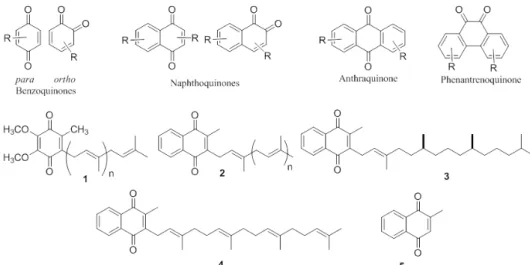
commonly occurring quinone in this family. Quinones are classified according to their aromatic carbon skeleton. The most common are the benzoquinones, naphthoquinones, anthraquinones and phenanthrenequinones (Figure 1). In organisms, these compounds are
related to chemical defenses and have specific biological functions in electron transfer in various oxidative processes in aerobic metabolism, such as photosynthesis, oxidative phosphorylation and other electron transfer reactions (Castro et al. 2008, Silva Júnior et al. 2008, Coates et al. 2013). Some of these quinones are involved in multiple metabolic processes in animals, microorganisms and plants. Among these compounds, benzoquinones stand out; these compounds include coenzyme Q (ubiquinones, 1) and the vitamin K family (Figure 1), which participates in electron transport during cellular respiration and blood coagulation (Mahler and Lordes 1971). Plastoquinones (2, Figure 1) are found in the chloroplasts of plants and algae and are involved in the transport of electrons during photosynthesis (Goodwin and Mercer 1972). Vitamins K1 (3), K2 (4) and K3 (5) (Figure 1) are synthesized by intestinal bacteria, but they are also present in several plants consumed as food and are essential for the formation of prothrombin, a chemical necessary for blood clotting (Dowd et al. 1995). The vitamin K family is large, and most of the members are commercially available as therapeutic treatments. From an ecological point of view, quinones are considered protective not only for plants but also for insects. The best-known example of this is the strategy adopted by some beetles to combat predators wherein they emit a spray consisting of vaporized benzoquinones and oxygen.
MECHANISMS OF BIOLOGICAL ACTION OF QUINONES
more prevalent than the others. Synthetic analogs can be modulated to act selectively via one of several mechanisms of action, especially when the structures of the synthesized molecules are similar to the natural or endogenous quinones of the organisms. In these cases, it is reasonable that these analogs can act in the same way and interfere in the same cellular biochemical processes. For example, quinones are involved in the intracellular transport of electrons, and it is generally accepted that exogenous quinones can transfer electrons in biological processes; this is an important cyclic property as oxidation is followed by reduction and vice-versa (Bendz 1948, Tanaka et al. 1975, Binkley et al. 1939). As electron transport via quinones is not inherent to the biochemical machinery, this process can initiate a cascade of unstable free radicals that react with other molecules generating free radicals that react rapidly with oxygen to generate reactive oxygen species (ROS), such as superoxide, hydroxyl radical, and hydrogen peroxide. These cascades of biochemical processes are catalyzed by NADPH-cytochrome
P450 reductase and other flavoproteins, which may
damage cellular components, such as DNA, lipids and proteins, causing direct damage to the cell (Castro et al. 2013, Kumagai et al. 2012).
Another possible mechanism for the cellular
cytotoxicity of some specific quinones is the in
situ generation of ortho- or para-quinone methides (QMs). QMs are very reactive intermediates that can be used in various chemical reactions (Ferreira et al. 2009), and they are generated in various biological processes, such as tree lignification, enzyme inhibition, phosphodiester reactions, alkylation and DNA cross-linking (Cao et al. 2014). This mechanism of action is quite common in quinones and has been used in nature as well as in prototypes and drugs in medicinal chemistry (Toteva and Richard 2011, Phillips et al. 1999, Wellington 2015, Lin et al. 1972, 1973, 1975). A classic example is the drug mitomycin C, which is a natural product isolated from Streptomyces coeruleorubidus and is commercially available as a therapeutic drug for use in humans against leukemia (Acton et al. 1979), several types of cancers (Kurylowicz 1981) and Trypanosoma rhodesiense (Wilhanson and Scott-Finningan 1981). Its mechanism of action is via its transformation into an intermediate capable of bioalkylating the DNA of tumor cells; however, other modes of action, such as the redox cycle and the inhibition of rRNA, can also contribute to the biological action of the drug (Paz et al. 2012). Troglitazone is an antidiabetic drug, and
its mechanism of action involves activation by cytochrome P450 leading to a QM. This drug was withdrawn from the market in March 2000 because of its hepatotoxicity possibly via the formation of covalent adducts with liver proteins that are deleterious to the liver (Kassahun et al. 2001). On the other hand, tamoxifen acts as a selective estrogen receptor modulator in breast tissue through its active metabolite hydroxitoxifene. This drug generates a QM through a P450-catalyzed two-step oxidation (Fan and Bolton 2001). However, the involvement of a quinone-methide intermediate generated in the in vivo from tamoxifen has never
been effectively demonstrated.
Recently, Silva et al. (2016) proposed that the mechanism of action of pterocarpanquinone (LQB-118), which is active against the promastigotes of Leishmania amazonensis, passes through a QM. This observation was made indirectly by electrochemical and spectroscopic studies in aprotic and protic media with and without oxygen and
through its interactions with DNA under different
conditions (Silva et al. 2016). New substances and new strategies for exploring these endogenous cellular mechanisms of QM action still need to be developed.
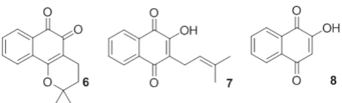
Another mechanism of action related to the cytotoxicity of naphthoquinones is their action on the enzymes topoisomerase I and II (Piyta et al. 1998). There are many published studies indicating that quinones, more specifically naphthoquinones, inhibit either one or both of these enzymes (Cunha et al. 2006, Coelho-Cerqueira et al. 2014, Karkare et al. 2013). Beta-lapachone (6) is the best-known quinone believed to inhibit topoisomerases I (Chiang et al. 1993), which is believed to inhibit topoisomerases I (Boothman et al. 1989, Frydman et al. 1997) and II (Krishnan and Bostow 2000). Direct incubation of this substance with topoisomerase I prior to the addition of
DNA dramatically increases the inhibitory effect,
suggesting β-lapachone directly interacts with
topoisomerase I. Another example that indicates the breadth of potential functions of naphthoquinones
is TU100. This naphthoquinone is able to efficiently
inhibit topoisomerase I and II enzymes, which act on the cell cycle, acting on one or both strands of
DNA. Because of this dual inhibitor effect with a
unique mechanism of action, this quinone has great potential to become a chemotherapeutic agent (Kennedy et al. 2011). Estévez-Braun synthesized and evaluated compounds with moieties conjugated between a naphthoquinone and coumarin as inhibitors of topoisomerase II. All compounds tested were active at submicromolar concentrations (Hueso-Falcóna et al. 2017). It is important to note that these nuclear enzymes are critical to the normal function and integrity of DNA by repairing damage by binding to one or two helices and are therefore important targets for the design of new naphthoquinones. The substances that act on topoisomerases can be divided into two classes: 1) stabilizers of the topoisomerase-DNA complex and 2) catalytic topoisomerase inhibitors (Bassi and Palitti 2000). Inhibitors of topoisomerases I and II by mechanism 1 are more numerous.
Considering the structural diversity of naphthoquinones, the similarities of the biochemistry of endogenous naphthoquinones and the diversity of their mechanisms of action, it is not surprising that these substances may have the capacity to treat and cure human cellular degenerative diseases or diseases caused by viruses or pathogenic microorganisms. These naphthoquinones can be synthesized or isolated from natural products in the form of extracts, teas, powders, oils, roots, etc. Many of these extracts are widely used in folk medicine and are recognized in traditional medical practices. In addition, the natural products produced by these plants, fungi, viruses and bacteria have been a source of inspiration
for structural modifications by rational synthetic
most important diseases affecting humanity (Coseri
2009, Gordaliza 2007, Soejarto and Farnsworth 1989, Raskin et al. 2002, Melo et al. 2011). For example, approximately 74 % of the drugs used in cancer chemotherapeutics are natural products, their derivatives or their synthetic analogs (Butler 2008, Tan et al. 2006).
It is important to note that the selection of these two subjects was because synthetic naphthoquinones have been widely screened against many biological targets (Asche 2005, Skibo et al. 2001, Gafner et al. 1996), such as the HIV-1 virus reverse transcriptase enzyme (Stagliano et al. 2006), and they have been explored for use as anticancer (Liu et al. 2004, Lu et al. 2013, Murakami et al. 2010, Montenegro et al. 2010), leischmanicidal (Kayser et al. 2000, Jernigan et al. 1996, Teixeira et al. 2001), antimalarial (Hooker and Richardson 1948), toxoplasmosis (Ferreira et al. 2002) and trypanocidal (Diogo et al. 2013, Pinto et al. 2000, Pinto and Castro 2009, Salas et al. 2011) agents.
Naphthoquinones are the most commonly occurring type of quinones. They are a diverse family of secondary metabolites that occur naturally in plants, lichens and various microorganisms (Morton 1965, Patai 1974, Patai and Rappoport 1988, Thomson 1997). This subgroup is constantly being expanded through the discovery of new natural products and new compounds that are synthesized via innovative methods. The interest in quinones and the search for quinones with new
biological activities has intensified in recent years
due to the pharmacological relevance of three
important natural naphthoquinones: β-lapachone
(6), lapachol (7) and lawsone (8) (Figure 2)(Silva and Ferreira 2016).
CANDIDIASIS: A WORLDWIDE GLOBAL HEALTH PROBLEM
Fungi are an important group of pathogens even though fungal infections are neglected by the public
health and global health communities (Nature Microbiology 2017). Over 300 million people are acutely or chronically infected by fungi, leading to death, long term illness and reduced work capacity in more than 140 countries around the world. It is estimated that 1.6 million deaths occur annually due to fungal infections (LIFE 2017a). Although there are approximately 2 million fungal species, approximately 600 are potentially pathogenic to humans, and of those, only 30 species are commonly isolated from human patients (LIFE 2017b).
Candida spp. are the most common cause of fungal infections in humans. They are a natural member of the human microbiota and usual live in harmony with humans in the gut and on mucosal surfaces, but when the natural balance is disrupted, infections can occur. In these cases,
Candida spp.infections can range from superficial to systemic and disseminated forms; however, systemic and disseminated infections occur mainly in immunocompromised patients (Gow and Hube 2012). Candida spp. is the fungal species responsible for the fourth most hospital-acquired
bloodstream infections (Wisplinghoff et al. 2004),
and invasive candidiasis has a high mortality rate ranging from 45 to 75 % (Brown et al. 2012). Thus,
Candida spp. are classified by the U.S. Centers
for Disease Control and Prevention (CDC) as a serious threat to human health due to its increasing resistance to antifungal drugs (CDC 2017a).
Although Candida albicans is still the most prevalent fungus, there has been a shift in distribution of species. This mean that of the invasive candidiasis species, the non-Candida albicans species are now more prevalent than they were a few years ago, and several of these species display high rates of antifungal resistance (Benedict et al. 2017). Candida glabrata, Candida parapsilosis, Candida tropicalis and Candida krusei are the most common species outside of non-Candida albicans, and their prevalences vary
is the second most common fungal species in the USA, UK, northern Europe and Australia; C. parapsilosis is ranked second in southern Europe, and C. tropicalis is second in India. C. tropicalis
and C. parapsilosis are also the second and third most prevalent species in Brazil. An important characteristic of C. glabrata and C. krusei is that these species are intrinsically less susceptible to
fluconazole than other Candida spp. (Colombo et al. 2017).
Three major classes of antifungal agents are available to treat invasive fungal infections, namely, polyenes, echinocandins and triazoles, and the main fungal targets are the fungal plasma membrane and the cell wall. An example of a polyene-type antifungal agent is amphotericin B, which destabilizes membranes due its binding to ergosterol allowing it to act as a sponge. Echinocandins, such as caspofungin, micafungin and anidalafungin, block cell wall biosynthesis
by blocking the catalytic subunit of the
β-1,3-glucan synthase. While triazoles inhibit the biosynthesis of ergosterol by blocking lanosterol
at the 14α-demethylation step. Triazoles are the
most used antifungal in medical practices, and
fluconazole is the most common triazole-derived
antifungal in clinical settings (Sanglard 2016). The main antifungal used to treat invasive infections due to Candida is fluconazole (intravenous or oral). In some cases, echinocandins can be used during initial therapies with a posterior
transition to fluconazole. Amphotericin B (lipid
formulation administered intravenously) is the only alternative in patients with azole- and
echinocandin-resistant Candida infections (Pappas et al. 2016). In addition to fluconazole, topical formulations of clotrimazole, miconazole, both are imidazole antifungals) or nystatin, a polyene antifungal (CDC
2017b) could be used to treat superficial Candida
spp. infections.
However, over the years, the use of antifungal agents for the treatment of fungal diseases has led to a new problem: antifungal resistance to fungi. The main mechanisms of antifungal
resistance identified in Candida spp. are decreases in the effective antifungal concentration and
antifungal target alteration. In the first case, the
intracellular concentrations of antifungals can
be decreased by active efflux mediated by efflux
transport systems (ATP-binding cassette (ABC) transporter or transporters of the major facilitator superfamily (MFS)). The antifungal target can
also be overexpressed causing the effective drug
concentration to decrease. In addition, the antifungal can be sequestered in extracellular compartments formed by Candida biofilms. In the second case,
the enzyme that the antifungal binds can change due to mutations (Sanglard 2016).
The increase in the incidences of fungal infections, the high rates of morbidity and mortality, the limitations in antifungal options and the occurrence of antifungal resistance highlight the importance of the development of new antifungal strategies (Scorzoni et al. 2017). However, both fungi and their human hosts are eukaryotic, thus their inherent similarities make the search for new antifungal molecules or antifungal targets much more challenging.
Attempts to elucidate the activity of small natural and synthetic molecules against infections caused by microorganisms have been frequently conducted for more than one hundred years to meet the health, social, environmental and economic needs of mankind. Many researchers have devoted a substantial amount of their time to understanding the biochemical functions of microorganisms
and their relationships with the human species. Today, diseases caused by microorganisms, such as those caused by fungi, remain neglected despite
the substantial amount of effort invested by the scientific community to understand these diseases.
To identify new molecules with high degrees of specificity and lower incidences of side effects, much more research needs to be done to understand the mechanisms controlling the biochemistry of these diseases and their relationship with their host. On the other hand, due to economic concerns, profitable diseases remain the focus of the pharmaceutical industry. In summary, new drugs are needed to meet the demands for both new and old diseases that are still not fully understood, and as we will discuss in this review, naphthoquinones belong to a class of natural and synthetic molecules with great potential for antifungal activity.
MATERIALS AND METHODS
This study is an integrative review of the scientific
literature involving antifungal naphthoquinones. The assembly of this review was conducted via the following steps: formulation of the objective and the research scope, determination of the inclusion and exclusion criteria, data analysis (including ordering, coding, categorization and summarizing), data interpretation and reporting the findings.
(Whittemore and Knafl 2005)
The selection of publications was guided by two
questions: (1) what is the scientific evidence for the
antifungal activity of the naphthoquinone against
Candida spp.; and (2) which naphthoquinones presented in the literature show the most promising antifungal activity against Candida spp. The survey was conducted using the Scopus® and SciFinder® electronic databases with “naphthoquinone” and “antifungal” as the controlled descriptors. The data were collected from June to July 2017. To be included a paper had to meet the following criteria:
they had to be scientific papers dealing with studies
on the antifungal activity of naphthoquinones, they had to be reporting research using fungi related to human or animal health, they had to be available in English, and they had to have been published between 2000 and June 2017. Editorials and reviews were excluded.
Initially, all the publications obtained in the literature search (n = 451) were examined for relevance and the inclusion criteria. Those that did
not meet the criteria specified above or duplicates were excluded. The first selection stage resulted in
a sample of 79 publications.
The selected publications were fully and systematically analyzed to classify the antifungal activity and origin (synthetic or natural products) of the quinones. We also investigated the methodology used in the antifungal evaluation and which species were tested.
From this selection, it was observed that 86 % of the publications (n = 68) evaluated the antifungal activity of the naphthoquinones against several
Candida spp. These publications were selected for a more detailed study of the method used to test the antifungal activity to allow a more precise
and efficient selection of the antifungal activity of
the most promising prototype compounds. In this regard, two other inclusion criteria were added: (1) the antifungal activity as indicated by the minimum inhibitory concentration (MIC) had to be determined by the microdilution method and (2) the activity of the quinones had to be compared to the activities of standard antifungal drugs used in medical clinics.
RESULTS
Approximately 57 % of the relevant papers (68) for this review were published between 2012 and 2017. The authors are from 30 countries, with India (29.4 %), Ukraine (19 %), Turkey (16 %) and Brazil (8.8 %) being the most represented countries. A
significant number of publications (23.5 %) involved
the participation of diverse research groups from
different universities and research centers and from different countries, highlighting the importance of
national and international partnerships.
Most of the publications discussed in this review reported the activities of synthetic naphthoquinones (57); only 11 publications evaluated naturally occurring naphthoquinones. These publications revealed that 691 naphthoquinones were screened for antifungal activity against Candida spp. It is worth noting that 98.5 % of the publications involved synthetic 1,4-naphthoquinones derivatives.
Methods for studying antifungal effects in vitro and in vivo have differed considerably over
time and among investigators, and the logic behind these interpretive categories is worth discussing. The antimicrobial activities of naphthoquinones in the publications evaluated in this review were
determined using the disc diffusion method (DM)
and/or by the microdilution method (MD). Most publications expressed the results of antifungal activity in MIC (57); a smaller fraction expressed the activity in terms of the diameter of the zone of inhibition (11). It should be noted that some of these reports expressed antifungal activities by both methods. Only 32 publications directly
compared the efficiencies of their quinones to the
those of standard antifungal drugs. Eight standard antifungal drugs were used: amphotericin B,
nystatin, ketoconazole, clotrimazole, fluconazole,
griseofulvin, itraconazole and miconazole. Publications that used reference compounds that are not commonly used clinically against Candida
spp. were discarded.
The following Candida spp. were used in these studies: C. albicans, C. dubliniensis, C. glabrata, C. kefir, C. krusei, C. parapsilosis, C. tenuis, and
C. tropicalis. The specimens were obtained from Culture Collection of microorganisms as well as from species isolated from patients at local hospitals. The naphthoquinones used in this review are presented in Table I, and their origins, the techniques used for the measurement of antifungal activity (MD or DM) and type of Candida spp. are shown.
A SELECTION OF RESULTS OBTAINED WITH THE MOST ACTIVE NAPHTHOQUINONES
Plumbagin (9, Figure 3) is a natural naphthoquinone investigated as an antifungal compound that can be isolated from plants of the families Plumbaginaceae (Paiva 2003), Droseraceae and Ebenceae. It has potent biological activities including
anti-inflammatory, antiparasitic and anticancer (Padhye
et al. 2012) activities. This naphthoquinone was tested against C. albicans ATCC 10231 and showed
a MIC of 0.78 μg/mL and a minimum fungicidal concentration (MFC) of 1.56 μg/mL. It is well
established that if the ratio MFC/MIC is lower than 4, the microorganism will die (the compound acts as a fungicide), and if MFC/MIC is greater than 4, the compound is fungistatic (Hazen 1998, Elansary et al. 2016). The MFC/MIC ratio for 26
is 2, indicating that plumbagin (9) can be classified
as a fungicide against C. albicans ATCC 10231. In addition, Gwee et al. (2014) obtained plumbagin (9) from fresh leaf extracts of Nepenthes gracilis Korth. (Nepenthaceae) and found MIC (MFC) values of
2 μg/mL (2 μg/mL) for C. albicans ATCC90028
and 8 μg/mL (16 μg/mL) for C. parapsilosis using amphotericin B as a standard drug. They calculated selectivity indices (SIs) of 0.30 for C. albicans and 0.08 for C. parapsilosis.
TABLE I
List of naphthoquinones, their origins, the techniques used for the measurement of their antifungal activity (disc diffusion
method (DM) and/or the microdilution method (MD)) and the Candida spp used (Legend: NQ = Naphthoquinone; NP =
Natural product; S = Synthesis).
Core NQ Origin of NQ Techniques Used species Reference
1,4-NQ NP MD C. albicans Elansary et al. 2016
1,4-NQ NP MD C. albicans, C.
parapsilosis Gwee et al. 2014
1,4-NQ NP MD C. albicans (7 strains) Hassan et al. 2016
1,4-NQ NP MD/DM C. albicans Inouye et al. 2006
1,4-NQ NP MD/DM C. albicans Ioset et al. 2000
1,4-NQ NP MD C. albicans, C. tropicalis,
C. parapsilosis Naira et al. 2016
1,4-NQ NP MD C. albicans Paiva et al. 2003
1,4-NQ S MD C. albicans (6 strains), C.
glabrata, C. krusei Allochio Filho et al. 2016
1,4-NQ S MD C. albicans Arasoglu et al. 2016
1,4-NQ S MD C. albicans Bayrak et al. 2016
1,4-NQ S MD
C. parapsilosis, C. krusei, C. lusitaniae, C. tropicalis, C. albicans (2
strains)
Castro et al. 2013
1,4-NQ S MD C. krusei, C. paraosilosis Sanchez-Calvo et al. 2016
1,4-NQ S MD C. albicans Tandon et al. 2004b
1,4-NQ S DM C. albicans Pawar et al. 2012
1,4-NQ S DM C. albicans Shinde and Wadekar 2015
1,4-NQ S DM C. albicans Sritrairat et al. 2011
1,4-NQ S MD/DM C. tenuis Ibis et al. 2014
1,4-NQ S MD/DM C. tenuis Ibis et al. 2013a
1,4-NQ S MD/DM C. tenuis Ibis et al. 2011
1,2-NQ S MD C. albicans Khasiyatullina et al. 2009
Dihydrofuran-1,2 and
Dihydrofuran-1,4-NQ S MD / DM
C. albicans (2 strains), C. krusei, C. kefyr, C. parapsilosis, C. tropicalis,
C. dubliniensis
Freire et al. 2010
Amino-1,4-NQ NP DM C. albicans Zhang et al. 2007
Amino-1,4-NQ S MD C. albicans, C.
parapsilosis Tandon et al. 2009a
Amino-1,4-NQ S MD C. albicans, C.
parapsilosis Tandon et al. 2006
Amino-1,4-NQ S MD C. albicans, C.
parapsilosis Tandon et al. 2005a
Amino-1,4-NQ S MD C. albicans, C.
parapsilosis Tandon et al. 2005b
Amino-1,4-NQ S MD C. albicans Tuyun et al. 2015
Amino-1,4-NQ S MD C. albicans Leyva et al. 2016
Core NQ Origin of NQ Techniques Used species Reference
Amino-1,4-NQ and
Thio-1,4-NQ S MD
C. albicans, C.
parapsilosis Tandon et al. 2009b
Amino-1,4-NQ S MD C. tenuis Rutkauskas et al. 2013
Amino-1,4-NQ S MD / DM C. tenuis Deniz et al. 2015
Amino-1,4-NQ S MD / DM C. albicans, C. tropicalis Errante et al. 2006
Amino-1,4-NQ S MD/DM C. tenuis Ibis et al. 2015
Amino-1,4-NQ S MD/DM C. tenuis Ibis et al. 2013b
Amino-1,4-NQ S MD/DM C. tenuis Mickeviciene et al. 2015
Amino-1,4-NQ S MD/DM C. albicans, C. tropicalis Pawar et al. 2014
Amino-1,4-NQ S MD/DM C. albicans Sreelatha et al. 2014
Amino-1,4-NQ S MD/DM C. tenuis Vaickelioniene et al. 2011
Amino-1,4-NQ S DM C. tenuis Voskiene et al. 2012
Amino-1,4-NQ S DM C. tenuis Voskiene et al. 2011
Amino-1,4-NQ S DM C. tenuis Buchkevych et al. 2012
Amino-1,4-NQ S DM C. tenuis Figurka et al. 2015
Furan-1,4-NQ S MD C. albicans, C. tropicalis,
C. krusei Ryu and Chae 2005
Furan-1,4-NQ S MD
C. albicans (2 strains), C. krusei, C. glabrata C. parapsilosis (2 strains)
Santos et al. 2010
Furan-1,4-NQ S MD C. albicans Yamashita et al. 2009
Aryl-1,4-NQ S MD/DM C. albicans, C krusei; C.
glabrata, C. tropicalis Louvis et al. 2016
Aryl-1,4-NQ S MD C. albican, C. parapsilosis Tandon et al. 2011
Alkyl-1,4-NQ NP MD
C. albicans, C. glabrata, C. krusei, C. tropicalis; C.
parapsilosis
Sasaki et al. 2002
Alkyl-1,4-NQ NP DM C. albicans (4 strains) Sidjui et al. 2014
Alkyl-1,4-NQ S MD C. tenuis Choudhari et al. 2017
Pyran-1,2-NQ S MD C. albicans (2 strains) Curvelo et al. 2015
Pyran-1,2- NQ S MD C. albicans (11 strains), C.
parapsilosis (9 strains) Ferreira et al. 2014
Pyran-1,4-NQ NP MD/DM C. albicans Lu et al. 2016
Pyran-1,4-NQ S MD C. albicans Jardosh and Patel 2013
Pyran-1,4-NQ S MD C. albicans Rao et al. 2016
Pyrrolidine-1,4-NQ S MD/DM C. albicans Bhaskar et al. 2012
Halogen-1,4-NQ S MD/DM C. albicans (2 strains) Tran et al. 2009
Thio-1,4-NQ S MD C. albicans Huang et al. 2002
Thio-1,4-NQ S MD C. albicans, C. tropicalis,
C. krusei Ryu et al. 2005
Thio-1,4-NQ S MD C. albicans Tandon et al. 2004a
Thio-1,4-NQ S MD C. albicans Yildirim et al. 2017
Thio-1,4-NQ S MD/DM C. tenuis Ibis et al. 2016
albicans. The results showed that both S. aureus
and C. albicans showed viability, and 26 broke up
mono- and polymicrobial biofilms in glass tubes as well as in urinary catheter silicon tubes. Biofilm
disruption was also observed when S. aureus and
C. albicans were allowed to adhere to silicon urinary catheter tubes and were then treated with plumbagin, but the effect was stronger in glass tubes (Naira et al. 2016).
Interestingly, many metal complexes have been designed and tested as therapeutic targets. Recently, Shinde and Wadekar, in an effort to
discover effective antifungal agents, synthesized a
plumbagin Sm(III) complex and tested it against C. albicans NCIM – 3471 (Shinde and Wadekar 2015). Plumbagin (9) and its Sm(III) complex showed significant antifungal activity, but plumbagin alone showed a higher activity than the plumbagin Sm(III) complex.
Antifungal combination is a challenging but promising strategy for inhibiting the proliferation of fungi and other microorganisms. Very recently, Hassan and co-workers (Hassan et al. 2016) reported the in vitro antimicrobial effect of a combination of plumbagin (9) and amphotericin B against clinical isolates of C. albicans from patients with vaginal yeast infections and C. albicans ATCC 10231. Plumbagin exhibited inhibitory activities against all tested strains of C. albicans with values
ranging from 7.41 to 11.24 μg/mL. An additive
antifungal activity was observed when 26 was used in combination with amphotericin B against
C. albicans ATCC 10231 and in four clinical
isolates with MICs ranging from 0.62 to 0.91 μg/
mL. Based on the results, the authors suggest that the proportion of plumbagin to amphotericin B plays an important role in the additive antifungal interaction.
Bis-naphthoquinone 10 (Figure 3), which has two naphthoquinones moieties, was isolated from
Ceratostigma plumbaginoides and showed potent activity against C. albicans ATCC 25555 (MIC
of 0.09 μg/mL and MFC of 0.17 μg/mL) using fluconazole and ketoconazole as standard drugs
(Elansary et al. 2016).
Hybrid naphthoquinone-anthraquinone 11
(Figure 3), newbouldiaquinone A, was isolated from the bark of Newbouldia laevis SEEM root. (Bignoniaceae). The antimicrobial activities of this compound against C. albicans, C. glabrata and C. krusei were evaluated. It showed potent activities with C. gabrata being the most sensitive yeast. Newbouldiaquinone A is 13-times more active against C. gabrata than nystatin (Eyong 2006).
The naphthoquinones deoxyshikonin (12), acetylshikonin (13), β-hydroxyisovalerylshikonin (13) and shikonin (14) were isolated from the chloroform extract of the roots of Lithospermum erythrorhizon (Boraginaceae, Figure 3). These compounds were assayed against C. albicans
YFC497, YFC803 (azole-resistant), C. glabrata
YFC 501, C. krusei YFC 827, C. tropicalis YFC 052, and C. parapsilosis YFC 826. Shikonin (15) was found to have a four-fold stronger fungicide activity
(MIC 4 μg/mL) than fluconazole. Deoxyshikonin
(12) also exhibited four-fold stronger activity against C. krusei (MIC 4 μg/mL). Acetylshikonin
(13) and β-hydroxyisovalerylshikonin (14) showed lower activities against all fungal pathogens
Core NQ Origin of NQ Techniques Used species Reference
Thio-1,4-NQ S MD/DM C. albicans, C. tropicalis Wittebolle et al. 2006
Thio-1,4-NQ S DM C. albicans Rău et al. 2016
Anthraquinone NP MD C. albicans, C. krusei, C.
gabrata Eyong et al. 2006
Anthraquinone S MD C. albicans (2 strains) Shrestha et al. 2017
than fluconazole except for C. krusei. 13 and
14 were also assayed against azole-resistant C. albicans YFC 830, but they presented the same
antifungal activity as that of fluconazole (Sasaki
et al. 2002). Recently, a series of compounds including 34 naphthoquinones were synthesized, and their in vitro antifungal activities against C. krusei and C. parapsilosis (Sánchez-Calvo el al. 2016) were screened and compared to the activity of the standard drug Amphotericin B. Some of the compounds showed potent activity against C. krusei. Among them, 2-chloro-5,8-dihydroxy-1,4-naphthoquinone (16) was the most active compound against C. krusei with an MIC of 2 μg/mL, which
is similar to that of amphotericin B. It is worth noting that 5,8-dimethoxy-1,4-naphthoquinone (17), 5-pentanoyl-juglone (18), 2,3-dichloro-5,8-diacethoxy-1,4-naphthoquinone (19) and 2,3-dibromo-5,8-dihydroxy-1,4-naphthoquinone (20) also have moderate activity against the same fungus (Figure 3).
Lawsone (13) is a naturally occurring naphthoquinone that can be obtained from the
extract of dried powdered leaves of henna Lawsonia
spp. It is commercially available and has been used as a starting material for the synthesis of many new compounds (Jordão et al. 2015). A series of
thirteen Mannich adducts and different aldehydes
were prepared from lawsone and evaluated as antifungal agents (Allochio Filho et al. 2016) against three Candida strains (C. albicans ATCC 10231, C. glabrata ATCC 2001 and C. krusei
ATCC 34135) and five clinical C. albicans isolates from patients. Among all the compounds, the best antifungal activities were observed against C. albicans ATCC 10231 with the compounds 21, 22
and 23 (Figure 4). They showed activities (MICs) in the 20 to 330 µg/mL range. Compounds 21 and
23 showed fungistatic activity against C. glabrata,
and compound 23 presented an MIC of 330 µg/mL against C. krusei. In addition, regarding clinical strains, compound 21 showed the best fungistatic activity against clinical C. albicans with MICs ranging from 40 to 670 µg/mL.
Following the premise that compounds with a 1,4-naphthoquinone core have promising antifungal activity, a series of halogenated naphthoquinone derivatives was synthesized, and their in vitro antifungal activities against C. albicans ATCC 10231 and C. albicans 955 were evaluated using an MD assay (Tran et al. 2009), and the results were compared with clotrimazole. The results showed that three compounds were quite active against C. albicans ATCC 10231. It should be noted that compounds 24 (MIC 1 μg/
mL), 25 (MIC 4 μg/mL) had better activities than
compound 26 (MIC 8 μg/mL), and they had similar
antifungal profiles as clotrimazole. Compounds 24
and 25 (Figure 4) had potent antifungal activities against C. albicans 955 (MIC 0.25 μg/mL and 32
μg/mL, respectively). Notably, the substituents
on the 1,4-naphthoquinone core are also impact in the activity, probably by modifying the redox potential of the naphthoquinone or changing its physical-chemical properties (Pawar 2014). These compounds were evaluated against C. tropicalis
NCIM 3548 and C. albicans NCIM 3557. All the compounds (24-30, Figure 4) exhibited fungicidal activity towards all the tested fungi with MICs ranging from 0.025 µg/mL to 1.25 µg/mL for all strains. Compound 27 (Figure 4) showed better antifungal activity than amphotericin B (MIC 1.25 µg/mL for C. albicans and C. tropicalis) and nitrofurantoin (31, MIC 1.25 µg/mL for C. albicans
and C. tropicalis).
As noted abo
ve, plumbagin (9) is an important natural naphthoquinone with well-established antifungal activity. Following these studies, another natural product known as juglone (66, 5-hydroxy-1,4-naphthoquinone, Figure 5), which is isolated from plants of the Juglandaceae family, particularly black walnut (Juglans nigra), was investigated. 66has potential uses in medicine and as a biocide for organic farming and pest control. Juglone can be synthesized by several methods and can be used as a starting material for many synthetic
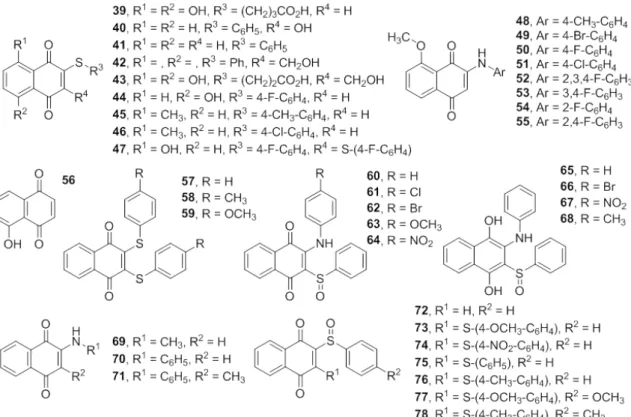
transformations. Reactions of juglone (56) with arylamines, arylthiols or halogens occur easily on the naphthoquinone moieties, producing several derivatives such as 39-55. These substituents can
enhance the biological efficacies of the compounds.
Tandon and coworkers demonstrated the synthesis of novel 1,4-naphthoquinone derivatives with alkyl or arylthio substituents at the 2 position, such as 1,4-naphthoquinone 39 and 2,3-disubstituted 1,4-naphthoquinone 40,and tested against various strains of pathogenic fungi, such as C. albicans, C. neoformans, S. schenckii, T. mentagraphytes, M. cannis and A. fumigatus. The antifungal activities of these compounds were compared with the activities of miconazole, nystatin and amphotericin B. Compounds 39 (MIC 3.125 µg/mL) and 40 (MIC 25 µg/mL, Figure 5) presented better activities against C. albicans than miconazole (MIC 25 µg/ mL for C. albicans). Comparison of the activities of the compounds to that of nystatin (MIC 7.8-7.9 µg/mL for C. albicans) showed that compound 39
was more active against C. albicans than nystatin (Tandon et al. 2004a). In another study, Tandon and co-workers (Tandon et al. 2004b) described a series of sulfur-containing 1,4-naphthoquinone derivatives that inhibit fungal growth with MICs in the same range as well-established antifungal drugs. The antifungal properties of the compounds were determined against the same strains of pathogenic fungi. The results showed that compounds 41, 42
and 43 possess pronounced antifungal activities (Figure 5). The comparison of the activities of
41, 42 and 43 to that of miconazole (MIC 25 µg/ mL for C. albicans) revealed that compounds 41
(MIC 12.5 µg/mL) and 43 (MIC 3.125 µg/mL) are
more effective against C. albicans,and compound
42 (MIC 1.56 µg/mL) is more active against C. neoformans and possesses the same activity profile
as that of miconazole against C. albicans. In this work, the antifungal activities of compounds 41,
C. albicans more than twice that of nystatin. In conclusion, the promising inhibitory effects of
sulfide 1,4-naphthoquinones were demonstrated,
and the activity of compound 43 against a number of fungi was found to be substantial. In addition to this, Ryu et al. (2005) prepared a series of 5-hydroxy- or 5-methoxy-1,4-naphthoquinones and tested those classes of compounds against
C. albicans and C. tropicalis using amphotericin B as the reference drug (MIC 0.25 µg/mL for C. albicans and 0.5 µg/mL for C. tropicalis, Figure 5). The results showed that the most active compounds were substituted arylthio derivatives of juglone
(44-47). Compounds such as 44 (MIC 2 µg/mL),
45 (MIC 4 µg/mL), 46 (MIC 0.5 µg/mL) and 47
(MIC 0.5 µg/mL) have activities comparable to
that of amphotericin B against C. tropicalis. It is worth noting that compounds 44 and 47, which
contain 4-fluoro-phenylthio substituents, exhibited
potent antifungal activity. In addition to this, 3-arylamino-5-methoxy-1,4-naphthoquinones 48-55 (Ryu and Chae 2005) showed potent antifungal activity against all tested fungi species, but their activities against C. tropicalis were highest. Many of these compounds had more potent antifungal activities against C. tropicalis than ketoconazole. It is interesting to note that compound 51 was more active against C. albicans KCCM 50235,
C. tropicalis KCCM 50662 and C. krusei KCCM
11655 (MIC 6.3, 0.8 and 6.3 μg/mL, respectively) than ketoconazole (MIC 6.3, 6.3 and 12.5 μg/mL,
respectively).
In their continuing effort to find new potent
1,4-naphthoquinone analogs, Tandon and co-workers developed new routes for preparing 1,4-naphthoquinones substituted at C2 and C3. The antifungal activities of these compounds (Figure 4) have been evaluated against C. albicans and C.
parapsilosis ATCC 22019. The antifungal activity was compared with standard drugs like miconazole. The research published in 2005 evaluated the antifungal activity of 10 naphthoquinones and compound 35 was found to have the most potent activity against C. albicans (MIC 6.26 μg/mL) (Tandon et al 2005a). Years later, the authors present the results of a new research with other group of Amino-1,4-naphthoquinones and Thio-1,4- naphthoquinones. Compound 32 (MIC 6.25 µg/mL) was found to have better activity against C. albicans than miconazole (MIC 25 µg/mL), and 33
showed the same activity as miconazole (Tandon
et al. 2009b). In the same work, they reported the synthesis of novel nitrogen- and sulfur-containing 1,4-naphthoquinones and the evaluation of their antifungal activities. Among the most promising antifungal compounds of this series, only 34
showed the same antifungal activity against C. albicans (MIC 1.56 µg/mL) as that of miconazole. Additionally, compounds 36 (MIC 3.00 mmol/ mL), 37 (MIC 5.38 mmol/mL), and 38 (MIC 5.66 mmol/mL) were prepared and showed good antifungal activity against C. albicans (Tandon et al. 2010). Compounds 36 (MIC 6.02 mmol/mL) and 38 (11.32 mmol/mL) also showed pronounced antifungal activities against C. parapsilosis.
Errante et al. (2006) reported a synthetic route to disulfidequinone-quinones (57-59), amino-sulfoxide-quinones (60-68) and
sulfidesulfoxide-quinones (73-78) and studied their activities
were screened in vitro for their growth inhibitory
activity against different fungi strains such as C. albicans CIP 884.65 and C. tropicalis CIP 1275.81. The MIC values were determined by comparison to amphotericin B as a standard. Amphotericin B was more active than all the synthesized compounds against C. albicans; however, compounds 57-68
(Figure 5) were all very active against C. tropicalis. The MICs of the synthesized molecules against C. albicans were substantial compared to amphotericin B. The MICswere lower than 0.2 μg/mL except for compound 81 which had an MIC between 0.2-0.3
μg/mL for C. tropicalis. No clear structure-activity relationship seems to exist with these series. As an expansion of this fruitful area of research, Wittebolle et al. (2006) reported the synthesis of several derivatives of 2-benzenesulfinyl-(1,4)-naphthoquinone (72) and evaluated them as antifungal agents against C. albicans and C. tropicalis, and they used amphotericin B as a reference. The results indicated that the compounds presented higher MICs than amphotericin B against C. albicans. Conversely, for C. tropicalis,
60-64 and 72-78 (Figure 5) all had MICs below
0.2 μg/mL, except compound 77, which had
an MIC of 0.3 μg/mL. As a further extension of
these studies, a conjugate addition of primary and secondary amines to 1,4-naphthoquinones to obtain 2-amino-1,4-naphtoquinones was developed, and the resulting compounds were tested in vitro as antifungal agents (Sharma et al. 2013). Compounds
69, 70 and 71 exhibited strong activity against C. albicans (MIC 15.6 µg/mL), but nystatin, which was used as a standard, was much more potent (MIC 7.8 µg/mL for C. albicans).
Xanthene and chromene are synthetic
heterocyclic scaffolds that exhibit a broad spectrum
of biological properties (Forezi et al. 2017a). Several preparative approaches for this class of compounds have been described in the literature and mainly center around two or three component reactions. In their search for new antimicrobial
agents, Jardosh and Patel (2013) prepared a series of xanthene-naphthoquinones and assayed them against Gram-positive and Gram-negative bacteria and tested their antifungal properties against C. albicans MTCC 227. The antifungal testing results revealed that compounds 79 and 80 (MIC 250 μg/ mL, Figure 6) had moderate activity against C. albicans compared with nystatin. Spirooxindoles are synthetic or naturally occurring heterocyclic compounds with a wide range of biological activities. Combining the spirooxindole structure with a naphthoquinone can produce compounds with enhanced antifungal activities. Indeed, Bhaskar et al. (2012) reported the synthesis of
1,4-naphthoquinones fused to isatin scaffolds and
sarcosine through 1,3-dipolar cycloadditions. The antimicrobial activities of these compounds were screened against eight bacteria and three fungi
in vitro using the disc DM. The results revealed that most of the synthesized compounds exhibited antimicrobial activities against Staphylococcus aureus, S. aureus (MRSA), Enterobacter aerogens,
Micrococcus luteus, Proteus vulgaris, Klebsiella pneumonia, Salmonella typhimurium, Salmonella paratyphi-B, Malassesia pachydermatis, C. albicans and Botyritis cinerea. Significant MICs were found for 1’-acetyl-2-methyl-2,3- dihydrospiro[benzo[f]isoindole-1,3’-indoline]-2’,4,9-trione (81) against C. albicans MTCC 227.
(Forezi et al. 2017b), it was possible to prepare eight naphthoquinone derivatives and evaluate their antifungal activities against C. albicans ATCC 90028, C. albicans PYCC 3436T, C. krusei PYCC 3341, C. parapsilosis PYCC 2545, C. parapsilosis
ATCC 22019, C. glabrata PYCC 2418T. Among them, two naphtho[2,3-d ]isoxazole-4,9-dione-3-carboxylates, 86 and 87, are more potent than amphotericin B against C. albicans, C. krusei, C. parapsilosis and C. glabrata. It is also important to note that in previous studies on this group of compounds, they were found to be non-cytotoxic to human cells (Santos et al. 2009). The authors suggest that the naphtho[2,3-d]isoxazole-4,9-dione
scaffold has the potential to be developed into novel
and safe therapeutic antifungal agents. Recently,
Mickevičienė et al. (2015) reported the synthesis
and biological activity of derivatives of aromatic, heterocyclic and/or naphthoquinone fragments containing p-amino acids. The antifungal activities of the obtained compounds were evaluated against
C. tenuis VKM Y-70 by means of DM and MD. The activities were compared to that of the antifungal
agent nystatin. The 3-(2-substituted-6,11-dioxo-6,11-dihydro-12H-benzo[b]phenoxazin-12-yl) butanoic acids showed the best results in this evaluation; compounds 83-85 (Figure 6) showed MICs of 15.6 μg/mL, 31.2 μg/mL and 31.2 μg/mL, respectively, against C. tenuis VKM Y-70.
An interesting series of quaternary ammonium salts of 1,2,3-triazoles fused to naphthoquinones were synthesized and screened as antifungal agents. It is well known that amphiphilic molecules are the most commonly used antimicrobial disinfectants. Shrestha et al. (2017) synthesized cationic amphiphilic units based on aminoglycosides and 1,4-naphthoquionones. Thus, the antifungal activities of a series of synthetic dimeric cationic naphthoquinone analogs were evaluated against
A. flavus, C. albicans 64124 (azole-resistant),
Candida albicans MYA2876 (azole-susceptible),
Cryptococcus neoformans and Rhodotorula pilimanae using Kanamycin A (K20) and
fluconazole as the standards. Compounds 88 and 89
(Figure 7) exhibited excellent antifungal activities against all tested fungi, and both compounds had
Figure 6 - a (Entry 79 – 81)– Examples of 1,4-benzoxanthenes with antifungal activity; b
MICs of 3.91 µg/mL for both C. albicans 64124 (azole-resistant) and C. albicans MYA2876 (azole-susceptible). These values are at least 17 times higher than those of the standard drugs for treating
C. albicans. A green fluorescence assay showed
that compound 89 exerts its antifungal activity via a pore formation mechanism.
Freire et al. (2010) reported the synthesis
and antifungal activities of substituted α- and β-dihydrofuran naphthoquinones prepared via
the reaction of lawsone (8) with olefins in the presence of cerium (IV) ammonium nitrate (CAN). The antifungal activities these compounds were evaluated against 89 fungal cultures, including
29 opportunistic yeasts, 40 filamentous fungi and
20 opportunistic dermatophytes. The compound designated IVS320 (90, Figure 8) presented the lowest MICs for all the tested cultures and was particularly active against dermatophyte fungi
(MICs ranged from 5-28 μg/mL) and yeasts from
the genus Cryptococcus (MICs ranged from 3-5
μg/mL, Ferreira et al. 2014). IVS320 induced
changes in the fungal membrane by interacting with ergosterol, but it did not alter the structure of the fungal cell wall. However, it changed the permeability of the cell membrane (as evidenced by
the efflux of K+ and leakage of substances), which
was not related to binding with ergosterol. These results are very relevant, and this compound could be used as a starting point for the development of new antifungal agents.
Among the publications discussed in this review, only two reports involved the development of naphthoquinone-derived pharmaceutical products for the treatment of candidiasis. Several studies have already described the preparation, characterization and in vitro activity formulations of natural molecules with antifungal activity such as juglone (56) and lawsone methyl ether. Arasoglu et al. (2016) synthesize and characterize juglone (56)-entrapped Poly(lactic-co-glycolic acid) nanoparticles (PLGA nanoparticles) and
compared the antifungal properties of free juglone (56) with the PLGA nanoparticle formulation. The antifungal activity was evaluated by two
different methods with A. flavus, C. albicans and
Fusarium spp. The antifungal studies using agar dilution and top agar dilution methods indicated that the juglone-encapsulated nanoparticles were more effective than free juglone (56). Sritrairat et al. (2011) determined the antifungal activity of lawsone methyl ether (LME) in mouthwash and compared it with that of chlorhexidine mouthwash
in vitro and in vivo. The antifungal activity of LME has been reported, and it has the potential to be used after successful treatment of oral candidiasis caused by Candida spp. to delay relapse and reduce antifungal drug use. The antifungal activity of
0.25 % LME was significantly greater than that of
0.12 % chlorhexidine mouthwash (P < 0.05). As an ideal antimycotic for treating oral candidiasis is not yet available, LME may be an appropriate alternative mouthwash for the prophylaxis of oral candidiasis caused by Candida spp. Despite the well-documented antifungal activity of several naphthoquinones, the real-world application of these compounds has encountered some challenges, mainly due to their low solubility in water. These studies showed that formulated products are an indication of promising antifungal activity, and product formulations may be an important tool for the successful application of these materials, especially the hydrophobic compounds, in many areas.
DISCUSSION
other Candida species and microorganisms other than C. albicans have become involved in such infections. The balance between C. albicans and non-Candida albicans determines the virulence profiles. Some fungal species can also form biofilms with other species which impacts their virulence (Martins et al. 2014). No new classes of antifungal drugs have been approved in the past ten years. This is problematic because current drugs
are not sufficiently active, their oral administration
is restricted, they have specific drug or class toxicities, or they have notable drug interactions (Denning and Bromley 2015).
The current literature confirms the urgent need for new drugs despite the effective drug treatments
on the market. One strategy for the development of new drugs is the screening of antifungal drugs using samples from compound libraries (Roemer and Krysan 2014, Calderone et al. 2014). A review
of scientific publications may assist in the selection
of promising targets for inclusion in more detailed stages of the evaluation of biological activity.
Amphotericin B is a broad spectrum antifungal and is used for the treatment of severe infections caused by Candida species and especially for
fluconazole resistant strains (Pianalto and Alspaugh
2016). The results discussed in this review indicate that three naphthoquinones are more active than amphotericin B against different strains of C. albicans: 27, 28 and 29 (Pawar et al. 2014). Two compounds are noteworthy due to their promising results against C. albicans and non-C. albicans. The naphtho[2,3-d]isoxazole-4,9-diones 86 and
87 were much more active than amphotericin B
against 6 different Candida species. (Santos et al. 2010)
Regarding C. tropicalis, it has become dramatically more prevalent in several countries. It is a potential opportunistic fungus in patients with neutropenia, the suppression of bacterial
flora due to the use of antibiotics, or damage to the gastrointestinal mucosa. The sulfidesulfoxide-quinones, disulfidequinones and
amino-sulfoxide-quinones (57-68 and 72-78) presented potent activity against C. tropicalis with MICs less than
0.2 μg/mL. (Errante et al. 2005) A similar result
was found for a series of 2-benzenesulphinyl-(1,4)-naphthoquinone derivatives, 72-78 (Wittebolle et al. 2006).
Azoles are the most commonly prescribed drugs to treat Candida spp. infections, but some
Candida spp. are less susceptible to azoles, and some species develop resistance during prolonged therapy (WHO 2016). Fluconazole is the most
widely prescribed azole and is very effective in the
treatment of infections caused by Cryptococcus
and Candida spp. Resistance to fluconazole is a
significant clinical problem, as some Candida spp., such as C. krusei, are intrinsically resistant to this drug. Health authorities worldwide are warning
of accurate identification of the infectious species
and the use of antifungal susceptibility tests for clinically relevant isolates (Pianalto and Alspaugh 2016).
Figure 8 - (Entry 90) – Structure of IVS320.
Figure 7 - (Entry 88 – 89)– Dimeric cationic naphthoquinone analogs with activities against A. flavus, C. albicans 64124, Candida albicans
MYA2876, Cryptococcus neoformans and
The evaluation of antifungal activities against azole-resistant C. albicans species allows the selection of compounds that may become very promising drugs for the solution of this major public health problem. Naphthoquinones 88 and
89 are noteworthy for their excellent activities against strains of azole-resistant C. albicans. The 1,2-naphthoquinone IVS320 (90) is one of the most promising compounds among the naphthoquinones discussed in this work. It has potent activity for all
Candida spp., including C. albicans, C. krusei, C. parapsilosis, C. kefyr, and C. tropicalis (Freire et al.
2010). Advances in this field have begun to build
a foundation for understanding the mechanism of action of these compounds(Ferreira et al. 2014).
C. krusei is an important pathogen among patients receiving bone marrow transplants and
is intrinsically resistant to fluconazole. C. krusei
is recognized as a potentially multidrug resistant pathogen (MDR) because of its intrinsic resistance
to fluconazole combined with both flucytosine and
amphotericin B. Even with its intrinsic resistance to fluconazole, C. krusei may be susceptible in vitro to voriconazole. C. glabrata and C. krusei
are considered indicative species and should be monitored for the development of antifungal resistance (Pfaller et al. 2008). The screening of the antifungal activities of naphthoquinones against C. krusei suggests new directions for the search for new antifungal compounds. The naphthoquinones isolated from roots of Lithospermum erythrorhizon, deoxyshikonin (12) and shikonin (15), showed potent activity against C. krusei. These compounds seem potentially useful for clinical applications (Sasaki et al. 2002).
Different mechanisms of action have already
been described for these compounds in eukaryotic cells, but the main mechanism of action is probably by the formation of reactive oxygen species in the cells (Salas et al. 2011) through interactions with NADPH-cytochrome P450 reductase (Salas et al. 2011). By searching the BLAST database
(https://blast.ncbi.nlm.nih.gov/Blast.cgi), it was found that the sequences of NADPH-cytochrome P450 reductase in C. albicans (sequence ID: KHC77605.1) and C. glabrata (sequence ID:
KTA97668.1) are quite different from that of the
human enzyme (sequence ID: P16435), showing 36 % and 35 % of identity, respectively. Thus, it is possible to develop molecules selective and active toward fungi. Through the evaluation of toxicity using different models, some naphthoquinones that were more selective for Candida spp. than mammalian cells (CC50>MIC) were identified (Castro et al. 2013, Louvis et al. 2016, Ryu et al. 2005, Shrestha et al. 2017, Tandon et al. 2009a).
Increasing the content of intracellular reactive oxygen species in Candida spp. can cause oxidation of intracellular lipids, proteins and nucleic acids leading to problems such as (i) DNA damage; (ii) protein function alteration; (iii) disruption of mitochondrial activity; (iv) enzymatic inhibition; and (v) membrane damage (Holmström and Finkel 2014). It was shown that naphthoquinones can alter the integrity of the plasmatic membrane. Allochio Filho et al. (2016) described that C. albicans treated with 21 showed increased plasmatic membrane permeability. Additionally, Shrestha at al. (2017) demonstrated that 81 induced a substantial loss of plasmatic membrane integrity in C. albicans. This molecule also had a large alkyl chain, which might allow it to interact directly with the membrane.
CONCLUSIONS
During the period covered by this review, many research groups have searched for new compounds with antifungal activity. The findings discussed in this review confirm that there is strong
scientific evidence for the antifungal activity of
on studies with C. albicans. However, only a few studies were devoted to clarifying the mechanism of action of the naphthoquinones on these fungi. Additional studies are needed to explore the mechanism behind the antifungal activities of these substances to design new and more active compounds. By evaluating the results of antifungal activity studies using the methodology proposed in this review, it was possible to identify 30 naphthoquinones that presented very promising activities; their activities were actually higher than the standard drugs. Only preliminary studies have been run on some of these compounds, so further studies are needed to clarify their mechanisms of action, metabolism and cytotoxicities on normal cells. Thus, many more biological and physical-chemical studies on these compounds are needed to develop them into new antimicrobial drugs.
REFERENCES
ACTON EM, TONG GL, MASHER CW, SMITH TH AND HENRY DW. 1979. 7(Aminoethyl) ether and thioether of daunomycinone. J Med Chem 22: 922-926.
ALLOCHIO FILHO JF ET AL. 2016. Synthesis, in vitro antifungal activity and molecular modeling studies of new Mannich bases derived from lawsone. J Braz Chem Soc 27: 2127- 2140.
ARASOGLU T, MANSUROGLU B, DERMAN S, GUMUS B, KOCYIGIT B, ACAR T AND KOCACALISKAN I. 2016. Enhancement of Antifungal Activity of Juglone (5-Hydroxy-1,4-naphthoquinone) Using a PLGA Nanoparticle System. J Agric Food Chem 64: 7087-7094. ASCHE C. 2005. Antitumour Quinones. Mini Rev Med Chem
449-467.
BASSI L AND PALITTI F.
2000. Anti-topoisomerase drugs as potent inducers of chromosomal aberrations. Genet Mol Biol 23: 1065-1069.
BAYRAK N, YILDIRIM H, TUYUN AF, KARA EM, CELIK BO AND GUPTA GK. 2016. Synthesis, biological, and computational study of naphthoquinone derivatives containing heteroatoms. J Chem Soc Pak 38: 1211-1221. BENDZ ZG. 1948 An antibiotic agent from Marasmius
graminum. Acta Chem Scand 2: 192-192.
BENEDICT K, RICHARDSON M, VALLABHANENI S, JACKSON BR AND CHILLER T. 2017. Emmerging issues, challenges, and changing epidemiology of fungal disease outbreaks. Lancet Infect Dis 17: 30443-30447
BHASKAR G, ARUN Y, BALACHANDRAN C, SAIKUMAR C AND PERUMAL PT. 2012. Synthesis of novel spirooxindole derivatives by one pot multicomponent reaction and their antimicrobial activity. Eur J Med Chem 51: 79-91.
BINKLEY S B, MAC CORQUODALE DW, CHENWY LC, THAYER SA AND DOISY EA. 1939. The constitution and synthesis of Vitamin K1. J Biol Chem 131: 357-370. BOOTHMAN DA, TRASK DK AND PARDEE AB. 1989.
Inhibition of a potencially lethal DNA damage repair in human tumor cells by β-Lapachone, an activator of topoisomerase I. Cancer Res49: 605-612.
BROWN GD, DENNING DW, GOW NAR, LEVITZ SM, NETEA MG AND WHITE TC. 2012. Hidden Killers: Human Fungal Infections. Sci Transl Med 4: 165rv13. BUCHKEVYCH I, STASEVYCH M, CHERVETSOVA
V, MUSYANOVYCH R AND NOVIKOV V. 2012. Synthesis, computational and antimicrobial studies of new 1,4-naphthoquinone aminothiazole derivatives. Chem Technol 3: 62-69.
BUTLER MS. 2008. Natural products to drugs: natural product-derived compounds in clinical trials. Nat Prod Rep 25: 475-516.
CALDERONE R, SUN N, GAY-ANDRIEU F, GROUTAS W, WEERAWARNA P, PRASAD S, ALEX D AND LI D. 2014 Antifungal drug discovery: the process and outcomes. Future Microbiol. 9: 791-805.
CAO S AND PENG X. 2014. Exploiting endogenous cellular process to generate quinone methides in vivo. Curr Org Chem 18: 70-85.
CASTRO FAV, MARIANI D, PANEK AD, ELEUTHERIO ECA AND PEREIRA MD. 2008. Cytotoxicity Mechanism of Two Naphthoquinones (Menadione and Plumbagin) in
Saccharomyces cerevisiae. PLoS One 3: e3999.
CASTRO MA, GAMITO AM, TANGARIFE-CASTAÑO V, ZAPATA B, CORRAL JMMD, MESA-ARANGO AC, BETANCUR-GALVIS L AND FELICIANO AS. 2013. Synthesis and antifungal activity of terpenyl-1,4-naphthoquinone and 1,4-anthracenedione derivatives. Eur J Med Chem 67: 19-27.
CASTRO SL, EMERY F S AND DA SILVA JUNIOR EN. 2013. Synthesis of quinoidal molecules: strategies towards bioactive compounds with an emphasis on lapachones. Eur J Med Chem 69: 678-700.
CDC - CENTER FOR DISEASE CONTROL AND PREVENTION. 2017a. https://www.cdc.gov/fungal/ diseases/candidiasis/index.html. Accessed 20 September 2017.