Aorticopulmonary septal defect in a dog: case report
[Defeito do septo aórtico-pulmonar em um cão: relato de caso]
J.P.E. Pascon1, A.C. Ondani1, D.P. Junior1, J.N.M. Andrade2, A.A. Camacho1
1
Faculdade de Ciências Agrárias e Veterinárias - UNESP Via de Acesso Prof. Paulo Donato Castellane
14870-000 – Jaboticabal, SP
2
Programa de Mestrado em Medicina Veterinária de Pequenos Animais - UF – Franca, SP
ABSTRACT
A 10-month-old intact female mixed breed dog was referred for evaluation of exercise-induced dyspnea and a low grade II/VI systolic murmur was detected. The communication between ascending aortic and pulmonary trunk was observed by detecting a continuous flow just above the semilunar valves on echoDopplercardiography and attested by surgery. After the surgical procedure, the dog was presented in good clinical conditions without exercise-induced dyspnea, reflecting the importance of an early and accurate diagnostic for the therapeutic success. This is the first Brazilian report of this rare congenital disease and the unique well succeed surgery in the veterinary literature.
Keywords: dog, aorticopulmonary window, aorticopulmonary fenestration
RESUMO
Na avaliação da dispneia pós-exercício em uma cadela de 10 meses de idade, não castrada e sem raça definida, foi detectado sopro sistólico leve grau II/VI. A comunicação entre a aorta ascendente e o tronco pulmonar, observada pela presença de fluxo contínuo logo abaixo das valvas semilunares, à ecoDopplercardiografia, foi confirmada pela cirurgia. Após o procedimento cirúrgico, a cadela apresentou boa condição clínica e ausência de dispneia mesmo ao exercício. Ressalta-se a importância do diagnóstico precoce e preciso para o sucesso terapêutico. Este é o primeiro relato brasileiro dessa rara doença e a única cirurgia, bem sucedida, descrita na literatura veterinária consultada.
Palavras-chave: cadela, janela aórtico-pulmonar, fenestração aórtico-pulmonar
INTRODUCTION
Aorticopulmonary septal defect (APSD), also called aorticopulmonary window or fenestration and aortic septal defect, is a rare congenital malformation resulting from abnormal separation of the truncus arteriosus into the aorta and pulmonary artery (Tkebuchava et al., 1997). This condition has previously been reported in five dogs (Eyster et al., 1975; Lombard et al., 1978; Nelson, 1986; Guglielmini et al., 2001; Mucha et al., 2002) and in a cat (Will, 1969). However, only one reference described the echoDopplercardiographic findings (Guglielmini et al., 2001) in a late stage of APSD presenting general cardiac enlargement plus systolic
Recebido em 26 de novembro de 2009 Aceito em 16 de junho de 2010 E-mail: jpep23@yahoo.com.br
dysfunction, and another unsuccessfully surgical repair (Lombard et al., 1978). Therefore, the aim of this study is to describe early diagnostic findings and a successful surgical treatment in an affected dog, improving the knowledge about this rare congenital cardiac disease.
CASE REPORT
had normal mucous membrane color, capillary refill time, and corporal score. Thoracic auscultation revealed a grade II/VI systolic murmur over the left cardiac base (aortic and pulmonary valves).
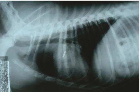
On lateral and dorsoventral thoracic radiographs (Figure 1AB), the heart tended to be normal with discrete right side enlargement, without evidences of pulmonary trunk abnormalities. The electrocardiography (ECG) confirmed the respiratory sinusal arrhythmia detected on auscultation. The heart rate was 112 beats per minute. Values of hematological and biochemical analyses were normal at the first day of evaluation.
Two-dimensional real time (2D) and M-mode echocardiography revealed normal dimensions of cardiac chambers, 39% of fractional shortening (FS), and 71% of eject fractions (EF). However, the color-flow (CF) Doppler image from the right parasternal short-axis view, at pulmonary level, showed a communication between ascending aortic and pulmonary trunk by detecting a continuous flow just above the semilunar valves (Figure 2), in a different location in which patent ductus arteriosus (PDA) is usually seen. The communication measured 0.58cm and showed a low turbulent blue flow in a CF Doppler, indicating that the flow was moving away from the transducer producing a left-to-right shunt (Figure 2). The pulsed-wave and the CF Doppler tracing of mitral, tricuspid,
pulmonic, and aortic valves showed normal flow velocities without turbulence, excluding other congenital heart diseases.
The dog was submitted to a left thoracotomy and the APSD diagnostic was confirmed. As observed in the echocardiogram, no PDA was found. There was a palpable thrill on the pulmonary trunk, close to the right ventricle outflow tract. Due to difficult anatomical location and surgical risks, the surgeon chose for external definitive clipping without arterial opening. After clipping, the pulmonary thrill considerably decreased; however, it did not disappear.
The immediate post-operative echocardiography revealed a sensible aorticopulmonar communication size reduction (from 0.58cm to 0.31cm – Figure 3AB) and the radiographic evaluation showed the clip in the APSD location (Figure 4). Seven and sixty days after the surgical, procedure, the dog was presented in good clinical conditions without exercise-induced dyspnea. Seven days after the procedure, radiographies demonstrated a normal heart size. The electrocardiogram on day sixty showed a ST segment depression, wide QRS complexes and increment in Q wave voltage in leads I and II, suggesting biventricular enlargement. In the last echocardiographic exam, performed sixty days after treatment, the APSD was reduced to 0.21cm.
Figure 1. Thoracic radiographs in a 10-month-old mixed breed dog with aorticopulmonary septal defect. (A) Right lateral thoracic radiograph. Discrete right side enlargement. (B) Ventrodorsal thoracic radiograph. None evidences of pulmonary trunk abnormalities.
A
Figure 2. Color-flow Doppler echocardiogram of right parasternal short-axis view, at pulmonary level, in a 10-month-old mixed breed dog. The Doppler indicates that blood flows from aorta (AO) to the pulmonary arteries (AP), through an aorticopulmonary septal defect (white arrows), during the systolic period. VD= right ventricle; AE= left atrium.
A
B
B
Figure 4. Right lateral thoracic radiograph in a 10-month-old mixed breed dog with aorticopulmonary septal defect, after surgery. Note the presence of the clip.
At the time of submission (seventy days after the surgical event), the dog presented normal life receiving only enalapril maleate (0.25mg/kg) once a day. One week before surgery, diuretic therapy was included with 2.0mg per kilogram of furosemide gradually retreated 15 days after.
DISCUSSION
Even sharing the same pathophysiology of PDA, some important clinical differences can be noted from APSD. A loud continuous machinery murmur is not always present (Eyster et al., 1975; Lombard et al., 1978; Nelson, 1986). It can occur in a systolic period and in a smaller grade (Eyster et al., 1975; Lombard et al., 1978), as observed in the present case. Probably, the larger opening of this congenital disease does not create so much blood flow turbulence allowing this distinct auscultation. Otherwise, changes in defect shape, location, or illness evolution can product a loud murmur (Guglielmini et al., 2001; Mucha et al., 2002) increasing the difficulty on diagnosticating without additional studies.
In early stages or in cases of left-to-right shunt, exercise intolerance and dyspnea seems to be frequent, but cyanosis was detected on physical examination just in one dog (Nelson, 1986), after an exercise challenge in two dogs (Eyster et al., 1975; Lombard et al., 1978), and was
undetectable in another (Guglielmini et al., 2001). In the present report, cyanosis was not seen during ambulatory examinations and at home, according to the owners, even during exercise situations, indicating adequate blood oxygen saturation in a left-to-right shunt.
The initial normality of the radiographies and ECG parameters, found in the dog described here, supports the precocity of the diagnostic and make the detection of heart changes after surgery procedure possible. Dogs portrayed in the literature had generalized cardiomegaly (Guglielmini et al., 2001), right side enlargement (Eyster et al., 1975; Lombard et al., 1978), or no obvious heart changes (Nelson, 1986) on a survey of thoracic radiography, but most of them had evidences of some intensity of lung hypervascularization or enlargement of main pulmonary artery (Eyster et al., 1975; Lombard et al., 1978; Nelson, 1986; Guglielmini et al., 2001), due to from right side overload.
other hand, atrial fribilation (Guglielmini et al., 2001), supraventricular bigeminy, and trigeminy (Nelson. 1986) were described in dogs with APSD, besides the suggestions of right (Eyster et al., 1975; Lombard et al., 1978; Nelson, 1986) or left (Guglielmini et al., 2001) ventricular enlargement.
The 2D and color-flow Doppler echocardiography were mandatory to the diagnostic of APSD, as described by another reference (Guglielmini et al., 2001). According to the experience of the authors, the right parasternal short-axis view, at pulmonary level, was especially important because it allowed the detection of communicating flow in a distinct place of PDA and a two-dimensional measurement of the defect.
Despite being limited, the measurement of communication was beneficial for the continuous evaluations. It also seems to be inversely correlated with the improvement of the dog, which indicates that the smaller the defect, the better the clinical improvements and prognostics. The decrease in an APSD orifice probably happened due to the local fibrous reaction, retracting the communication.
Unlike expected and previously reported (Nelson, 1986; Guglielmini et al., 2001), 2D and M-mode echocardiography demonstrated normal dimensions of cardiac chambers. Typically, PDA (Kaplan, 1991; Goodwin and Lombard, 1992) and APSD are characterized by left-side cardiac enlargement and eccentric hypertrophy of the left ventricle by volume overload according to Frank-Starling Law. All these findings express the precocity of diagnostic and the sensibility of this auxiliary method of cardiac investigation, increasing the surgical correction success percentage.
Angiography would allow the precise location of an anomalous communication between the pulmonary artery and the aorta to be seen, which has been used in previously reported cases of APSD (Eyster et al., 1975; Lombard et al., 1978; Nelson, 1986). However, this method is invasive and requires general anesthesia, which was avoided here and sufficiently substituted by Doppler echocardiography, as describe by others (Guglielmini et al., 2001). Additionally, the congenital abnormality was confirmed by
surgery, reinforcing the effectiveness of this non invasive method.
Many techniques are useful as a therapeutic approach in pediatric cardiac surgery for this congenital disease (Gross, 1952; Tkebuchava et al., 1997; Backer and Mavroudis, 2002). However, only one dog was surgically treated and it died during this procedure (Eyster et al., 1975). The chosen technique depends on the surgeon experience, availability of material, equipment, associated abnormalities, and the anatomical access (Tkebuchava et al., 1997). Although the surgical method chosen in this case and reported here had only partial success, the life of the dog is being maintained and improved as wanted. To the best of the knowledge of the authors, this is the first well succeed surgery in a dog with APSD, attested by the remission of the clinical signs.
Most recently, a classification scheme for APSD was proposed by the Society of Thoracic Surgeons Congenital Heart Surgery Database Committee (Jacobs et al., 2000). According to this classification, the APSD reported here bellow to the Type I, as most frequent observed in pediatric patients (Tkebuchava et al., 1997). Nevertheless, the rare incidence or poor diagnostic in Veterinary Medicine turns unlikely the creation of a specific classification, hindering the development of more efficient surgical techniques and proving the importance of reports like this.
In this matter, early and accurate diagnosis improve the therapeutic success, for which the echoDopplercardiography seems to have a crucial importance in addition to the clinical signs and others complementary exams, like ECG and angiography. The decrease or total closure of the APSD, when possible, is essential for life quality and longevity. For the reason that must be tried before shunt reversal or high pulmonary hypertension be present, which can be inferred by catheterization or by echoDopplercardiography.
REFERENCES
EYSTER, G.E.; DALLEY, J.B.; CHAFFEE, A. et al. Aorticopulmonary septal defect in a dog. J. Am. Vet. Med. Assoc., v.167, p.1094-1096, 1975. GOODWIN, J.K.; LOMBARD, C.W. Patent ductus arteriosus in adult dogs: clinical features in 14 cases. J. Am. Anim. Hosp. Assoc., v.28, p.349-354, 1992.
GROSS, R.E. Surgical closure of an aortic septal defect. Circulation, v.5, p.858-863, 1952.
GUGLIELMINI, C.; PIETRA, M.; CIPONE, M. Aorticopulmonary septal defect in a German Shepherd dog. J. Am. Anim. Hosp. Assoc., v.37, p.433-437, 2001.
JACOBS, J.P.; QUINTESSENZA, J.A.; GAYNOR, J.W. et al. Congenital heart surgery nomenclature and database project: aortopulmonary window. Ann. Thorac. Surg., v.69, Suppl., p.S44-S49, 2000.
KAPLAN, P.M. Congenital heart disease. Probl. Vet. Med.,v.3,p.500-519,1991.
LOMBARD, C.W.; KNIGHT, D.H.; BUCHANAN, J.W. Clinico-pathologic conference. J. Am. Vet. Med. Assoc., v.172, p.75-80, 1978.
MUCHA, C.J.; BELERENIAN, G.; PIELLA, M. et al. Ventana aorticopulmonar tipoI en un canino. Rev. Med. Vet., v.83, p.90-92, 2002.
NELSON, A.W. Aorticopulmonary window in a dog. J. Am. Vet. Med. Assoc., v.188, p.1055-1058, 1986.
TKEBUCHAVA, T.; VON SEGESSER, L.K.; VOGT, P.R. et al. Congenital aortopulmonary window: diagnosis, surgical technique and long-term results. Eur. J. Cardio-Thoracic Surg., v.11, p.293-297, 1997.


