Case Report
Key words
Pulmonary hypertension; arteriovenous fistula; pulmonary artery.
The coronary fistula is an anomaly characterized by the communication between a coronary artery and a cardiac chamber, pulmonary artery, coronary sinus and pulmonary veins. It represents 0.2 to 0.4% of the congenital cardiopathies and 0.1% to 0.2% of the adult population submitted to coronary angiography. We report the clinical case of a 64-yr-old female patient, whose anomaly was diagnosed during a clinical investigation due to chest discomfort, dyspnea and syncope; the surgical correction was indicated, with opening of the pulmonary artery through extracorporeal circulation.
Pulmonary Hypertension Secondary to Coronary-to-Pulmonary
Artery Fistula
José Ramos Filho, Otávio Andrade Carneiro da Silva, Diego Oliveira Vilarinho, Filipe Giordano Guilherme, Jomara
Custódio Ferreira, Aline Miranda de Souza
Universidade São Francisco, Bragança Paulista, SP - Brazil
Mailing Address: José Ramos Filho•
Rua Roterdam, 57 - Residencial Euroville – 12917-040, Bragança Paulista, SP - Brazil
E-mail: [email protected]
Manuscript received July 27, 2007; revised manuscript received September 27, 2007; accepted October 08, 2007..
Introduction
Described in 1865 by Krause1, the coronary fistula is
a congenital or acquired anomaly, characterized by the abnormal communication between a coronary artery and a cardiac chamber or at the pulmonary artery2.
The occurrence of coronary fistulae is an uncommon fact; however, it becomes significant when the blood flow is of a certain magnitude. Cieslinski et al3, in a retrospective study of
4,016 coronary angiographies performed during a five-year period, only found one case of coronary fistula. Most of the reported cases are congenital or secondary to complications caused by an invasive procedure. They represent 0.2% to 0.4%4-5 of the congenital cardiopathies and have a man-woman ratio of 1.9:13.
Case Report
A. F. M., a 64-year-old female patient, sought medical attention complaining of indistinct chest discomfort in the last five years, with dyspnea and syncope in the last three months. At the physical examination, BP was 140x80 mmHg, pulse was 78 ppm and arrhythmic, due to isolated ventricular extrasystoles and heart auscultation showing 3+/+++
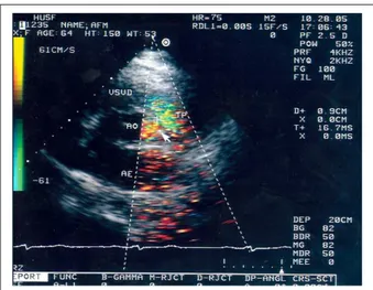
systolic murmur in the tricuspid area. At the complementary examinations, the ECG showed unspecific alterations of ventricular repolarization in the anteroseptal region; the bidimensional echocardiogram showed the presence of an aneurysm in the interatrial septum, increased dimension of the right ventricle and the right atrium. The color-flow mapping showed a regurgitating turbulent flow through the tricuspid valve and a turbulent systodiastolic flow between the aorta and the pulmonary trunk artery (Figure 1), indicating the possibility of a fistula communicating the two large vessels.
The left ventricular function was preserved with signs of relaxation alteration and the systolic pressure of the pulmonary artery was estimated by the tricuspid reflux at 58 mmHg.
The left cardiac catheterism demonstrated the topic origin of the coronary ostia and the selective angiography of the coronaries showed fistulae in both arteries, communicating with the pulmonary artery trunk (Figures 2 and 3). The anterior descending artery was displaced by the pulmonary artery trunk, as the latter was markedly dilated (Figure 2). The cardiac catheterism of the right chambers showed dilation of the pulmonary trunk and branches and increased dimension of the right cardiac chambers. The manometry showed pulmonary systolic pressure of 56 mmHg and mean pulmonary pressure of 32 mmHg.
Figure 1 -Transthoracic bidimensional echocardiogram with color low;
Transversal parasternal view at the aorta level showing turbulent low between the aorta and the pulmonary trunk; LA - left atrium; AO - aorta; PT - pulmonary trunk; RVOT - right ventricle outlow tract.
Case Report
Ramos Filho et al Secondary pulmonary hypertension due to fistulas
Arq Bras Cardiol 2008; 91(2) : e19-e21 Figure 2 -Selective angiography of the left coronary in caudal right anterior
oblique (RAO) view, showing the anterior descending artery with anomalous branch (arrow) emptying into the pulmonary artery trunk.
Figure 3 -Selective angiography of the right coronary in caudal left anterior oblique
(LAO) view, with anomalous branch emptying into pulmonary artery trunk
Discussion
Approximately 50% of the coronary artery fistulae originate from the right coronary artery and 45% from the left coronary artery; less than 5% of them originate from both2.
The hemodynamic effect depends on the site of origin and end and the size of the fistula. Several available diagnostic methods, such as angioresonance and angiotomography can be used for the diagnosis of arteriovenous fistulae. However, our investigation was started based on the echocardiographic findings with color flow mapping , which showed a turbulent flow between the aorta artery and the pulmonary artery trunk and, subsequently, cardiac catheterism of the left and right chambers. Most of the coronary fistulae is small and generally have no hemodynamic effect. The high output fistulae, as in the case reported here, can result in pulmonary hypertension and symptoms caused by chronic myocardial ischemia, such as chest angina, heart failure, cardiomyopathy, myocardial infarction, endocarditis, arrhythmia, thrombosis and/or fistula rupture, which is a rare event, although it has been described in the literature6.
Our patient showed clinical symptoms compatible with RV dysfunction due to chronic pulmonary hypertension, as well as symptoms caused by probable reduction in the coronary flow reserve. Additionally, as a consequence of the coronary flow reduction in the systole as well as in the diastole, there is a reduction in the oxygen supply, with myocardial perfusion impairment and increased blood volume in the lungs, which can increase the intrapulmonary pressure due to the magnitude of the fistulae7.
The treatment for the closing of the fistula in some regions can be performed by interventionist catheterism; however, we chose the surgical treatment due to the lack of experience of our hemodynamicists in this type of procedure. In the case reported here, chronic pulmonary hypertension and the presence of symptoms compatible with decreased coronary flow reserve were demonstrated and the surgical correction was indicated, with opening of the pulmonary artery through extracorporeal circulation, an approach that has also been proposed by other authors8-10.
The patient was submitted to surgical treatment in January 2006; she is currently clinically well and still maintain a slight degree of pulmonary hypertension (pulmonary systolic pressure estimated by Doppler at 34 mmHg). She is presently using 40 mg of telmisartan, 40 mg of nifedipine retard and 200 mg of acetylsalicylic acid.
Case Report
References
1. Krause W. Uber den Ursprung einer akzessorischen coronaria aus der pulmonalis. Z Rati Med. 1865; 24: 225-7 apud Groppo AA, Coimbra LF, Santos MVN. Fístula da artéria coronária: relato de três casos operados e revisão da literatura. Rev Bras Cir Cardiovasc. 2002; 17 (3): 271-5.
2. Groppo AA, Coimbra LF, Santos MVN. Fístula da artéria coronária: relato de três casos operados e revisão da literatura. Rev Bras Cir Cardiovasc. 2002; 17 (3): 271-5.
3. Cieslinski G, Rapprich B, Kuber G. Coronary anomalies: incidence and importance. Clin Cardiol. 1993; 16: 711-5.
4. Abundis A, Rangel A, Muñoz Castellanos L, Marin G, Chavez-Pérez E, Baduí E. Correlacion morfofuncional en las anomalias congenitas de las arterias coronarias y fistulas arteriales coronarias. Arch Inst Cardiol Mex. 1994; 64: 161-74.
5. Gasul BM, Arcilla RA, Fell EH, Lynfield J, Bicoff JP, Luan LL. Congenital coronary arteriovenous fistula. Pediatrics. 1960; 25: 531-60.
6. Ito M, Kodama M, Saeki M, Fukunaga H, Goto T, Inoue H, et al. Rupture of a giant saccular aneurysm of coronary arteriovenous fistulas. Jpn Heart J. 2000; 41: 659-64.
7. Okei K, Horita Y, Shimizu M, Mabuchi H. A quantitative evolution of coronary steal phenomenon in coronary artery-pulmonary artery fistula: case report. J Cardiol. 1995; 26: 305-12.
8. Neufeld HN, Lester RG, Adams P Jr, Anderson RC, Lillehei CW, Edwards JE. Congenital communication of a coronary artery with a cardiac chamber or the pulmonary trunk (“coronary artery fistula”). Circulation. 1961; 24:171-9.
9. Buxton B, Ventimeglia R, Reul GJ, Cooley DA. Congenital coronary-to-pulmonary artery fistula: diagnosis and treatment. Cardiovasc Dis. 1976; 3 (2): 202-9.
10. de Nef JJ, Varghese PJ, Losekoot G. Congenital coronary artery fistula: analysis of 17 cases. Br Heart J. 1971; 33: 857-62.
Ramos Filho et al
Secondary pulmonary hypertension due to fistulas
Arq Bras Cardiol 2008; 91(2) : e19-e21