Image
Eustachian Valvula: Real-Time Three-Dimensional Transthoracic
Echocardiographic Imaging
Marcelo Luiz Campos Vieira and Antônio José Sproesser
Hospital Israelita Albert Einstein - São Paulo, SP - Brazil
Mailing address: Marcelo Luiz Campos Vieira •
Rua Cardoso de Melo, 463/21 - 04548-002 - São Paulo, SP - Brazil E-mail: [email protected]
Manuscript received January 09, 2007; revised manuscript received January 01, 2007; accepted March 13, 2007.
The use of three-dimensional echocardiography for the analysis of cardiac structures allows the viewing of planes that make the anatomic identification more realistic1-3. We present
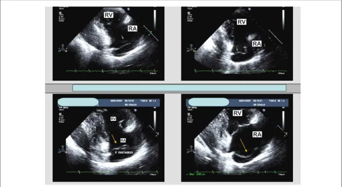
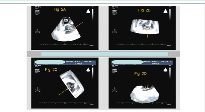
the case of a 41-year-old male patient, a high-performance athlete (marathoner) in whom an Eustachian valvula was identified. In the two-dimensional echocardiographic study (Figure 1), an elongated, filiform structure is observed. Three-dimensional echocardiography shows an elongated, but not filiform valvula which appears as a structure with a wide base, as identified in the short-axis view, which enables anatomic observation from a depth or elevation plane (Figure 2).
Potential Conflict of Interest
No potential conflict of interest relevant to this article was reported.
Sources of Funding
There were no external funding sources for this study.
Study Association
This study is not associated with any graduation program.
Key words
Echocardiography; heart valves, Eustachian valvula, anatomy.
Fig. 1 - Two-dimensional transthoracic echocardiogram. Parasternal view of right chambers. A prominent Eustachian valvula is observed (arrows). RA - right atrium; RV - right ventricle; V - valvula.
RV
RV
RV
RV RA
RA
RA
RA
Image
Vieira & Sproesser Eustachian valvula
Arq Bras Cardiol XXXX; XX(X) : XXX-XXX
References
1. Li J, Sanders SP. Three-dimensional echocardiography in congenital heart disease. Curr Opin Cardiol. 1999; 14: 53-9.
2. Kisslo J, Firek B, Takahiro O, Kang DH, Fleishman CE, Stetten G, et al. Real-time volumetric echocardiography: the technology and the possibilities.
Echocardiography. 2000; 17: 773-9.
3. Ahmad M. Real-time three-dimensional echocardiography in assessment of heart disease. Echocardiography. 2001;18 (1): 73-7.
Fig. 2 - Real-time three-dimensional transthoracic echocardiography. Parasternal view of right chambers. The Eustachian valvula is observed (arrows) by rotating the parasternal view of right chambers. A) Conventional parasternal view of right chambers. B) Parasternal view of right chambers in atrial translation (viewing from the RV). C) Parasternal view of right chambers in clockwise rotation (viewing from the RV). D) Parasternal view of right chambers in ventricular translation (viewing from the RA). RA - right atrium; RV - right ventricle.
RV