Research Article Open Access
Bioactive form of resveratrol in glioblastoma cells and its
safety for normal brain cells
Xiao-HongShu1,2, Hong Li1, Xiao-Xin Sun1, Zheng Sun1, Li-Li Wang1, Xue Song1, Shun Shi1, Mo-Li Wu1, Xiao-Yan Chen1, Qing-You Kong1, Liu Jia1
1
Liaoning Laboratory of Cancer Genetics and Epigenetics and Department of Cell Biology,
Dalian Medical University, Dalian 116044, China; 2Department of Medicinal Chemistry,
College of Pharmacy, Dalian Medical University, Dalian 116044, China
Corresponding Author: Liu Jia, PhD, Professor, and Xiao-Hong Shu, PhD, Professor,
Liaoning Laboratory of Cancer Genetics and Epigenetics and Department of Cell Biology,
Dalian Medical University, Dalian 116044, China
Submission date: April 24, 2013; Acceptance date: May 30, 2013; Publication date: May 31, 2013
ABSTRACT
Background: Resveratrol, a plant polyphenol existing in grapes and many other natural
foods, possesses a wide range of biological activities including cancer prevention. It has been
recognized that resveratrol is intracellularly biotransformed to different metabolites, but no
direct evidence has been available to ascertain its bioactive form because of the difficulty to
maintain resveratrol unmetabolized in vivo or in vitro. It would be therefore worthwhile to
elucidate the potential therapeutic implications of resveratrol metabolism using a reliable resveratrol-sensitive cancer cells.
Objective: To identify the real biological form of trans-resveratrol and to evaluate the safety
of the effective anticancer dose of resveratrol for the normal brain cells.
Methods: The samples were prepared from the condition media and cell lysates of human
glioblastoma U251 cells, and were purified by solid phase extraction (SPE). The samples
were subjected to high performance liquid chromatography (HPLC) and liquid
chromatography/tandem mass spectrometry (LC/MS) analysis. According to the metabolite(s),
trans-resveratrol was biotransformed in vitro by the method described elsewhere, and the
resulting solution was used to treat U251 cells. Meanwhile, the responses of U251 and
Results: The results revealed that resveratrol monosulfate was the major metabolite in U251
cells. About half fraction of resveratrol monosulfate was prepared in vitro and this
trans-resveratrol and resveratrol monosulfate mixture showed little inhibitory effect on U251
cells. It is also found that rat primary brain cells (PBCs) not only resist 100μM but also
tolerate as high as 200μM resveratrol treatment.
Conclusions: Our study thus demonstrated that trans-resveratrol was the bioactive form in
glioblastoma cells and, therefore, the biotransforming activity of trans-resveratrol would be
reversely correlated with the chemosensitivity of the treated cells. The findings from PBCs
suggest that an effective anti-glioblastoma dose of resveratrol may not exert side-effect on normal brain cells, providing a strong evidence for practical use of resveratrol in the
management of human brain malignancies.
Key words: Resveratrol, glioblastoma, drug metabolism
BACKGROUND:
Glioblastoma multiforme (GM) is the most common primary brain malignancy in human
adults [1]. Irrespective to the combination of surgical operation with improved external
radiotherapy and adjuvant chemotherapy, the prognosis of GMB remains very poor due to its
highly aggressive biological behavior and frequent recurrence rate [2]. Therefore, exploring
effective and less toxic chemotherapeutic approaches would be of clinical values in better management of this sort of lethal disease.
Resveratrol, a plant polyphenol existing in grapes and many other natural foods [3],
possesses a wide range of biological activities including cancer prevention [4,5]. More
importantly, resveratrol has little cytotoxic effect and is able to penetrate blood-brain barrier
[6], suggesting its potential therapeutic values in the management of glioblastoma. It has been
recognized that resveratrol is intracellularly biotransformed to different metabolites, but no
direct evidence has been available to ascertain its bioactivity form, and how normal brain
cells respond to this agent. So it would be worthwhile to identify the real biological form of
trans-resveratrol and evaluate the resveratrol’s safety in the normal glial cells with effective
anticancer dose.
MATERIALS AND METHODS:
Primary rat brain cell culture and treatment. A 1-day-old Wistar rats were obtained from the Experimental Animals Center of Dalian Medical University. The rat brains were freshly
removed and minced with a scalpel and triturated in high glucose DMEM (Gibico, Invitrogen
Corporation, NY, USA). After centrifugation at 2000 rpm for 5 minutes, the brain cells were
washed with DMEM and centrifugated for 5 minutes. The cell suspensions were plated to
supplemented with 10% FBS under 37oC and 5% CO2 condition, then treated with resveratrol.
All experimental protocols had been reviewed and approved by the ethics committee of
Dalian Medical University (ECDMU-09066) for protection of human subjects and
experimental animals before conducting the project.
Sample preparation and purification. After 100μM resveratrol treatment, the primary brain
cells and glioblastoma U251 cells were collected, washed three times with PBS (pH 7.4), and
lysed by sonication. Meanwhile, cell-free media were harvested directly by the end of
48-hour resveratrol treatment or after an additional 24-hour normal culture. The collected
media and cell lysates were centrifuged at 10,000g for 5 minutes, and the supernatants were
purified by SPE [7]. The eluate was dried by nitrogen spraying, and the residues were
dissolved in 500μL methanol and a 10μL aliquot was injected onto the liquid chromatography
column for HPLC and LC/MS analysis.
Structural identification of resveratrol metabolite(s). The HPLC system (Waters Co.,
Milford, MA, USA) is consisted of a Waters 1525 binary pump and 2487 dual wavelength
UV-VIS detector. The detection was carried out at 306nm [8]. Chromatographic separation of
resveratrol and its metabolite(s) was performed on a Cosmosil 5C18-AR-Ⅱ column (4.6
ID×250 mm, NACALAI TESQUE, INC. Japan) containing a C18 guard column (5μm,
4.6×10mm), using the mobile phase of 5mM ammonium acetate (phase A) and methanol
(phase B) with a flow rate of 1.0 ml/min. The gradient solution conditions were referred
elsewhere [9]. Further LC-MS/MS analysis was performed on a Agilent 1200 liquid
chromatography series (Agilent Technol Inc., Santa Clara, CA, USA) coupled to an Applied Biosystems API 3200 QTrap tandem mass spectrometer (Applied Biosystem/MDS SCIEX,
Foster City, CA, USA) equipped with an ESI source, and operated by Applied
Biosystem/MDS SCIEX analyst software (Version 1.4.1) to obtain the MS and MS/MS data
in a negative ion mode [9]. Further identification and confirmation of the metabolites was
performed on a Shimadzu LC/MS-IT-TOF (Shimadzu Co., Kyoto, Japan) instrument
equipped with an ESI source in negative ion mode at a resolution of 10000 FWHM. MS data
were processed with LC/MS solution ver. 3.4 software (Shimadzu, Japan).
Resveratrol biotransformation and cell treatment. One female specific pathogen free
Wistar rat was cultured in a wire cage in a room maintained at 22°C with a 12-hour light/dark
period at the Experimental Animal Center of Dalian Medical University. It was sacrificed by
an animal expert in accordance with approved Ministerial procedures appropriate to the species. The brain tissue was rapidly obtained and treated to prepare cytosolic
sulfotransferase.
For resveratrol sulfonation, 5mM resveratrol, 100μL cytosolic sulfotransferase, 2mM
PAPS (Sigma Chem Co., St. Louis, MO, USA), 1mM DTT and 20mM Mops buffer were
admixed and incubated at 37°C for 2h. After the product was centrifuged, the supernatant
to treat U251 cells in the total concentration of 100μM. The cells treated by the chemical
solution for brain lysate preparation, the brain lysate alone and the combination of resveratrol
and brain lysate (without PAPS supplementation) were respectively used as background
controls.
RESULTS:
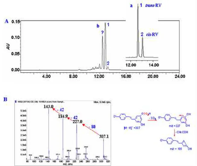
Resveratrol monosulfate as major metabolite. According to the HPLC and LC-MS/MS
analysis, the major metabolite in human glioblastoma U251 cells was resveratrol monosulfate
Figure 1A. The [M-H]- ion of the compound (tR=12.92min) showed the deprotonated
molecule ions of m/z 307 and 227, respectively, and the ion corresponding to resveratrol (m/z
227) through the neutral loss of the sulfate unit (m/z 80) from the resveratrol monosulfate, then the m/z 227 was fragmented to m/z 185 for the further loss of 42 amu (C2H2O) from
resveratrol (Figure 1B), which was concluded to resveratrol monosulfate as reported by other
investigators [10]. The compound was further confirmed by HRMS that gave the [M-H]
-molecular ion exact mass as 307.0261 (C14H11SO6, calculated m/z 307.0276), which
corresponded to resveratrol monosulfate.
Figure 1. LC-MS analyses of resveratrol metabolites in glioblastoma U251 cells
(A) HPLC-UV chromatography analysis on the metabolites of U251 cells treated with 100μM resveratrol for
48h. 1, 2 represent trans-resveratrol and cis-resveratrol, respectively. (B) indicates the MS1 and MS2 analysis of
Metabolic pattern of resveratrol in glioblastoma cells. Furthermore, the results showed that the resveratrol monosulfate concentration increased to the platform level at 12-hour time
point, but the U251 cells reached apoptotic peak at 48-hour time point, which suggested that
the metabolic mechanism pre-existed in the tumor cells apoptosis and its efficiency was
enhanced in response to resveratrol treatment. It was also noticed that the amounts of
trans-resveratrol elevated at 24- and 48-hour time points presumably due to the hydrolytic
reaction that reversed resveratrol monosulfate to trans-resveratrol as proposed by Walle T. et
al [11,12]. Several additional small peaks were observed from the HPLC spectrum, but their
chemical features were not further analyzed here because of their low amounts.
trans-Resveratrol as the bioactive form in glioblastoma cells. Resveratrol monosulfate was
prepared by incubating trans-resveratrol with the brain lysates for 2h, and then was analyzed
by HPLC and LC/MS. It was also revealed that the monosulfate was the major metabolite and
more than 1/2 of parent trans-resveratrol was biotransformed to resveratrol monosulfate. In
difference with the situation of 100μM trans-resveratrol treatment, U251 cells treated by the same concentration of this mixture for 48 hours showed neither distinct growth suppression
nor apoptosis signs (Figure 2).
Figure 2. Biotransformation and bioacitve form evaluation in U251 cells (A) The chemical
structures of trans-resveratrol and resveratrol monosulfate; (B) Morphologic evaluation of U251 cells incubated
with 100μM of trans-resveratrol and trans-resveratrol/resveratrol monosulfate mixture for 48 h by H&E staining
(40X).
No side-effect of resveratrol on normal rat brain cells. After 100μM resveratrol treatment,
apoptosis and death, even incubated with 200μM resveratrol. Trypan blue staining revealed
that the percentage of nonviable cells was 0.11%, 12.26%, 31.79%, 34.96%, and 52.73% in
U251 cells, but 0.17%, 1.09%, 0.95%, 1.61%, and 1.99% in PBC cells after 100μM
resveratrol treatment for 0, 12, 24, 36, and 48 hours, respectively. FCM analyses further
demonstrated that G1 and S fractions were 37.7% and 25.7% in normal U251 cells and
changed to 37.9% and 47.2% in the resveratrol-treated cells. In PBC cells, G1 and S fractions
were 54.5% and 32.9% in normal cells and remained almost unchanged (53.7% and 35.0%)
after resveratrol-treatment. The percentages of apoptotic cells in 100μM resveratrol-treated
U251 and PBC cells were 24.3% and 1.1%, respectively.
DISCUSSION:
An ideal cancer therapeutic agent should have minimal cytotoxicity to normal tissues,
meanwhile, exerts crucial effects on cancer cells. Our results showed that resveratrol could
induce glioblastoma U251 cells growth arrest and apoptosis, on the contrary resveratrol was
insensitive to the PBC cells. Which suggested that resveratrol had no side effect on normal
brain cells, but exerted significantly anti-glioblastoma bioactivity, furtherly, resveratrol could
penetrate the blood-brain-barrier (unpublished data). Therefore, resveratrol would be an ideal
cancer-therapy drug for clinical brain tumors therapy.
trans-Resveratrol has been recognized as an ideal cancer preventive and therapeutic
agent, and it can generate one or more metabolites in cells [10,13]. However, the
pharmaceutical potentials of those metabolic products have not yet been well ascertained.
Some researchers proposed that resveratrol metabolite(s) such as resveratrol sulfates or
resveratrol glucuronides were the bioactive forms because of the low bioavailability of parent
trans-resveratrol in vivo [11], while others considered that trans-resveratrol by itself was
sufficient to cause vital cellular and molecular consequences in the treated cancer cells
[14,15]. Apparently, determination of the bioactive form(s) of resveratrol in individual cell
and cancer types becomes a fundamental issue for the successful application of resveratrol in
clinical therapy.
In this study, it was found that the metabolite of trans-resveratrol was mainly
resveratrol monosulfate, and rather than its metabolite, resveratrol parent form exhibited
anti-tumor activity in the human glioblastoma U251 cells. It is therefore demonstrated that
the efficiency of metabolic reduction of trans-resveratrol is the major determinant of the fate
of resveratrol-treated glioblastoma cells. These findings would be of translational values in
exploring tumor-selective and personalized application of resveratrol in cancer prevention
and treatment.
Acknowledgments: This work was supported by the grants from National Natural Science Foundation of China (No. 30971038, 81071971, 81072063 and 81272786), Program for
Liaoning Excellent Talents in University (LJQ2012078) and the creative research team
REFERENCES:
1.Van Meir EG, Hadjipanayis CG, Norden AD, Shu HK, Wen PY, Olson JJ. Exciting new
advances in neuro-oncology: the avenue to a cure for malignant glioma. CA Cancer J
Clin. 2010;60:166-193.
2.Parsons DW, Jones S, Zhang X, Lin JC, Leary RJ, Angenendt P, Mankoo P, Carter H, Siu
IM, Gallia GL, Olivi A, McLendon R, Rasheed BA, Keir S, Nikolskaya T, Nikolsky Y,
Busam DA, Tekleab H, Diaz LA Jr, Hartigan J, Smith DR, Strausberg RL, Marie SK,
Shinjo SM, Yan H, Riggins GJ, Bigner DD, Karchin R, Papadopoulos N, Parmigiani G,
Vogelstein B, Velculescu VE, Kinzler KW. An integrated genomic analysis of human
glioblastoma multiforme. Science. 2010;321:1807-1812.
3.Jang M, Cai L, Udeani GO, Slowing KV, Thomas CF, Beecher CW, Fong HH,
Farnsworth NR, Kinghorn AD, Mehta RG, Moon RC, Pezzuto JM. Cancer
chemopreventive activity of resveratrol, a natural product derived from grapes. Science.
1997; 275:218–220.
4.Smoliga JM, Baur JA, Hausenblas HA. Resveratrol and health – A comprehensive review
of human clinical trials. Mol Nutr Food Res. 2011;55:1129-1141.
5.Dixon RA. Natural products and plant disease resistance. Nature.2001; 411:843-847.
6.Wang Q, Xu J, Rottinghaus GE, Simonyi A, Lubahn D, Sun GY, Sun AY. Resveratrol
protects against global cerebral ischemic injury in gerbils. Brain Res 2002; 958:439-447
7.Mercolini L, Addolorata Saracino M, Bugamelli F, Ferranti A, Malaguti M, Hrelia S,
Raggi MA. HPLC-F analysis of melatonin and resveratrol isomers in wine using an
SPE procedure. J Sep Sci. 2008; 31:1007-1014
8.Qian G, Leung SY, Lu G, Leung KS. Optimization and validation of a chromatographic
method for the simultaneous quantification of six bioactive compounds in Rhizoma et
Radix Polygoni Cuspidati. J Pharm Pharmacol. 2008; 60:107-113.
9.Shu XH, Li H, Sun Z, Wu ML, Ma JX, Wang JM, Wang Q, Sun Y, Fu YS, Chen XY,
Kong QY, Liu J. Identification of metabolic pattern and bioactive form of resveratrol in
human medulloblastoma cells. Biochem Pharmacol. 2010; 79:1516-1525.
10.Murias M, Miksits M, Aust S, Spatzenegger M, Thalhammer T, Szekeres T, Jaeger W.
Metabolism of resveratrol in breast cancer cell lines: impact of sulfotransferase 1A1
expression on cell growth inhibition. Cancer Lett. 2008; 261:172-181
11.Walle T, Hsieh F, DeLegge MH, Oatis JE, Walle UK. High absorption but very low
bioavailability of oral resveratrol in humans. Drug Metab Dispos. 2004; 32:1377-1182.
12.Gescher AJ, Steward WP. Relationship between mechanisms, bioavailibility, and preclinical chemopreventive efficacy of resveratrol: a conundrum. Cancer Epidemiol
Biomarkers Prev. 2003; 12:953-957.
13.Lançon A, Hanet N, Jannin B, Delmas D, Heydel JM, Lizard G, Chagnon MC, Artur Y,
Latruffe N. Resveratrol in human hepatoma HepG2 cells: metabolism and inducibility
of detoxifying enzymes. Drug Metab Dispos 2007; 35:699-703
Rev Drug Discov. 2006; 5:493-506.
15.Howitz KT, Bitterman KJ, Cohen HY, Lamming DW, Lavu S, Wood JG, Zipkin RE,
Chung P, Kisielewski A, Zhang LL, Scherer B, Sinclair DA. Small molecule activators