BASIC RESEARCH
The effects of antidepressants and pilocarpine on rat
parotid glands: an immunohistochemical study
Tatiana Maria Folador Mattioli,ISilvana da Silva,IAna Maria Trindade Gre´gio,IIMaria Aˆ ngela Naval Machado,IIIAntoˆnio Adilson Soares de Lima,IIILuciana Reis Azevedo-AlanisII
IDepartment of Stomatology, School of Dentistry, Pontifical Catholic University of Parana´, Curitiba, Brazil.IISchool of Dentistry, Pontifı´cia Universidade
Cato´lica do Parana´, Brazil.IIIDepartment of Stomatology, School of Dentistry, Universidade Federal do Parana´, Brazil.
OBJECTIVES:To evaluate the effects of antidepressants and pilocarpine on the quantity of myoepithelial cells and on the proliferation index of the epithelial cells of rat parotid glands.
INTRODUCTION:Hyposalivation, xerostomia, and alterations in saliva composition are important clinical side effects related to the use of antidepressants.
METHODS:Ninety male Wistar rats were allocated to nine groups. The control groups received saline for 30 (group C30) or 60 days (group C60) or pilocarpine for 60 days (group Pilo). The experimental groups were administered fluoxetine (group F30) or venlafaxine for 30 days (group V30); fluoxetine (group FS60) or venlafaxine (group VS60) with saline for 60 days; or fluoxetine (group FP60) or venlafaxine (group VP60) with pilocarpine for 60 days. Parotid gland specimens were processed, and the immunohistochemical expression of calponin and proliferating cell nuclear anti-antigen on the myoepithelial and parenchymal cells, respectively, was evaluated. Analysis of variance (ANOVA), Tukey HSD and Games-Howell tests were applied to detect differences among groups (p,0.05).
RESULTS:Compared with the controls, chronic exposure to antidepressants was associated with an increase in the number of positively stained cells for calponin. In addition, venlafaxine administration for 30 days was associated with an increase in the number of positively stained cells for proliferating cell nuclear anti-antigen. Fluoxetine and pilocarpine (group FP60) induced a significant decrease in the number of positively stained cells for calponin compared with all other groups.
CONCLUSIONS: The number of positively stained cells for calponin increased after chronic administration of antidepressants. The proliferation index of the epithelial cells of rat parotid glands was not altered by the use of antidepressants for 60 days.
KEYWORDS: Antidepressants; Immunohistochemistry; Salivary glands; Rats; Saliva.
Mattioli TMF, Silva S, Gre´gio AMT, Machado MAN, Lima ADS, Azevedo-Alanis LR. The effects of antidepressants and pilocarpine on rat parotid glands: an immunohistochemical study. Clinics. 2011;66(9):1605-1610.
Received for publication onMarch 10, 2011;First review completed onApril 5, 2011;Accepted for publication onMay 23, 2011 E-mail: [email protected]
Tel.: 55 41 3271-2592
INTRODUCTION
The current generation of antidepressants includes drugs that only act on one neurotransmitter, such as the serotonin (fluoxetine) or the noradrenaline (reboxetine) selective reuptake inhibitors, and drugs that that act on multiple neurotransmitters (venlafaxine) without targeting other cerebral receptors that are not related to depression, such
as histamine and acetylcoline.1-2Venlafaxine is a serotonin
and noradrenaline reuptake inhibitor, and it exerts a weak activity as a dopamine reuptake inhibitor, which is
only clinically significant at high doses.3,4 The current
antidepressants differ from the classic tricyclic antidepres-sants and from the monoamine oxidase inhibitors, which are irreversible, because of their enhanced pharmacological selectivity and diminished side effects.5
In a Cochrane systematic review (1966-2004), the adverse effects of fluoxetine (dry mouth sensation, dizziness, and sudoresis) were compared with the adverse effects of the most recent antidepressants (venlafaxine, reboxetine, phe-nelzine, and nefazodone), and the adverse effects were
shown to be less pronounced in the more recent drugs.6
Xerostomia is defined as a subjective sensation of dry mouth reported by the patient. It can result from a reduction in saliva secretion, but it can also occur in the presence of a
normal salivary flow rate.7 Stimulated salivary flow rate
(SSFR) values of #0.7 mL/min are considered to indicate
hyposalivation.8 Hyposalivation, xerostomia, and
altera-tions in the saliva composition are important side effects related to the use of psychotropic medications, including
antidepressants.7,9-14 The mechanism by which these side
effects occur has still not been completely clarified; how-ever, histomorphometric and gravimetric studies have contributed to a better understanding of the cytotoxic effects
of psychotropic drugs on the salivary glands.12-15
Gre´gio et al.,12studied the effects of chronic administra-tion of a benzodiazepine (diazepam) and an antidepressant (amitryptiline) on the parotid glands of rats and observed hyposalivation and hypertrophy of the serous cells. These findings suggested a possible inhibition of the activity of the myoepithelial cells (originating from nervous stimulation), a decrease in the number of myoepithelial cells following chronic administration of psychotropic drugs, or an altera-tion in the number of acinar and ductal cells.
The myoepithelial cells, which are located between the basal lamina and the plasmatic membrane of the acinar cells and intercalated ducts, are nonmuscular cells that exert contractile functions and contribute to the emptying of the
secretion from the secretory units and the ducts.16,17
Calponin (C) is a protein located in myoepithelial cells that is involved in the regulatory system of smooth muscle
contraction.18 Acinar and ductal cells are epithelial cells
with proliferative potential for maintenance and regenera-tion.19-23
The aim of this study was to evaluate the quantity of myoepithelial cells and the proliferation index of acinar and ductal cells of the parotid glands of rats treated with fluoxetine and venlafaxine using immunoreactions with calponin and proliferating cell nuclear anti-antigen (PCNA).
MATERIALS AND METHODS
This study was approved by the Research Ethics Committee at Universidade Tuiuti do Parana´ (CEP-UTP n. 55/2003).
Parotid glands from male Wistar rats (Rattus norvegicus
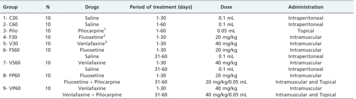
albinus,Rodentia, mammalian) with an approximate weight of 250 g were embedded in paraffin blocks from Laborato´rio de Patologia Experimental, Pontifı´cia Universidade Cato´lica do Parana´. Ninety animals were allocated to nine groups, and each group was composed of ten animals. The animals received different treatments, which are described in Table 1.15
We used tissue microarrays (TMAs) containing ten cylinders of paraffinized salivary glands (3 mm in diameter)
organized in lines and columns.24,25 Each TMA exhibited
ten specimens of salivary glands from the rats of each group.
For immunohistochemistry, anti-calponin, and anti-PCNA
(DakoCytomationH; Dako North America, Carpinteria, CA,
USA) antibodies were used. The secondary antibody was
EnVisionH+Dual Link/Peroxidase (DakoCytomationH), and
the antigen-antibody reactions were revealed with a DAB
substrate-chromogen system (DakoCytomationH). The slides
were counterstained with Harris hematoxylin.
We used an anti-calponin (C) monoclonal antibody (Dako Corporation) for myoepithelial cell staining and an anti-PCNA antibody (Dako Corporation) for the staining of proliferating acinar and ductal cells. PCNA is a monoclonal antibody that allows the study of cell kinetics. The immunoreactions with C and PCNA used antibody dilu-tions of 15800 and 15400, respectively.
Slides were visualized by only one examiner using an
OlympusHBX50 optical microscope (Olympus Corporation,
Ishikawa, Japan) with a 40X objective. The immunohisto-chemical expression of C in the myoepithelial cells and PCNA in the acinar and ductal cells was analyzed by interpreting the entire TMA area.
The presence or absence of PCNA and C antibody staining was evaluated. Cells that exhibited any expression for PCNA or for C in the glandular epithelium were considered positive (represented by a brownish staining) regardless of the staining intensity.
There were 24 histological fields in each cylinder of paraffinized tissue. Before counting the number of stained cells, we verified the number of fields (out of 24) that could be evaluated. Therefore, the integrity and quality of the tissue and the presence of technical artifacts were taken into consideration. After evaluating the 24 histological fields of all cylinders, the numerical indices for the positive staining of cells for C and for PCNA were obtained.
To determine the average value of stained cells for C and PCNA from each cylinder, the average value of stained cells in a cylinder was represented by the sum of the number of stained cells for the antibody in each evaluated field divided by the number of evaluated fields. After obtaining the
Table 1 -Control and experimental groups according to the drugs used.
Group N Drugs Period of treatment (days) Dose Administration
1- C30 10 Saline 1-30 0.1 mL Intraperitoneal
2- C60 10 Saline 1-60 0.1 mL Intraperitoneal
3- Pilo 10 Pilocarpine1 1-60 0.05 mL Topical
4- F30 10 Fluoxetine2 1-30 20 mg/kg Intramuscular
5- V30 10 Venlafaxine3 1-30 40 mg/kg Intramuscular
6- FS60 10 Fluoxetine 1-30 20 mg/kg Intramuscular
Saline 31-60 0.1 mL Intraperitoneal
7- VS60 10 Venlafaxine 1-30 40 mg/kg Intramuscular
Saline 31-60 0.1 mL Intraperitoneal
8- FP60 10 Fluoxetine 1-30 20 mg/kg Intramuscular
Fluoxetine+Pilocarpine 31-60 20 mg/kg/0.05 mL Intramuscular and Topical
9- VP60 10 Venlafaxine 1-30 40 mg/kg Intramuscular
Venlafaxine+Pilocarpine 31-60 40 mg/kg/0.05 mL Intramuscular and Topical
N = sample size.
1Gel base prepared with 1% pilocarpine hydrochloride (Gerbras Quı´mica e Farmaceˆutica Ltda., Sa˜o Paulo, Brazil). 2Injectable fluoxetine (20040625, Galena Quı´mica e Farmaceˆutica Ltda., Campinas, Brazil).
average value for each cylinder, the values were added together and divided by ten (equivalent to the total number of TMAs), which resulted in the average value of stained cells for each group.
In addition to the C and PCNA variables, this study also used the average values for the SSFR and cellular volume
(CV) determined in a study by da Silva et al.15Because the
present study investigated the same sample as the da Silva et al. study, the SSFR and CV findings were compared with the immunohistochemical staining results for C and PCNA.
Statistical analysis
The data were analyzed using Statistical Package for Social Sciences (SPSS) software version 15.0 for Windows.
The normality analysis was performed using the
Kolmogorov-Smirnov test, and the Levene test was used to analyze the variance of homogeneity. For the groups with a normal distribution, analysis of variance (ANOVA) at one criterion was performed. When ANOVA at one criterion showed differences among the groups and treatment, the Tukey HSD multiple comparison test was used for the variables that presented variance of homogeneity among the groups. For the variables that did not present variance homogeneity, the Games-Howell test was used. The level of significance for all the statistical tests was set at 5% (p,0.05).
RESULTS
Three out of nine groups (C30, F30, and V30) were treated for 30 days, and six groups (C60, Pilo, FS60, VS60, FP60, and VP60) were treated for 60 days. The studied variables were C and PCNA, and they were compared with the CV and SSFR variables.15
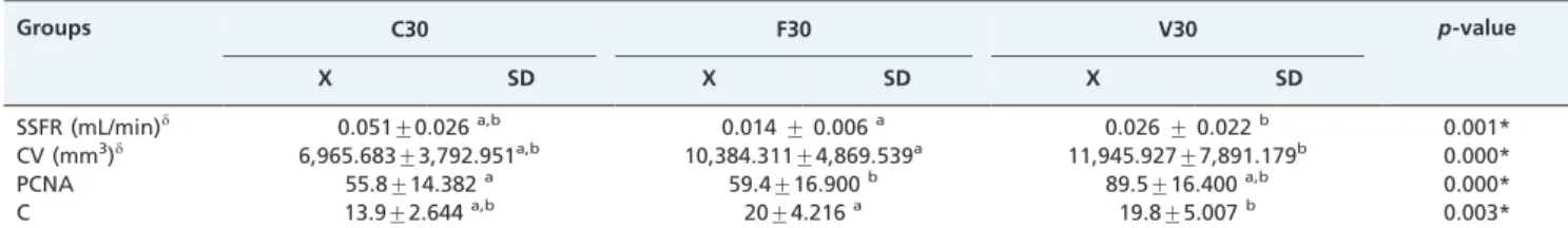
Groups treated for 30 days
Table 2 shows the average values, standard deviations,
andp-values for the studied variables in the groups treated
for 30 days (C30, F30, and V30). There were significant differences among the groups treated for 30 days for the
SSFR, CV, PCNA, and C variables (p,0.05).
Compared with the control group (C30), there was a significant increase in the number of myoepithelial cells (positively stained cells for C) in the groups treated with
antidepressants (V30 and F30) (p,0.05). In addition, there
was a significant increase in the number of positively stained cells for PCNA in the group treated with
venlafax-ine (V30) compared with the C30 and F30 groups (p,0.05).
SSFR was significantly decreased in the experimental (V30 and F30) groups compared with the control group
(p,0.05). Conversely, the highest average values for CV
were observed in groups F30 and V30.
Groups treated for 60 days
Table 3 shows the average values, standard deviations
andp-values for the studied variables in the groups treated
for 60 days (C60, Pilo, FS60, VS60, FP60, and VP60). There were significant differences among the groups treated for 60 days for the CV, SSFR, and C variables (p,0.05).
There was a significant increase in the number of positively stained cells for C in the group treated with fluoxetine and saline (FS60) compared with the control group (C60) (p= 0.0258). In addition, there was a significant decrease in the number of myoepithelial cells in group FP60
compared with all groups (p,0.05).
SSFR was significantly increased in the group treated with venlafaxine and pilocarpine (VP60) compared with the
group treated with venlafaxine and saline (VS60)
(p= 0.0214). In addition, the highest SSFR average value
was found in the Pilo group. Moreover, the lowest average value for CV was found in the Pilo group, whereas the
highest CV value was found in the VP60 group (p,0.05).
Figures 1-4 illustrate photomicrographs of the parotid glands of the rats in the experimental and control groups.
DISCUSSION
The anticholinergic actions of psychotropic drugs have not been completely clarified. Many studies have suggested that central nervous system (CNS) drugs have pharmaco-logical actions on the salivary glands. The attempts to clarify the xerostomic effects of such drugs have been of great scientific value, both in explaining the physiopharmacolo-gical effects and in bringing additional benefits to patients using these drugs.
In the present study, the chronic use of fluoxetine and venlafaxine for 30 days was associated with a decrease in SSFR, an increase in CV and an increase in the number of myoepithelial cells in rats. In addition, venlafaxine was associated with an increase in acinar and ductal cell proliferation in the parotid glands. Interestingly, the use of pilocarpine with venlafaxine for 60 days re-established a normal SSFR in rats. The number of myoepithelial cells remained stable in the parotid glands of rats that were treated for 60 days with venlafaxine irrespective of whether it was administered with saline or pilocarpine. Pilocarpine used in association with fluoxetine for 60 days did not induce changes in SSFR, but it was associated with a severe
Table 2 -Mean values (X) and standard deviations (SD) of the studied variables in the groups treated for 30 days with saline (C30), fluoxetine (F30), or venlafaxine (V30).
Groups C30 F30 V30 p-value
X SD X SD X SD
SSFR (mL/min)d
0.051¡0.026a,b 0.014¡0.006a 0.026¡0.022b 0.001*
CV (mm3)d
6,965.683¡3,792.951a,b 10,384.311¡4,869.539a 11,945.927¡7,891.179b 0.000*
PCNA 55.8¡14.382a 59.4¡16.900b 89.5¡16.400a,b 0.000*
C 13.9¡2.644a,b 20¡4.216a 19.8¡5.007b 0.003*
d
Values obtained from the study by da Silva et al. (2009)15.
SSFR – stimulated salivary flow rate; CV – cellular volume; PCNA – proliferating cell nuclear antigen; C – calponin.
a,bGroups followed by the same letter differed statistically from each other. *Statistically significant difference among groups (p
decrease in the number of myoepithelial cells in the parotid glands of rats. Considering these results, three main points must be discussed: the mechanisms of action of the medications on the CNS, physiological adaptation to the use of psychotropic medications, and alterations in receptor sensitivity due to the chronic use of medication.
Fluoxetine and venlafaxine have anticholinergic effects of different intensities. Because fluoxetine is a selective serotonin reuptake inhibitor, it may cause greater serotonin availability in the synaptic gap, which alters the binding of acetylcholine to the muscarinic receptors (M3) present in the salivary glands. Thus, fluoxetine may decrease the quantity of salivary secretion. Venlafaxine is an atypical antidepres-sant and acts as a reuptake inhibitor of serotonin,
noradrenaline, and dopamine.11Venlafaxine does not show
affinity for a1 adrenergic receptors, M3 muscarinic
recep-tors, or histamine receptors. Interestingly, the actions of venlafaxine become unpredictable because it can act through multiple targets, which can elicit results ranging from a decrease in salivary secretion to alterations
in salivary composition.13,21 Although antidepressant
medications do not seem to block saliva production, they interfere with the binding of acetylcholine to M3 receptors.
Thus, saliva is produced, but it cannot be excreted.12
Compared with the control groups (C30), the increase in
CV values in the parotid glands of rats that received antidepressants for 30 days (groups F30 and V30) appeared to represent acinar and ductal cell hypertrophy. Serous cell hypertrophy, which is characterized by widened acinar cells and secretory granule accumulation, has previously been reported following the chronic use of psychotropic drugs.12,26
The present study showed the anticholinergic effects of chronic fluoxetine and venlafaxine administration for 30 days . With 60-day treatment, the SSFR reduction was not significantly different than the control group. Studies have shown that the prolonged use of psychotropics causes alterations in receptor sensitivity.24In addition, therapeutic
effects and side effects gradually decrease after continuous or repeated psychotropic administration, which may cause desensitization, resistance, or tolerance. Physiological adap-tation may also occur, and many side effects of drugs tend
to diminish over time despite continuous use of a drug.27
Therefore, the least pronounced SSFR decrease in rats treated for 60 days may be a consequence of tolerance or adaptation to psychotropic drugs.
The absence of a significant difference in the mean SSFR values among the C60, FS60, and VS60 groups suggested that the normal SSFR of the rats was re-established 30 days after suspending the medication (Table 3). These data must be carefully analyzed, however, because a decrease in SSFR
Table 3 -Mean values (X) and standard deviations (SD) of the studied variables in the groups treated for 60 days with saline (C60), pilocarpine (Pilo), fluoxetine and saline (FS60), venlafaxine and saline (VS60), fluoxetine and pilocarpine (FP60), or venlafaxine and pilocarpine (VP60).
Groups C60 PILO FS60 VS60 FP60 VP60 p- value X SD X SD X SD X SD X SD X SD
SSFR (mL/min)d
0.052¡0.029 0.067¡0.028a,b 0.036¡0.017 0.020¡0.004a,c 0.034¡0.014b 0.055¡0.026c 0.000*
CV (mm3)d
6,505.564¡ 3,343.475d
5,825.418¡ 1,968.070ab
6,809.347¡ 3,189.246e
7,525.112¡ 3,196.085ac
7,519.797¡ 4,272.808f
10,194.315¡ 4,456.345b,c,d,e,f
0.000*
PCNA 50¡17.531 46.8¡13.506 36.7¡16.687 52.5¡18.710 52¡17.153 43.6¡18.007 0.296ns
C 13.6¡4.789c,f 22.1¡7.385a 24.2¡7.955b,c 19.7¡4.322d 7¡1.563a,b,d,e,f 18.2¡6.250e 0.000*
d
Values obtained from the study by da Silva et al. (2009)15.
SSFR – stimulated salivary flow rate; CV – cellular volume; PCNA – proliferating cell nuclear antigen; C – calponin.
a,b,c,d,e,fGroups followed by the same letter differed statistically from each other. *Statistically significant difference among the groups (p
,0.05).
ns– No statistically significant difference among the groups (p.0.05).
Figure 1 - Rat parotid gland with normal parenchyma. Immunohistochemical staining of PCNA on epithelial cells (C60, original magnification 400X).
greater than 50% brings relevant prejudicial consequences (C60 – 0.052 mL/min; VS60 – 0.020 mL/min).
SSFR reduction in rats undergoing chronic treatment with fluoxetine may still be an indirect consequence of serotonin action at the 5-HT (5-hydroxytryptamine) receptors present in peripheral microcirculation. Indeed, fluoxetine increases serotonin availability, and serotonin may bind to 5-HT receptors, which would alter the blood flow in the salivary glands and, consequently, the quantity and composition of the salivary flow.28
In the present study, pilocarpine exerted a significant secretagogue effect, which showed its cholinergic agonist capacity and corroborated the findings of Davies and
Shorthose.29Pilocarpine was associated with an increase in
SSFR and efficiently treated the hyposalivation caused by venlafaxine (groups VS60 and VP60). Conversely, when pilocarpine was used in association with fluoxetine (group FP60), there was no SSFR increase compared with the group that did not receive pilocarpine (group FS60 – Table 3). Pilocarpine is a parasympathomimetic agent that acts as a nonselective muscarinic receptor agonist.30,31Thus, pilocar-pine promotes stimulation of the exocrine glands and salivary secretion, which is efficient for patients without
extensive glandular parenchymal destruction.30
In the present study, chronic administration of either fluoxetine or venlafaxine for 30 days was associated with an increase in the number of myoepithelial cells in the parotid glands of rats. These results contradict Gre´gio et al.12who
suggested that the hyposalivation and serous cell hyper-trophy observed after chronic treatment with benzodiaze-pines and antidepressants in rats could be explained by a
decrease in the number of myoepithelial cells.12 We
hypothesized that a deficit in saliva elicits a compensatory activity that increases the number of myoepithelial cells, which have contractile functions, to assist in releasing the retained saliva. The myoepithelial cells accelerate the initial saliva emptying flow rate and promote the drainage of any adjacent extracellular fluid. The dendrites of the myoepithe-lial cells fold around the duct system, and their function is to compress the acinar and ductal cells.16,17
The parenchymatous cells (i.e., acinar, myoepithelial, and ductal) show a proliferative potential for maintenance and regeneration of the adult parenchymatous cell population in normal glands. In addition, parenchymatous cells show a
proliferative capacity when submitted to physical injury, such as ductal obstruction.19,20,32,33
This study was performed because there were no immunohistochemical studies of myoepithelial, acinar and ductal cells following the use of psychotropic drugs. There have been immunohistochemical studies on the proliferative capacity of parenchymatous cells in induced glandular
atrophy,19,22,23,33 and a comparative analysis between the
present study and the studies of induced glandular atrophy may be established. Interestingly, an animal model of induced atrophy of the parotid gland’s main duct did not prevent the gland from producing saliva. This saliva, however, was not excreted due to a physical obstruction in the main excretory duct. In an animal model of chronic antidepressant administration, we suggested that the saliva was continuously produced, but it was not secreted because of neurochemical interference from the psychotropic drugs. In animal models submitted to injuries (physical and neurochemical), there were significant increases in the number of myoepithelial cells.22,23,33
Because calponin is not a cellular proliferation marker, this study determined the numerical index of positively stained cells for calponin, which specifically identifies the myoepithelial cells in the glandular parenchyma. Due to the methodology that was employed, it was not possible to directly infer the cell proliferation, but it was possible to suggest that there was an alteration in the myoepithelial cellular proliferation rate among the studied groups.
In the groups of rats treated for 30 days, venlafaxine (V30) seemed to induce an increase in acinar and ductal cell proliferation compared with saline (control group). In an attempt to compensate for the significant SSFR reduction that resulted from the chronic use of venlafaxine (V30), mitoses may have occurred to create new acini and ducts (Table 2). The intense proliferative capacity of acinar and ductal cells has already been reported in studies of
glandular atrophy induced by obstruction.19Moreover, the
V30 group showed the highest mean CV value, which suggested the presence of cellular hypertrophy. It is worth emphasizing that there is a critical size for each cell, above which the stimulus for hypertrophy does not cause adaptative reactions, and the cell goes through mitosis to
compensate for the excessive cytoplasm.34We believe that
the proliferative capacity of the acinar and ductal cells and the presence of cellular hypertrophy may have contributed
Figure 3 - Immunohistochemical staining of calponin on myoepithelial cells (Pilo, original magnification 400X).
to the increase in the mean value of positively stained cells for PCNA in the V30 group.
For the groups treated for 60 days, there were not any significant differences in PCNA. Generally, chronic use of antidepressants has not been associated with significant aggression to the acinar and ductal cells. We believe that the proliferative potential of acinar and ductal cells assists in maintaining and regenerating the adult parenchymatous cellular population.
Although rat models have some limitations, diseases and/or therapeutic simulations in animal models are important tools for examining disease prognoses and investigating the side effects of drugs.
CONCLUSIONS
The number of positively stained cells for C was shown to be increased with the chronic use of venlafaxine or fluoxetine. The proliferation index of the epithelial cells of rat parotid glands, however, was not altered by the use of antidepressants for 60 days.
ACKNOWLEDGEMENTS
The authors would like to thank the employees of the Laborato´rio de Patologia Experimental, Pontifı´cia Universidade Cato´lica do Parana´. This study was supported by CNPq (grant 474790/2004-5).
REFERENCES
1. Sansone RA, Sansone LA. Pain, pain, go away: antidepressants and pain management. Psychiatry (Edgmont). 2008;5:16-9.
2. Cusack B, Nelson A, Richelson E. Binding of antidepressants to human brain receptors: focus on newer generation compounds. Psycho-pharmacology (Berl). 1994;114:559-65, doi: 10.1007/BF02244985. 3. Dhir A, Kulkarni SK. Antidepressant-like effect of 17 beta-estradiol:
involvement of dopaminergic, serotonergic, and (or) sigma-1 receptor systems. Can J Physiol Pharmacol. 2008;86:726-35, doi: 10.1139/Y08-077. 4. Spina E, Santoro V, D9Arrigo C. Clinically relevant pharmacokinetic drug interactions with second-generation antidepressants: an update. Clin Ther. 2008;30:1206-27, doi: 10.1016/S0149-2918(08)80047-1. 5. Stahl SM. Psychopharmacology of antidepressants. London: Martin
Dunitz. 1997;114.
6. Cipriani A, Brambilla P, Furukawa TA, Geddes J, Gregis M, Hotopf M, et al. Fluoxetine versus other types of pharmacotherapy for depression. Cochrane Database of Systematic Reviews. In: The Cochrane Library. 2006;3.
7. Guggenheimer J, Moore PA. Xerostomia: etiology, recognition and treatment. J Am Dent Assoc. 2003;134:61-9.
8. Tenovuo J. Salivary parameters of relevance for assessing caries activity in individuals and populations. Community Dent Oral Epidemiol. 1997;25:82-6, doi: 10.1111/j.1600-0528.1997.tb00903.x.
9. Ferguson MM. Pilocarpine and other cholinergic drugs in the manage-ment of salivary gland dysfunction. Oral Surg Oral Med Oral Pathol. 1993;75:186-91, doi: 10.1016/0030-4220(93)90092-I.
10. Scully C. Drug effects on salivary glands: dry mouth. Oral Dis. 2003;9:165-76, doi: 10.1034/j.1601-0825.2003.03967.x.
11. Keene JJ Jr, Galasko GT, Land MF. Antidepressant use in psychiatry and medicine: importance for dental practice. J Am Dent Assoc. 2003;134:71-9.
12. Gre´gio AMT, Durscki JRC, Lima AAS, Machado MAN, Igna´cio SA, Azevedo LR. Association of amitriptyline and diazepam on the histomorphometry of rat parotid glands. Pharmacologyonline. 2006;2:96-108.
13. de Almeida P del V, Gre´gio AM, Brancher JA, Igna´cio SA, Machado MA, de Lima AA, et al. Effects of antidepressants and benzodiazepines on stimulated salivary flow rate and biochemistry composition of the saliva.
Oral Surg Oral Med Oral Pathol Oral Radiol Endod. 2008;106:58-65, doi: 10.1016/j.tripleo.2007.11.008.
14. Zaclikevis MV, D’Agulham AC, Bertassoni LE, Machado MA, de Lima AA, Gre´gio AM, et al. Effects of benzodiazepine and pilocarpine on rat parotid glands: histomorphometric and sialometric study. Med Chem. 2009;5:74-8, doi: 10.2174/157340609787049262.
15. da Silva S, de Azevedo LR, de Lima AA, Igna´cio SA, Machado MA, ZacliKevis MV, et al. Effects of fluoxetine and venlafaxine and pilocarpine on rat parotid glands. Med Chem. 2009;5:483-90, doi: 10. 2174/157340609789117868.
16. Klein RM. Development, structure and function of the salivary glands. In: Avery JK. Oral development and histology. USA: New York. 2001;292-331.
17. Berkovitz BKB, Holland GR, Moxham BJ. Oral anatomy, embryology and histology. Chicago: Mosby. 2002;255-67.
18. Zarbo RJ, Prasad AR, Regezi JA, Gown AM, Savera AT. Salivary gland basal cell and canalicular adenomas: immunohistochemical demonstra-tion of myoepithelial cell participademonstra-tion and morphogenetic considera-tions. Arch Pathol Lab Med. 2000;124:401-5.
19. Burford-Mason AP, Cummins MM, Brown DH, MacKay AJ, Dardick I. Immunohistochemical analysis of the proliferative capacity of duct and acinar cells during ligation-induced atrophy and subsequent regenera-tion of rat parotid gland. J Oral Pathol Med. 1993;22:440-6, doi: 10.1111/j. 1600-0714.1993.tb00122.x.
20. Norberg L, Dardick I, Burford-Mason AP. Differentiating myoepithelial and acinar cells in rat neonatal parotid gland and histogenetic concepts for salivary gland tumors. J Oral Pathol Med. 1996;25:474-80, doi: 10. 1111/j.1600-0714.1996.tb00300.x.
21. Denys D, van der Wee N, van Megen HJ, Westenberg HG. A double blind comparison of venlafaxine and paroxetine in obsessive-compulsive disorder. J Clin Psychopharmacol. 2003;23:568-75, doi: 10.1097/01.jcp. 0000095342.32154.54.
22. Takahashi S, Schoch S, Walker NI. Origin of acinar cell regeneration after atrophy of the rat parotid induced by duct obstruction. Int J Exp Pathol. 1998;79:293-301, doi: 10.1046/j.1365-2613.1998.710405.x.
23. Burgess KL, Dardick I, Cummins MM, Burford–Mason AP, Bassett R, Brown DH. Myoepithelial cells actively proliferate during atrophy of rat parotid gland. Oral Surg Oral Med Oral Pathol Oral Radiol Endod. 1996;82:674-80, doi: 10.1016/S1079-2104(96)80443-4.
24. Rocha RM, Andrade VP, Nunes CB, Rocha GFS, Sanches FSF, Oliveira FS, et al. Construc¸a˜o de arrays de tecido com equipamento alternativo e de baixo custo para estudo imuno-histoquı´mico de tumores mama´rios. J Bras Patol Med Lab. 2006;42:477-82, doi: 10.1590/S1676-2444200 6000600012.
25. Schuler S, Gurmini J, Cecı´lio WA, Viola de Azevedo ML, Olandoski M, de Noronha L. Hepatic and thymic alterations in newborn offspring of malnourished rat dams. J Parenter Enteral Nutr. 2008;32:184-9, doi: 10. 1177/0148607108314387.
26. Martinez-Madrigal F, Micheau C. Histology of the major salivary glands. Am J Surg Pathol. 1989;13:879-99, doi: 10.1097/00000478-198910000-00008.
27. Gorenstein C, Scavone C. Avanc¸os em psicofarmacologia - mecanismos de ac¸a˜o de psicofa´rmacos hoje. Rev Bras Psiquiatr. 1999;2:64-73, doi: 10. 1590/S1516-44461999000100012.
28. Rang HP, Dale MM, Ritter JM, Flower RJ. Pharmacology. London: Churchill Livingstone. 2007;557-74.
29. Davies AN, Shorthose K. Parasympathomimetic drugs for the treatment of salivary gland dysfunction due to radiotherapy. Cochrane Database Syst Ver. 2007;18:19.
30. Grisius MM. Salivary gland dysfunction: a review of systemic therapies. Oral Surg Oral Med Oral Pathol Oral Radiol Endod. 2001;92:156-62, doi: 10.1067/moe.2001.116601.
31. Ship JA, Pillemer SR, Baum BJ. Xerostomia and the geriatric patient. J Am Geriatr Soc. 2002;50:535-43, doi: 10.1046/j.1532-5415.2002.50123.x. 32. Denny PC, Ball WD, Redman RS. Salivary glands: a paradigm for
diversity of gland development. Crit Rev Biol Med. 1997;8:51-75, doi: 10. 1177/10454411970080010301.
33. Miguel MC, Andrade ES, Taga R, Pinto LP, Souza LB. Hyperplasia of myoepithelial cells expressing calponin during atrophy of the rat parotid gland induced by duct ligation. Histochem J. 2002;34:499-506, doi: 10. 1023/A:1024761923303.