UNIVERSIDADE ESTADUAL PAULISTA “JÚLIO
DE MESQUITA FILHO”
FACULDADE DE MEDICINA
Carolina Mendonça Gorgulho
Terapia gênica com interferon-alfa no controle do câncer colorretal
Dissertação apresentada à Faculdade de
Medicina, Universidade Estadual Paulista “Júlio
de Mesquita Filho”, Câmpus de Botucatu, para
obtenção do título de Mestre em Patologia
Orientador: Prof. Dr. Ramon Kaneno
Carolina Mendonça Gorgulho
Terapia gênica com interferon-alfa no controle do câncer colorretal
Dissertação apresentada ao Programa de
Pós-Graduação em Patologia da Faculdade de
Medicina de Botucatu, Universidade Estadual Júlio
de Mesquita Filho – UNESP, para obtenção do
título de Mestre em Patologia.
Orientador: Prof. Dr. Ramon Kaneno
Ao Prof. Dr. Ramon Kaneno, meu orientador, por me receber em seu laboratório tão bem, pela paciência em me ensinar, por todo o investimento que fez em mim e por ter me dado a oportunidade de me encontrar dentro da imunologia e da pesquisa.
À minha amiga e professora Dra. Graziela Romagnoli, uma das pessoas mais competentes e generosas que já conheci, pela amizade, por todos os momentos passados no laboratório, por ter me acalmado diversas vezes durante o mestrado, por tudo o que me ensinou, por ser uma inspiração todos os dias.
Ao meu pai Rafael, que me passa imensa segurança e que para mim é um exemplo a ser seguido. À minha mãe Marisa, minha melhor amiga. Agradeço aos dois por todo o apoio, por me darem tranquilidade para fazer minhas escolhas e por sempre demonstrarem entusiasmo e orgulho pela minha vida acadêmica.
Ao meu irmão Caio, uma das pessoas mais inteligentes que eu conheço, pelas tentativas de me fazer entender Física, por todo o companheirismo e amizade.
Ao meu avô Dâmaso, pelo carinho, conversas e interesse por este trabalho.
À minha também amiga e professora Dra. Marcela Camargo, por ter me acolhido no laboratório há tantos anos atrás, por me levar ao meu primeiro congresso, por tudo que me ensinou e fez por mim.
Ao Prof. Dr. João Pessoa Araújo Junior e à Prof. Dra. Alexandrina Sartori pelas sugestões e pela contribuição no meu Exame de Qualificação.
Ao Prof. Dr. Bryan Strauss por ter aberto as portas de seu laboratório e ter gentilmente cedido o vetor lentiviral. A todo o seu grupo, especialmente Dra. Daniela e Marlous, por terem me acolhido no laboratório e pela dedicação a este trabalho.
Aos doadores de sangue que possibilitaram a realização dos experimentos.
À Sophia, por toda a amizade, ajuda, almoços apressados e incontáveis MLRs, e aos demais colegas do laboratório de Imunologia de Tumores, Bianca, Marina, Juliana, Rebeca, Jofer, Edson e Nathalia, pela ajuda, discussão de resultados, companheirismo e amizade.
Às minhas colegas da época da iniciação científica Cecília, Fabiana, Juliana, Victoria e Carol, com quem ainda discuto resultados e divido inseguranças. Obrigada pela amizade que dura até hoje.
Aos funcionários do departamento de Microbiologia e Imunologia Ana, Larissa, Aline, Ivana, Rafael, Luiz e Lula pela ajuda, pelas risadas e pelo apoio nesses últimos anos.
À secretária do programa de Patologia, Vânia, pela ajuda e por ser extremamente eficiente.
Às minhas queridas amigas Renata e Amanda, por todo o apoio, especialmente na fase final deste trabalho.
Ao meu amigo Renan, que por inúmeras vezes ouviu meus problemas e comemorou comigo minhas conquistas.
À minha prima Notomi, que sempre me aconselhou e orientou nas questões acadêmicas.
A todos os meus amigos de infância, graduação e pós, que torcem por mim e me acompanharam durante os bons e maus momentos.
Às minhas tias da família Gorgulho, que sempre demonstram interesse pelo trabalho que fazemos no laboratório.
Ao Conselho Nacional de Desenvolvimento Científico e Tecnológico (CNPq), pela concessão de bolsa de estudo.
À Fundação de Amparo à Pesquisa do Estado de São Paulo, pela concessão de auxílio financeiro.
A todos que não foram citados aqui, mas que de alguma forma participaram desta etapa da minha vida.
Resumo
O interferon alfa (IFN-α), um IFN do tipo I, se apresenta como uma citocina com
grande potencial terapêutico, pois atua no combate direto às células tumorais, além de agir
sobre a maturação de células dendríticas (DCs), que são células apresentadoras de
antígenos profissionais e peças chave na elaboração da uma resposta antitumoral.
Entretanto, a administração sistêmica de citocinas pode produzir toxicidade importante nos
pacientes, de modo que a indução de sua produção in situ poderia representar uma forma
de imunomodulação mais adequada. Assim, o objetivo deste estudo é verificar a ação de
vetores lentivirais carregando o gene do IFN-α para transdução de células tumorais,
permitindo assim a produção localizada de IFN-α, a fim de explorar, in vitro, seu potencial
lítico e imunomodulatório sobre DCs. Vetores lentivirais carregando o gene do IFN-α
humano (Lego-IFN) ou GFP (Lego-GFP) foram utilizados para a transdução in vitro de
células de câncer colorretal. A transdução foi feita com diferentes multiplicidades de infecção
(MOIs – 0.3, 1.0, 2.0, 4.0) para avaliarmos o efeito dose-dependente, seguido de co-cultura
com DCs derivadas de monócitos de doadores saudáveis (DC-0.3, DC-1.0, DC-2.0, DC-4.0).
Após 48h de co-cultura, as DCs foram avaliadas fenotípica e funcionalmente, através da
análise dos marcadores de membrana por citometria de fluxo, capacidade de aloestimulação
e de indução de linfócitos T citotóxicos (CTLs). Nós observamos que a transdução com
Lego-GFP, mas não com Lego-IFN, aumentou a imunogenicidade das células tumorais, com
aumento de expressão de CD54 e HLA-DR. A co-cultura de DCs com células tumorais
transduzidas com Lego-IFN aumentou discretamente seu perfil de ativação, mas não seu
potencial aloestimulatório in vitro. Observamos que linfócitos cultivados com DC-2.0
produziram níveis mais altos de IFN-γ, sugerindo a indução de um perfil Th1, enquanto que
DC-GFP induziu maior produção de IL-10 e IL-4. DC-4.0 foi mais eficiente na geração de
CTLs que o controle DC, entretanto, DC-GFP induziu ainda mais proliferação de linfócitos T
CD8+. O aumento de imunogenicidade das células tumorais e o potencial superior de
geração de CTLs por Lego-GFP e DC-GFP exigem uma maior investigação.
Abstract
Interferon alpha (IFN-α) is a type I IFN with great therapeutic potential, since it is able
to directly fight tumor cells and enhance the maturation of dendritic cells (DCs), the main
antigen-presenting cells, required for an effective antitumor response. However, the systemic
administration of cytokines can induce severe collateral effects. Therefore, the induction of
cytokine secretion in situ should represent a more adequate approach for cytokine-based
immunotherapy. Thus, the goal of this study was to induce IFN-α secretion by colon cancer
cells by transduction with a lentivirus vector carrying the human IFN-α gene, followed by
analysis of its immunomodulatory potential over DCs. Transduction was made with different
multiplicities of infection (MOIs – 0.3, 1.0, 2.0 and 4.0) to evaluate the dose-dependent
effects. Such cells were co-cultured with monocyte-derived DCs from healthy donors
(DC-0.3, DC-1.0, DC-2.0 and DC-4.0). Forty-eight hours later, DCs were evaluated for their
phenotype (surface activation/maturation markers) by flow cytometry, their ability to induce
allogeneic response in a mixed leukocyte reaction (MLR) and effectiveness to induce
cytotoxic T cells. We observed that transduction with Lego-GFP, but not Lego-IFN, increased
tumor cells’ immunogenicity with up-regulation of the markers CD54 and HLA-DR. Co-culture
of Lego-IFN-transduced tumor cells with DCs slightly enhanced their activation phenotype
but not their potential to stimulate T cell proliferation in vitro. Furthermore, we observed that
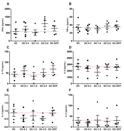
lymphocytes cultured with DC-2.0 produced higher levels of IFN-γ, suggesting an induction of
Th1 profile on T cells, while DC-GFP induced more IL-10 and IL-4. Additionally, DC-4.0 was
more efficient in generating cytotoxic T lymphocytes (CTLs) that the control DC, however
DC-GFP induced even more CD8+T cell proliferation. The enhancement of tumor cell
immunogenicity and the superior induction of CTLs by Lego-GFP and DC-GFP require
additional investigation.
12
Sumário
14
Interferons do tipo I
Os interferons (IFNs) foram descritos pela primeira vez em 1957, por Isaacs e
Lindenmann, que investigavam o fenômeno de interferência viral, no qual a exposição a um
vírus atenuado confere resistência contra infecções posteriores pelo mesmo vírus (1). Até
então, acreditava-se que o bloqueio de receptores na membrana da célula alvo pelo vírus
inativado era responsável por conferir resistência às células e que este era um fenômeno
relativamente “passivo”. Entretanto, Isaacs e Lindenmann não encontravam evidências de
que o vírus atenuado estivesse “impedindo a captura de vírus vivos pelas células (...) e que
para estabelecer o fenômeno de interferência, mais de 4h de incubação eram necessárias”.
Em seus experimentos, os autores utilizaram pedaços da membrana corioalantóide de ovos
de galinha cultivados in vitro tratados com o vírus influenza A atenuado pelo calor (o que
abolia sua infectividade, mas não suas propriedades de interferência), para depois serem
desafiados com vírus influenza A vivos. Observaram que o fenômeno de interferência
acontecia de fato nas membranas infectadas, o que foi chamado de “interferência inicial”,
mas que, além disso, ao transferir as membranas para uma cultura fresca, livre de vírus, o
novo sobrenadante também apresentava uma “interferência residual”, de magnitude
comparável à inicial. Este achado levou os autores a especularem que a própria membrana
estava envolvida no fenômeno de interferência e não somente as partículas virais. Os
autores cogitaram a possibilidade de que a membrana pré-imunizada produzia um “agente
de interferência” caracterizado como “uma macromolécula com propriedades distintas
daquelas dos vírus influenza A atenuados”. Este agente foi chamado de interferon. Em
humanos, existem 3 famílias distintas de IFNs. Os do tipo I, no qual se enquadram os IFNs
α, β, ω e ε (2); tipo II, representado pelo IFN-γ (3) e tipo III, no qual estão os mais
recentemente descobertos IFNs λ (4). Dada a amplitude do tema, a presente revisão é
focada na biologia do IFN tipo I, com ênfase no papel do IFN- na estimulação do sistema
imune e no combate ao câncer.
15 uma gama de estímulos externos que muito frequentemente envolve a ativação de
receptores de reconhecimento de padrão (PRR – pattern recognition receptors) por
componentes e/ou produtos microbianos (5), além de componentes endógenos
ectopicamente expressos, como RNA e DNA extracelulares (6). Os PRRs estão presentes
na superfície, citoplasma e compartimentos endossomais das células. Na superfície, os
receptores toll-like (TLR) reconhecem principalmente produtos microbianos, sendo que o
TLR4 é o mais potente indutor de IFNs do tipo I mediante ativação por lipolisacarídeos (LPS)
de origem bacteriana (5). O receptor RIG-I (RNA helicases retinoic acid-inducible gene I) é
um dos principais receptores citosólicos de RNA e está intimamente ligado com a produção
de IFNs do tipo I, assim como o receptor MDA5 (melanoma differentiation-associated
gene 5) (5). Em compartimentos endossomais, o TLR3 reconhece RNA de fita dupla e TLR7
e 8 reconhecem RNA de fita simples (6). Dependendo da natureza do patógeno (como sua
espécie e local de infecção) e da origem da célula alvo, os PRRs são diferencialmente
ativados, na tentativa de montagem de uma resposta adequada ao tipo de infecção (7).
Assim, ampliando o conceito primordial de indução de IFN tipo I por infecções virais,
sabe-se hoje que muitos outros estímulos podem sabe-ser responsáveis pela indução dessa citocina.
A família de IFNs do tipo I compartilha o receptor IFNAR, que comanda a indução de
diversos genes através da ativação de ISGF3 (IFN-stimulated gene factor 3) que induz a
subsequente fosforilação de fatores de transcrição como STAT1 e 2 e IRF9 (interferon
regulatory factor 9) (8), levando à sua ativação e entrada no núcleo, possibilitando a
transcrição de genes IFN-respondedores, em sua maioria com papel antiviral, envolvidos em
processos como síntese de proteínas, crescimento e sobrevivência celulares (9) (8).
TLRs e sinalização de IFNs do tipo I
Os TLRs são PRRs transmembrana do tipo I e respondem principalmente a estímulos
microbianos, tendo importante função na indução de imunidade inata. Em mamíferos, a
16 humanos e camundongos (9). Há uma estrutura básica entre os TLRs, composta por um
ectodomínio N-terminal envolvido no reconhecimento de padrões moleculares
patógeno-associados (PAMPs – pathogen associated molecular patterns), um domínio transmembrana
do tipo I e um domínio toll/receptor de interleucina 1 (IL-1) (TIR) intracelular responsável
pela transdução do sinal (6) (9).
Dependendo da sua localização celular e de seus ligantes, os TLRs podem ser
subdivididos em 2 grupos: o primeiro, composto pelos TLRs1,2,4,5,6 e 11, é expresso na
superfície das células e reconhecem principalmente componentes de membrana microbiana
como lipídeos e proteínas; o segundo engloba os TLRs3, 7, 8 e 9, que estão localizados em
vesículas citoplasmáticas e reconhecem material genético (6).
A ativação de TLR3 e TLR4 leva à indução direta do promotor do IFN-β, que
desencadeia o recrutamento de proteínas adaptadoras, como TRIF (TIR domain-containing
adaptor protein inducing IFNβ), que levam complexos sinalizadores até o núcleo, onde IRF3
(interferon regulatory factor-3) é ativado e promove resposta transcricional de IFN-β (9) (10).
Na maioria dos casos, IRF3 e 7 são fundamentais para a produção de IFN-α/β (5). TLR4
responde à ligação com LPS bacteriano através de um complexo formado na superfície da
célula com a proteína de diferenciação mielóide 2 (MD2 - myeloid differentiation protein 2)
(11). O complexo TLR4-MD2 recruta moléculas adaptadoras que irão para o núcleo iniciar a
transdução de sinal.
A produção de IFNs do tipo I também pode ser mediada pela ligação de TLR7, 8 e 9 e
indução da molécula adaptadora MyD88, que é recrutada para o domínio TIR do receptor e
orquestra a ativação do fator de transcrição IRF7 (12). TLR7 e 9 localizam-se no retículo
endoplasmático de células não estimuladas e migram para as vesículas endolisossomais
após ativação (13). TLR7 reconhece principalmente RNA retroviral de fita simples e é
altamente expresso em DCs plasmocitóides (pDCs), capazes de liberar altas doses de IFNs
do tipo I após infecção viral. Há uma alta expressão de TLR7 e 9 em pDCs (8). Nestas
17 para vesículas endolisossomais onde RNA de fita simples é reconhecido, iniciando a
resposta antiviral (6). Além disso, TLR7 quando expresso em DCs convencionais (cDCs) e
ativado por RNA bacteriano, também induz a produção de IFNs do tipo I (14). TLR8
reconhece RNA de fita simples e é expresso ubiquamente, especialmente em monócitos, e
é ativado após infecção bacteriana (6). TLR9 reconhece DNA microbiano, que contém ilhas
CpG não metiladas, forte indutoras de resposta imune (15).
A estimulação simultânea de dois ou mais TLRs, o que geralmente ocorre em
situações de infecção, pode desencadear atividades sinérgicas, agonistas ou antagonistas
entre os receptores (16). Foi demonstrado que na infecção por Mycobacterium tuberculosis,
a ativação de TLR2 pela bactéria inibe a produção de IFNs do tipo I por TLR9, através da
rápida degradação da molécula IRAK-1 (IL-1R associated kinase 1), necessária para a
sinalização de IFNs do tipo I via MyD88 (16)(17).
RIG-I e sinalização de IFNs do tipo I
Os receptores RIG-I e RIG-I-like (RLR) reconhecem vírus em replicação no
citoplasma, especialmente na fase inicial de infecção e são capazes de distinguir RNA
invasor de RNA endógeno (18). A família dos RLRs compreende os receptores RIG-I, MDA5
e LGP2. Eles possuem uma estrutura em comum, o domínio helicase DExD/Hbox, cuja
função é o desdobramento de RNA de fita dupla (19). Estes receptores também possuem o
domínio de recrutamento e ativação de caspase (CARD), responsável direto pela indução de
produção de INFs do tipo I (18). A ação dos RLRs está ligada à ubiquitinação de seus
domínios. O domínio CARD dos receptores RIG-I necessita de ubiquitinação para que haja
sinalização da produção de IFNs do tipo I. O mesmo ocorre com o receptor MDA5, porém
sob controle de outros fatores de ubiquitinação. Além da regulação positiva de sua atividade,
os RLRs também são negativamente controlados por ubiquitinação, evitando a ativação
descontrolada destes receptores (18).
18
presença de ATP, o domínio CARD interage com IPS-1 (interferon β promoter stimulator-1),
uma molécula imprescindível para a sinalização de IFNs do tipo I mediante infecção viral
(20), que se localiza na membrana externa da mitocôndria. MDA5 também sinaliza a
produção de IFNs através de IPS-1, que ativa IRF3 e IRF7 (21).
RIG-I é essencial para a produção de IFNs do tipo I, além de outras citocinas pró
inflamatórias, mediante infecção viral (18). Células que não possuem RIG-I tem produção
deficiente de IFNs do tipo I frente à infecção com diversas famílias de vírus (22). Células
com o gene do LGP2 nocauteado tem produção de IFNs diminuída, mas não totalmente
bloqueada, sugerindo que este receptor coopera com RIG-I e MDA5 no reconhecimento de
RNA viral (18).
Após ligação com RNA viral, RIG-I passa por uma mudança conformacional para
expor seu domínio CARD, na presença de ATP. O domínio CARD interage com proteínas
adaptadoras para a ativação de quinases de forma TRAF3 (tumor necrosis fator receptor
associated fator 3)-dependente. Estas quinases fosforilam IRF3 e 7 (23), resultando na sua
translocação para o núcleo e consequente transcrição de genes IFN-respondedores (24).
NLRs e sinalização de IFNs do tipo I
As proteínas NLRs (nucleotide binding domain and leucina rich repeat) são uma
família de proteínas citosólicas implicadas na resposta inata frente a infecções bacterianas e
indução de resposta anti-inflamatória e estão associadas à regulação de inflamassomas
(25). A composição destas proteínas é relativamente comum a todos os membros e consiste
em: um domínio amino-terminal EBD (effector-binding domain), um domínio central NOD
(nucleotide-binding oligomerization domain) responsável pela própria oligomerização e um
domínio LRD (carboxy-terminal ligand-recognition domain) (26).
Os NLRs podem ser subdivididos em 2 categorias, NLRC e NLRP. Os NLRC possuem
domínios CARD e compreendem NOD1 e NOD2, entre outros. Estes receptores estão
19 fragmentos de peptideoglicanos. Diferentes peptideoglicanos ativam receptores NOD
distintos, o que leva a diferentes vias de sinalização (27). O segundo grupo, NLRP, são
proteínas com um domínio PYR (pyrin) (28). A ligação de NLRs por DAMPs
(danger-associated molecular patterns) ou PAMPs leva à ativação de NF-κB (nuclear fator kappa B),
sinalização de MAPK ou inflamassomas (29).
O receptor NOD2 está envolvido na sinalização de IFNs do tipo I, especialmente de
IFN-β, através do reconhecimento de RNA viral de fita simples e ativação de IRF3.
Semelhantemente à sinalização por RIG-I, o domínio CARD de NOD 2 também necessita de
ligação com IPS-1 para induzir a produção de INFs (30).
O papel do IFN- na terapia antitumoral
O IFN-α é uma das citocinas mais amplamente exploradas no contexto da terapia
sistêmica, como revisto por Dranoff (31). A atividade antitumoral relevante do IFN-α vem
sendo amplamente testado para o tratamento de vários tipos de tumores (32). Por exemplo,
em um ensaio clínico realizado com pacientes com melanoma, o tratamento com IFN-α2b
em alta dosagem (1000 U/mL) produziu modulação positiva da ação de linfócitos T e células
NK e aumento na expressão de MHC de classe II pelas células tumorais (33). Em outro
ensaio do mesmo grupo, com pacientes de melanoma metastático, os autores observaram
que o tratamento sistêmico com IFN-α2b reduziu a incidência de recidiva da doença (34).
O IFN-α também se mostrou eficaz no tratamento de tumores hematológicos, a
exemplo da leucemia mielóide crônica, com uma melhora de sobrevida em 15% dos
pacientes tratados com quimioterapia e a citocina, versus aqueles submetidos somente ao
regime convencional de quimioterapia (35).
Medicamentos que têm como princípio ativo o IFN-α são atualmente comercializados
por diversos laboratórios farmacêuticos. Em 1986 o FDA aprovou as drogas Intron A
(IFN-α2b, Schering) e Roferon A (IFN-α2a, Hoffman-La Roche). Mais recentemente foram
20 organismo, a exemplo da droga Pegasys (PEG-IFN-α2a, Hoffman-La Roche), aprovada em
2002. Porém, assim como na terapia com IL-2, o uso sistêmico do IFN-α também oferece
reações adversas ao paciente, incluindo manifestações físicas e psiquiátricas, fato que pode
exigir a redução da dose, a implementação de intervalos no tratamento ou a interrupção do
uso do medicamento.
Além de sua ação direta sobre as células tumorais, o IFN-α tem sido amplamente
explorado pelo seu importante papel no desenvolvimento dirigido de DCs. Nesse sentido, foi
relatado que PBMCs (monócitos obtidos a partir de sangue periférico) cultivados em meio
rico em IFN-α e GM-CSF adquiriram características morfológicas de DCs, com alta
expressão de MHC-I e -II, da molécula co-estimulatória B7 e do marcador CD40, além de se
mostrarem boas apresentadoras de antígenos (36). Estudos in vitro demonstram a maior
eficiência de DCs geradas na presença de IFN-α, em comparação com DCs geradas na
ausência desta citocina, quanto à capacidade de sensibilizar linfócitos T CD8+ com alta
atividade citotóxica. Observou-se também geração de maior número linfócitos T CD4+
secretores de IFN- e menor expansão de linfócitos T CD4+CD25+Foxp3+ (regulatórios) (37).
Tais resultados indicam que preparações de DCs utilizando IFN-α ou a presença da citocina
no ambiente tumoral induzam DCs que apresentem resultados clínicos superiores aos
observados até hoje.
O sucesso das vacinas de DCs diferenciadas a partir de monócitos é dependente da
eficiência em se gerar DCs maduras para administração em animais portadores de tumor,
sendo que a presença de IFN-α no meio de cultura dessas células (38-40), é capaz de
direcionar essa maturação para que produzam IL-12 e adquiram o fenótipo α-DC1. Essas
DCs, por sua vez, têm a habilidade de dirigir a maturação de linfócitos T para um fenótipo
Th1, favorecendo a resposta antitumoral. A adição de prostaglandina E2 (PGE2) ao coquetel
de citocinas usadas na preparação de vacinas de DCs tem o propósito de induzir a
maturação destas células (37). Entretanto, esse mediador sozinho tende a reduzir a
21 IFN-γ. A adição de IFN-α, por sua vez, promove a secreção de IFN-γ pelos linfócitos de
modo dose-dependente, mesmo na presença de PGE2 (41).
Em estudo clínico de fase I/II observou-se que a administração de vacinas
terapêuticas de αDC-1 derivadas de monócitos do sangue a 22 pacientes com glioma
maligno recorrente não provocou mortes ou efeitos tóxicos de nível 3 ou 4. Os autores
demonstraram pela primeira vez a indução in vivo de linfócitos T CD8+ contra 3 dos 4
peptídeos encontrados em gliomas (42). Em outro estudo, as αDC-1 foram geradas com o
uso de um coquetel de citocinas contendo IFN-α e ácido poliosínico:policitidílico (poli I:C).
Sua comparação com DCs convencionais mostra que as αDC-1 são mais hábeis em induzir
a geração de linfócitos CD4+ produtores de IFN-γ e que a capacidade de induzir a produção
de IL-12 também é superior às DCs convencionais, com produção de níveis de IL-12 cerca
de 100 vezes maiores do que de IL-10 (43).
O uso de vetores virais na terapia gênica
Entretanto, embora diferentes preparações de vacinas de DCs tenham demonstrado
sua eficiência como vacina terapêutica, essa alternativa de tratamento continua a ter um
custo extremamente elevado, dificultando seu uso como terapia corrente. Assim, ainda na
tentativa de modular a resposta imune antitumoral utilizando o potencial imunomodulatório
das citocinas e o papel central das DCs na elaboração desta resposta, as vacinas gênicas
utilizando vetores virais têm sido cada vez mais investigadas no campo da imunoterapia
antitumoral. Nesse aspecto, a versatilidade dos vetores virais permite customizar as vacinas,
de forma que atinjam um alvo muito específico ou tenham ação localizada, minimizando os
efeitos adversos comuns produzidos por terapias sistêmicas. Deste modo, alguns estudos
têm relatado a inserção de genes codificadores de antígenos tumorais, visando potencializar
a atividade das células apresentadoras de antígenos (44, 45). Outros grupos têm trabalhado
com a inserção de genes codificadores de proteínas imunoestimulatórias, como as citocinas,
22 genes codificadores de moléculas com ação lítica direta sobre as células tumorais (47).
Um aspecto importante na construção das vacinas gênicas com vetores virais é que
não há necessidade de uso de células dos pacientes ou de qualquer material biológico
específico que restrinja demasiadamente sua preparação em laboratório ou sua utilização
terapêutica (47).
Muitos tipos de vetores virais têm sido avaliados para uso na imunoterapia, cujas
características específicas devem atender a diferentes demandas ou estratégias
moleculares. Os vetores adenovirais são provavelmente as ferramentas mais extensamente
exploradas no campo da vetorologia aplicada à imunoterapia do câncer (48), porém outros
vírus vêm apresentando ótima aplicabilidade na área e entre eles destacam-se os lentivírus.
Os lentivírus são membros da família Retroviridae (sendo o vírus da imunodeficiência
humana-1 [HIV-1] o mais bem estudado desta família) (49). Segundo revisto por Emeagi e
colaboradores (49), os lentivírus possuem características que os tornam ótimos candidatos
para aplicação na terapia gênica, como a) possibilidade de inserção de grande quantidade
de material genético (~10 kilobases); b) os lentivírus selecionam locais seguros de
integração com o genoma hospedeiro, o que proporciona uma expressão persistente do
gene de interesse e aumenta sua segurança de uso; c) são capazes de transduzir células
que se dividem e células quiescentes; d) em geral não há imunidade pre-existente a
esses vetores (50) e eles desencadeiam resposta imune neutralizadora menos intensa
quando comparada àquela induzida por vetores adenovirais, não comprometendo a
expressão do gene de interesse [36]. Além disso, vetores derivados de HIV-1 de terceira
geração tem pobre capacidade replicativa e contém apenas três (gag, pol e rev) dos nove
genes do vírus (51), garantindo integração estável no genoma do hospedeiro e ausência de
expressão de qualquer gene viral que não os transgenes (52).
Levando em conta estas informações, consideramos que a terapia gênica é uma
importante estratégia imunoterapêutica a ser estudada, para o desenvolvimento de
23 Considerando as DCs como elementos centrais da resposta imune antitumoral, nossa visão
é de que a estimulação in vivo das DCs, através da administração de citocinas ou
moduladores capazes de induzir a produção de mediadores como IL-12, IL-18, IFN- e
IFN-ou de inibir a geração in situ de citocinas regulatórias como IL-10, TFG-β e fatores
angiogênicos, poderia ser uma abordagem economicamente viável. Além disso, visto que
nesse caso não haveria restrição do tratamento pelo complexo principal de
histocompatibilidade (MHC), nem pela disponibilidade de DCs (outra limitação técnica
24
Objetivo
Assim a transdução de células tumorais in situ com gene de IFN-α seria capaz de
induzir a síntese desta citocina no sítio tumoral, favorecendo a maturação de DCs
produtoras de IL-12 e a consequente ativação de células Th1. Desse modo, o presente
estudo teve por objetivo avaliar a viabilidade de uso de um lentivírus como vetor para a
transferência do gene de IFN-α humano em células tumorais e o efeito da transdução sobre
25
Referências bibliográficas
1. Isaacs A, Lindenmann J. Virus interference. I. The interferon. Proceedings of the Royal Society
of London Series B, Biological sciences. 1957;147(927):258-67.
2. Pestka S, Krause CD, Walter MR. Interferons, interferon-like cytokines, and their receptors.
Immunological reviews. 2004;202:8-32.
3. Schoenborn JR, Wilson CB. Regulation of interferon-gamma during innate and adaptive
immune responses. Advances in immunology. 2007;96:41-101.
4. O'Brien TR, Prokunina-Olsson L, Donnelly RP. IFN-lambda4: the paradoxical new member of
the interferon lambda family. Journal of interferon & cytokine research : the official journal of the International Society for Interferon and Cytokine Research. 2014;34(11):829-38.
5. McNab F, Mayer-Barber K, Sher A, Wack A, O'Garra A. Type I interferons in infectious
disease. Nature reviews Immunology. 2015;15(2):87-103.
6. Kawai T, Akira S. The role of pattern-recognition receptors in innate immunity: update on
Toll-like receptors. Nature immunology. 2010;11(5):373-84.
7. Honda K, Taniguchi T. IRFs: master regulators of signalling by Toll-like receptors and cytosolic
pattern-recognition receptors. Nature reviews Immunology. 2006;6(9):644-58.
8. Uematsu S, Akira S. Toll-like receptors and Type I interferons. The Journal of biological
chemistry. 2007;282(21):15319-23.
9. Perkins DJ, Vogel SN. Space and time: New considerations about the relationship between
Toll-like receptors (TLRs) and type I interferons (IFNs). Cytokine. 2015;74(2):171-4.
10. Doyle S, Vaidya S, O'Connell R, Dadgostar H, Dempsey P, Wu T, et al. IRF3 mediates a
TLR3/TLR4-specific antiviral gene program. Immunity. 2002;17(3):251-63.
11. Akashi-Takamura S, Miyake K. TLR accessory molecules. Current opinion in immunology.
2008;20(4):420-5.
12. Kawai T, Sato S, Ishii KJ, Coban C, Hemmi H, Yamamoto M, et al. Interferon-alpha induction
through Toll-like receptors involves a direct interaction of IRF7 with MyD88 and TRAF6. Nature immunology. 2004;5(10):1061-8.
13. Kim YM, Brinkmann MM, Paquet ME, Ploegh HL. UNC93B1 delivers nucleotide-sensing
toll-like receptors to endolysosomes. Nature. 2008;452(7184):234-8.
14. Mancuso G, Gambuzza M, Midiri A, Biondo C, Papasergi S, Akira S, et al. Bacterial recognition
by TLR7 in the lysosomes of conventional dendritic cells. Nature immunology. 2009;10(6):587-94.
15. Akira S, Uematsu S, Takeuchi O. Pathogen recognition and innate immunity. Cell.
2006;124(4):783-801.
16. Liu YC, Simmons DP, Li X, Abbott DW, Boom WH, Harding CV. TLR2 signaling depletes IRAK1
and inhibits induction of type I IFN by TLR7/9. Journal of immunology. 2012;188(3):1019-26.
17. Simmons DP, Canaday DH, Liu Y, Li Q, Huang A, Boom WH, et al. Mycobacterium tuberculosis
and TLR2 agonists inhibit induction of type I IFN and class I MHC antigen cross processing by TLR9. Journal of immunology. 2010;185(4):2405-15.
18. Kato H, Takahasi K, Fujita T. RIG-I-like receptors: cytoplasmic sensors for non-self RNA.
Immunological reviews. 2011;243(1):91-8.
19. Yoneyama M, Fujita T. RIG-I family RNA helicases: cytoplasmic sensor for antiviral innate
immunity. Cytokine & growth factor reviews. 2007;18(5-6):545-51.
20. Kawai T, Takahashi K, Sato S, Coban C, Kumar H, Kato H, et al. IPS-1, an adaptor triggering
RIG-I- and Mda5-mediated type I interferon induction. Nature immunology. 2005;6(10):981-8.
21. Kumar H, Kawai T, Kato H, Sato S, Takahashi K, Coban C, et al. Essential role of IPS-1 in innate
26
22. Kato H, Takeuchi O, Sato S, Yoneyama M, Yamamoto M, Matsui K, et al. Differential roles of
MDA5 and RIG-I helicases in the recognition of RNA viruses. Nature. 2006;441(7089):101-5.
23. Hacker H, Redecke V, Blagoev B, Kratchmarova I, Hsu LC, Wang GG, et al. Specificity in
Toll-like receptor signalling through distinct effector functions of TRAF3 and TRAF6. Nature. 2006;439(7073):204-7.
24. Honda K, Yanai H, Negishi H, Asagiri M, Sato M, Mizutani T, et al. IRF-7 is the master regulator
of type-I interferon-dependent immune responses. Nature. 2005;434(7034):772-7.
25. Kufer TA, Sansonetti PJ. NLR functions beyond pathogen recognition. Nature immunology.
2011;12(2):121-8.
26. Inohara N, Nunez G. NODs: intracellular proteins involved in inflammation and apoptosis.
Nature reviews Immunology. 2003;3(5):371-82.
27. Chamaillard M, Hashimoto M, Horie Y, Masumoto J, Qiu S, Saab L, et al. An essential role for
NOD1 in host recognition of bacterial peptidoglycan containing diaminopimelic acid. Nature immunology. 2003;4(7):702-7.
28. Claes AK, Zhou JY, Philpott DJ. NOD-Like Receptors: Guardians of Intestinal Mucosal Barriers.
Physiology. 2015;30(3):241-50.
29. Franchi L, Warner N, Viani K, Nunez G. Function of Nod-like receptors in microbial recognition
and host defense. Immunological reviews. 2009;227(1):106-28.
30. Sabbah A, Chang TH, Harnack R, Frohlich V, Tominaga K, Dube PH, et al. Activation of innate
immune antiviral responses by Nod2. Nature immunology. 2009;10(10):1073-80.
31. Dranoff G. Cytokines in cancer pathogenesis and cancer therapy. Nature reviews Cancer.
2004;4(1):11-22.
32. Tarhini AA, Gogas H, Kirkwood JM. IFN-alpha in the treatment of melanoma. J
Immunol.189(8):3789-93.
33. Kirkwood JM, Richards T, Zarour HM, Sosman J, Ernstoff M, Whiteside TL, et al.
Immunomodulatory effects of high-dose and low-dose interferon alpha2b in patients with high-risk resected melanoma: the E2690 laboratory corollary of intergroup adjuvant trial E1690. Cancer. 2002;95(5):1101-12.
34. Kirkwood JM, Strawderman MH, Ernstoff MS, Smith TJ, Borden EC, Blum RH. Interferon
alfa-2b adjuvant therapy of high-risk resected cutaneous melanoma: the Eastern Cooperative Oncology Group Trial EST 1684. Journal of clinical oncology : official journal of the American Society of Clinical Oncology. 1996;14(1):7-17.
35. Interferon alfa versus chemotherapy for chronic myeloid leukemia: a meta-analysis of seven
randomized trials: Chronic Myeloid Leukemia Trialists' Collaborative Group. Journal of the National Cancer Institute. 1997;89(21):1616-20.
36. Paquette RL, Hsu NC, Kiertscher SM, Park AN, Tran L, Roth MD, et al. Interferon-alpha and
granulocyte-macrophage colony-stimulating factor differentiate peripheral blood monocytes into potent antigen-presenting cells. Journal of leukocyte biology. 1998;64(3):358-67.
37. Gigante M, Mandic M, Wesa AK, Cavalcanti E, Dambrosio M, Mancini V, et al.
Interferon-alpha (IFN-Interferon-alpha)-conditioned DC preferentially stimulate type-1 and limit Treg-type in vitro T-cell responses from RCC patients. Journal of immunotherapy. 2008;31(3):254-62.
38. Giermasz AS, Urban JA, Nakamura Y, Watchmaker P, Cumberland RL, Gooding W, et al.
Type-1 polarized dendritic cells primed for high IL-Type-12 production show enhanced activity as cancer vaccines. Cancer immunology, immunotherapy : CII. 2009;58(8):1329-36.
39. Lee JJ, Foon KA, Mailliard RB, Muthuswamy R, Kalinski P. Type 1-polarized dendritic cells
loaded with autologous tumor are a potent immunogen against chronic lymphocytic leukemia. Journal of leukocyte biology. 2008;84(1):319-25.
40. Yang DH, Kim MH, Lee YK, Hong CY, Lee HJ, Nguyen-Pham TN, et al. Successful
27
41. Sakakibara M, Kanto T, Inoue M, Kaimori A, Yakushijin T, Miyatake H, et al. Quick generation
of fully mature dendritic cells from monocytes with OK432, low-dose prostanoid, and interferon-alpha as potent immune enhancers. J Immunother. 2006;29(1):67-77.
42. Okada H, Kalinski P, Ueda R, Hoji A, Kohanbash G, Donegan TE, et al. Induction of CD8+ T-cell
responses against novel glioma-associated antigen peptides and clinical activity by vaccinations with {alpha}-type 1 polarized dendritic cells and polyinosinic-polycytidylic acid stabilized by lysine and carboxymethylcellulose in patients with recurrent malignant glioma. J Clin Oncol.29(3):330-6.
43. Hansen M, Hjorto GM, Donia M, Met O, Larsen NB, Andersen MH, et al. Comparison of
clinical grade type 1 polarized and standard matured dendritic cells for cancer immunotherapy. Vaccine.31(4):639-46.
44. Odunsi K, Matsuzaki J, Karbach J, Neumann A, Mhawech-Fauceglia P, Miller A, et al. Efficacy
of vaccination with recombinant vaccinia and fowlpox vectors expressing NY-ESO-1 antigen in ovarian cancer and melanoma patients. Proceedings of the National Academy of Sciences of the United States of America.109(15):5797-802.
45. Hermans IF, Chong TW, Palmowski MJ, Harris AL, Cerundolo V. Synergistic effect of
metronomic dosing of cyclophosphamide combined with specific antitumor immunotherapy in a murine melanoma model. Cancer research. 2003;63(23):8408-13.
46. Malvicini M, Rizzo M, Alaniz L, Pinero F, Garcia M, Atorrasagasti C, et al. A novel synergistic
combination of cyclophosphamide and gene transfer of interleukin-12 eradicates colorectal carcinoma in mice. Clin Cancer Res. 2009;15(23):7256-65.
47. Eisenberger A, Elliott BM, Kaufman HL. Viral vaccines for cancer immunotherapy.
Hematology/oncology clinics of North America. 2006;20(3):661-87.
48. de Gruijl TD, van de Ven R. Chapter six--Adenovirus-based immunotherapy of cancer:
promises to keep. Advances in cancer research.115:147-220.
49. Emeagi PU, Goyvaerts C, Maenhout S, Pen J, Thielemans K, Breckpot K. Lentiviral vectors: a
versatile tool to fight cancer. Current molecular medicine.13(4):602-25.
50. Kootstra NA, Verma IM. Gene therapy with viral vectors. Annual review of pharmacology and
toxicology. 2003;43:413-39.
51. Manilla P, Rebello T, Afable C, Lu X, Slepushkin V, Humeau LM, et al. Regulatory
considerations for novel gene therapy products: a review of the process leading to the first clinical lentiviral vector. Human gene therapy. 2005;16(1):17-25.
52. Miller AD, Miller DG, Garcia JV, Lynch CM. Use of retroviral vectors for gene transfer and
28
Potential of IFN-α-producing tumor cells on the activation and maturation of dendritic cells
Carolina M. Gorgulho1, 2, Graziela G. Romagnoli 2, Marlous V.G. Lana3, Bryan E. Strauss 3,
Ramon Kaneno 2
1 Department of Pathology, School of Medicine of Botucatu, UNESP - São Paulo State University; Brazil.
2 Department of Microbiology and Immunology, Institute of Biosciences, UNESP – São Paulo
State University; Brazil.
3 Viral Vectors Laboratory, São Paulo State Institute of Cancer – ICESP; Brazil.
Corresponding author: - Ramon Kaneno
Department of Microbiology and Immunology, Institute of Biosciences, UNESP – Sao Paulo State University; Brazil.
rskaneno@yahoo.com.br
Abstract: Interferon alpha (IFN-α) is a type I IFN with great therapeutic potential, since it is able to directly fight tumor cells and enhance the maturation of dendritic cells (DCs), the main antigen-presenting cells, required for an effective antitumor response. However, the systemic administration of cytokines can induce severe collateral effects. Therefore, the induction of cytokine secretion in situ should represent a more adequate approach for cytokine-based immunotherapy. Thus, the goal of this study was to induce IFN-α secretion by colon cancer cells by transduction with a lentivirus vector carrying the human IFN-α gene, followed by analysis of its immunomodulatory potential. Transduction was made with different multiplicities of infection (MOIs – 0.3, 1.0, 2.0 and 4.0) to evaluate the dose-dependent effects. Such cells were co-cultured with monocyte-derived DCs from healthy donors (DC-0.3, DC-1.0, DC-2.0 and DC-4.0). Forty-eight hours later, DCs were evaluated for their phenotype (surface activation/maturation markers) by flow cytometry, their ability to induce allogeneic response in a mixed leukocyte reaction (MLR) and effectiveness to induce cytotoxic T cells. We observed that transduction with Lego-GFP, but not Lego-IFN, increased tumor cells’ immunogenicity. Co-culture of Lego-IFN-transduced tumor cells with DCs slightly enhanced their activation phenotype but not their potential to stimulate T cell proliferation in vitro. Furthermore, we observed that lymphocytes cultured with DC-2.0 produced higher levels of IFN-γ, suggesting an induction of Th1 profile on T cells, while DC-GFP induced more IL-10 and IL-4. Additionally, DC-4.0 was more efficient in generating cytotoxic T lymphocytes (CTLs) that the control DC, however DC-GFP induced even more CD8+T cell proliferation. The enhancement of tumor cell immunogenicity and the superior induction of CTLs by Lego-GFP and DC-GFP required additional investigation.
Keywords: colorectal cancer, cytokines, interferon, dendritic cells
Précis: Transduction with Lego-GFP, but not Lego-IFN, increased tumor cells’
29
Abbreviations
7AAD – 7- aminoactinomycin D
ANOVA – analysis of variance
CCR – colorectal cancer
CD – cluster of differentiation
CFSE - carboxyfluorescein succinimidyl ester
CTLs – cytotoxic T lymphocytes
DCs – dendritic cells
DEX – dextramer
ELISA – enzyme linked immuno sorbent assay
FDA – U.S. Food and Drug Administration
GFP – green fluorescent protein
GM-CSF – granulocyte/macroohage colony stimulating factor
HLA – human leukocyte antigen
iDCs – immature dendrtitic cells
Influenza – FLU
IFN – interferon
IL – interleukin
MLR – mixed leukocyte reaction
MOI – multiplicity of infection
mDCs – mature dendritic cells
PBMCs – peripheral blood mononuclear cells
PE – phycoerytrin
PD-L1 – programed death ligand-1
PD-1 – programed cell death protein 1
PSA – prostatic specific antigen
30
Introduction
Cancer is a diverse group of diseases that have been acquiring an epidemiologic
character due to several causes, which include aging of the population and higher exposure
to risk factors associated with the contemporary lifestyle [1]. The Brazilian National Institute
of Cancer (INCA) has estimated that in the year 2014, more than half a million new cases of
cancer have emerged among the Brazilian population and that colorectal cancer (CCR) is
one of the most frequent in the country[2]. These data are quite similar to those observed in
other western countries as reported by [3].
Despite the extensive progress made in diagnostics and therapeutics, surgical
intervention still figures as the main choice for the treatment of colorectal cancer patients [4].
However, this type of cancer usually presents metastatic complications, which could
compromise the cure through surgery [5, 6]. Consequently, cytotoxic chemotherapy is very
frequently employed in the treatment of inoperable tumors or as neoadjuvant or adjuvant
therapy, administered before or after surgery, respectively. The traditional regimen of
cytotoxic chemotherapy is based on the administration of maximum tolerable doses of
chemotherapeutic drugs. Despite the massive attack against tumor cells, this therapeutic
schedule causes severe side effects, including the suppression of innate and adaptive
defense mechanisms, requiring a “resting period” between treatments for the normalization
of the patient’s hematological parameters, which implies that there is a lapse of reduced
serum levels of medication that could allow the selective growth of resistant tumor variants,
favoring relapse of the disease. CCR has a high rate of relapsing and of metastatic disease,
so immunotherapeutic approaches with selected cytokines can be important alternatives for
combined use with conventional treatments.
Systemic administration of interleukin (IL)-2 was the first cytokine-based therapy
approved by the FDA (Food and Drug Administration) for the treatment of renal cell
carcinoma and metastatic melanoma, with 20% and 8% of patients presenting total or partial
31 and severe toxicity among patients, most frequently with induction of a vascular permeability
syndrome, among other hematological alterations [8].
Another example of cytokines used for cancer treatment is the interferon (IFN)-α, one
of the most vastly explored cytokines for systemic therapy of cancer [9], since besides their
antiviral properties, type I IFNs also show relevant antitumor activity [10].
IFN-α is currently being tested for the treatment of several malignancies, such as chronic
myelogenous leukaemia, melanoma and renal cell carcinoma [9-11]. High dosage IFN-α2b in
the treatment of melanoma induces positive modulation of T and NK cells activity, while
enhancing tumor cell immunogenicity by up-regulating major histocompatibility complex
(MHC) class II expression [12]. In another clinical trial, with metastatic melanoma patients,
the authors observed that systemic treatment with IFN-α2b reduces relapsing rates [13].
IFN-α has also been vastly explored due to its important role in the differentiation and
maturation of dendritic cells (DCs). Peripheral blood mononuclear cells (PBMCs) cultivated in
the presence of IFN-α and GM-CSF display morphological characteristic equivalent of DCs,
with high expression of MHC class I and II and activation markers CD40 and CD86, as well
as increased antigen presenting capacity [14]. In vitro studies report that DCs that
differentiate in the presence of IFN-α have better potential to stimulate CD8+ and CD4+ T
cell, and lower ability to expand CD4+CD25+FoxP3+ regulatory T cells than those cultivated
without this cytokine [11].
The success of DC-based immunotherapies relies on the elaboration of proper
maturation/activation protocols so that DC can fulfill their therapeutic role. The presence of
IFN-α is able to drive DC maturation and activation, inducing them to produce high levels of
IL-12 and acquire the α-DC1 phenotype [15] [16] [17]. These DCs, in turn, have the capacity
to drive the activation of T cells to a Th1 profile, favoring the antitumor response. Such
results indicate that DC differentiation protocols that include IFN-α or the presence of this
cytokine in the tumor microenvironment might induce clinically superior DCs for their use in
32 However, one of the main limitations of IFN-α is the life-threatening side effects
elicited by the systemic treatment, which often requires interruption or dose reduction [18].
Side effects include neurological and hematological toxicity, flu-like symptoms such
as fever, chills, myalgia and headaches [19]. Chronic manifestations include autoimmune
and dermatological disorders and diabetes. Moreover, in the systemic administration
schedule, cytokine degradation occurs and a smaller concentration of the drug is able to
reach its end-point [18].
Taking these points into account, we hypothesized that sustained secretion of IFN-α
by tumor cells would be able to activate DCs and improve antitumor responsiveness, while
avoiding the side effects produced by systemic therapy and the degradation of the cytokine
in the blood, cytokine gene transfer therapy figures as an interesting alternative. Then, in this
study we aimed to evaluate the use of a lentiviral vector for IFN-α gene transfer to tumor cells
and their in vitro effect over the maturation and activation of DCs.
Materials and methods
Tumor cells
Human tumor cell line HCT 116 of colorectal carcinoma was maintained in complete culture
medium [RPMI 1640 medium (Gibco) supplemented with1% HEPES (Sigma), 10% FBS
(Fetal bovine serum), 1% sodium pyruvate, 1% non-essential aminoacids, 1%antibiotics and
antimycotics (Life Technologies)] at 37oC and under 5% CO
2 tension. Cells were detached by
treatment with trypsin-EDTA solution (Gibco) and washed with culture medium before use in
the proposed assays. Immunophenotyping of HCT 116 cells was performed by flow
cytometry, before and after transduction, using fluorochrome-conjugated monoclonal
antibodies for the markers CD54, CD63, CD81, HLA-DR and CD83 (BD Biosciences).
Lentiviral vector
33 fluorescent protein – Lego-GFP) were constructed at the Viral Vectors Laboratory, Center for
the Translational Investigation in Oncology, São Paulo State Cancer Institute, with the
collaboration of Dr. Bryan Strauss. A third generation lentiviral vector was used, Lego-iG2,
which was kindly provided by Dr. Kristoff Webber, University Medical Center
Hamburg-Eppendorf, Hamburg, Germany [20]. Construction, production and titration of the viral vector
was carried in accordance with protocols previously established in this laboratory [21].
Briefly, Lego-iG2 contains the SFFV (retroviral enhancer/promoter of spleen focus forming
virus) promoter, expressed ubiquitously by several cell types but especially by hematopoietic
cells. Since HCT 116 cells have epithelial origin, our first concern was that they would not be
susceptible to Lego-iG2, so in the transduction assays, we also used K562 cells, originated
from chronic myeloid leukaemia for purposes of comparison.
Transduction of HCT 116 cells with the lentiviral vector, confirmation of transduction
and determination of MOI curve
Transduction was carried for 6-8h and confirmed by detection of GFP expression by both
flow cytometry and fluorescence microscopy. To determine the multiplicity of infection (MOI)
curve, we quantified the percentage of GFP+ cells by flow cytometry were used.
Enzyme linked immuno sorbent assay (ELISA) for quantification of IFN-α production
by transduced HCT 116 cells
Supernatant of transduced HCT 116 cells was collected at different time points (24h, 48h and
72h) and quantified for IFN-α by ELISA (eBiosciences), according to the manufacturers’
instructions.
Peripheral blood collection
One hundred milliliters of peripheral blood were collected from healthy donors to obtain both
34 lymphocytes of one donor were used in the mixed leukocyte reactions. For the generation of
cytotoxic T lymphocytes (CTLs), monocytes from male HLA-A02+ (human leukocyte antigen
A02) donors were obtained by leukapheresis at the Hemotherapy and Hematology Unit of
Hospital Samaritano, São Paulo-SP, Brazil. All donors were informed about the purposes of
this study and have signed the Agreement Term. The study was approved by the Ethics
Committee of the School of Medicine of Botucatu under the registry 531.857.
Generation of monocyte-derived DCs
Mononuclear cells (PBMCs) were isolated from peripheral blood from healthy donors by
centrifugation on Ficoll gradient. PBMCs were then submitted to a centrifugation on Percoll
gradient for the enrichment of the monocyte population. Monocytes were seeded on 6-well
culture plates in serum-free AIM V culture medium (Gibco) and left for 1h30 to adhere on
plastic. After Percoll separation, the pellet of total lymphocytes was collected and
cryopreserved for posterior use. Adherent cells, mostly monocytes, were then cultivated in
the presence of IL-4 and GM-CSF (at 50ng/mL, Peprotech) for differentiation into immature
DCs (iDCs). IFN-a-producing HCT 116 cells were seeded in transwell inserts for 6-well plates
(BD Biosciences; membrane porosity of 0,4µm) and cultivated for 24h.
Lego-GFP-transduced HCT 116 cells were used as negative control. After that, the cells were
transferred into another 6-well culture plate containing iDCs to induce their maturation
(mDCs). As a positive control, DCs were treated with recombinant human IFN-α on day 5.
On day 7, mDCs were used for the proposed assays and were phenotyped to evaluate the
co-culture effects on DC maturation. The use of genetically modified cells was approved by
the Internal Commission of Biosafety of the Biosciences Institute of Botucatu (CQB
164/02-01).
Immunophenotyping
35 conjugated monoclonal antibodies for the surface markers CD9, CD63, CD81, CD54,
HLA-ABC, HLA-DR, CD40, CD80, CD86 and CD83 (BD Biosciences). DCs were stained for the
surface markers CD1a, CD14, CD83, HLA-DR, CD86, CD80, HLA-ABC and PD-L1 (BD
Biosciences), 48h after culture with transduced HCT 116 cells. DCs were initially gated
based on morphology and then doublets, 7- aminoactinomycin D+ (7AAD) and CD11c- events
were excluded from analysis. Both the percentage of positive cells and the median
fluorescence intensity of the markers were analyzed. Flow cytometry was performed in a
FACSCantoTM II flow cytometer with FACSDiva software (BD Biosciences) and all of the
analyses were performed using FlowJo software vX10.6 (Tree Stars Inc.).
Mixed leukocyte reaction (MLR)
On day 7 of culture (48h after culture with transduced HCT 116 cells), DCs were with
allogeneic total lymphocytes were stained with carboxyfluorescein succinimidyl ester (CFSE)
[22] at a 1:10 DC: lymphocyte ratio. Cultures were maintained for 5 days in “U”-bottomed
96-well culture plates and supernatants were collected and cryopreserved for cytokine
quantification. Proliferation was observed by CFSE dilution within CD8+ and CD4+
populations by flow cytometry. Phenotyping was carried by staining with fluorochrome
conjugated monoclonal antibodies for the molecules CD3, CD4, CD8, CD69 and PD-1.
Cytokine quantification
Supernatants of the MLR assays were collected for cytokine quantification by Cytometric
Beads Array (CBA) using the kit Human Th1/Th2/Th17 (BD Biosciences) according to
manufacturer’s instructions. CBA was carried in a FACSCanto II flow cytometer (BD
Biosciences) using the software FACSDiva (BD Biosciences) for acquisition of samples and
36
Specific cytotoxic T lymphocyte (CTL) stimulation
HLA-A02+ iDCs were differentiated as previously described and cultured with
IFN-α-producing HCT 116 cells on day 5. Twenty-four hours later DCs were pulsed with
HLA-A02-restricted Influenza peptide (FLU - GILGFVFTL). DCs were then cultured with autologous
total lymphocytes for an additional 7 days. Every 2 days culture medium was supplemented
with IL-7, IL-2 and IL-15 (5ng/mL, 25U/mL and 5ng/mL, respectively; Peprotech). On day 7,
lymphocytes were stained with fluorochrome conjugated monoclonal antibodies for the
surface markers CD3 and CD8, as well as with a FLU-specific HLA-A02 dextramer (DEX)
conjugated with phycoerytrin (PE). Analysis of CTL generation was performed by
quantification of the CD8+DEX+ population by flow cytometry. A prostatic specific antigen
(PSA) -specific HLA-A02 DEX served as irrelevant antigen control of the experiment.
Statistical analysis
Statistical analysis was carried by one way analysis of variance test (ANOVA) followed by
multiple comparison Tukey test. Differences were considered significant when the probability
of error was lower that 5% (p<0.05). GraphPad Prism 5 software was used for analyses and
graphic preparations.
Results
HCT 116 were successfully transduced by the lentiviral vector and expressed high
levels of IFN-α
In order to show the effectivity of our gene transference approach, we used an empty vector
encoding only GFP to transduce HCT 116 cells. This control vector was also used to
determine the curve of MOI. Success of transduction was confirmed by detection of GFP
expression on the target cells by flow cytometry (Fig. 1A-D) and fluorescence microscopy
37 were detectable even with the lowest MOI of 0.1 (Fig 1A). The MOI curve shows that
increase of MOI reflected in the increase in number of cells infected with at least one viral
particle, until reaching a plateau in the MOI 10.0 (Fig. 1G). Higher MOIs induces strong
decrease of cell numbers in the flow cytometric analyses, suggesting toxicity despite the
efficiency of transduction (data not shown). IFN-α quantification of culture supernatants by
ELISA revealed that even at the MOI of 0.3, Lego-IFN-transduced cells produced 944.35
pg/mL of IFN-α 72h after transduction (Table 1). Transduction with Lego-GFP yielded very
38
Fig. 1 – Success of transduction with Lego-GFP was demonstrated by quantification of GFP+
cells by flow cytometry (Fig. 1 A-D) after transduction with MOIs 0.1, 1.0, 2.0 and 4.0,
respectively, and by fluorescence microscopy (Fig. 1 E and F) of transduced HCT 116 cells
showing GFP expression in the MOIs 2.0 and 4.0, respectively. (G) MOI curve showing that
transduction rates reached a plateau after the MOI 10.0.
58% 77%
C D
5,4% 30%
A B
E F
39 Table 1 – ELISA IFN-α quantification of the supernatants of HCT 116 cells transduced with
Lego-IFN, Lego-GFP or non-transduced at different time points. WT (wild type); n.d. (not
determined).
Transduction with Lego-GFP renders HCT 116 tumor cells more immunogenic
Post transduction phenotyping showed that Lego-GFP-transduced HCT 116 cells suffered a
dramatic increase in the frequency of CD54+ and HLA-DR+. In addition, while
Lego-IFN-transduced tumor cells had a slight decrease in the frequency of CD63+ cells, that effect was
not seen in Lego-GFP-transduced cells. Both Lego-IFN and Lego-GFP-transduced cells
presented slight increase in the frequency of CD83+ and decrease in CD81+ cells.
Lego-IFN MOI
0.1
Lego-IFN MOI
3.0
Lego-GFP MOI
3.0
HCT 116 WT
24h
70.36 pg/mL
n.d.
n.d.
n.d.
48h
385.47 pg.mL
973.00 pg/mL
0
40 Fig.2 – Immunophenotyping of HCT 116 cells transduced with Lego-IFN and Lego-GFP,
showing expression of CD54, CD83, HLA-DR, CD63 and CD81 in Lego-IFN-transduced cells
(blue: MOI 1,0; orange: MOI 2,0; light green: MOI 4,0), Lego-GFP-transduced cells (dark
green) and non-transduced cells (red). (A) Gating of HCT 116 cells based on morphology.
Analysis CD54 (B), CD83 (C), HLA-DR (D) CD63 (E), and CD81 (F) in transduced and
non-transduced cells.
A B
C D
41
Indirect co-culture with IFN-α-producing HCT 116 slightly increases the activation but
not maturation of DCs
In order to verify whether IFN-α produced by HCT 116 cells would be able to modulate DC
activation and maturation, the former were indirectly co-cultured with iDCs, using transwell
inserts. After 48h, DCs were phenotypically analyzed by flow cytometry. The frequency of
CD1a+ cells, a typical marker for iDCs, was slightly lower in the groups DC-1.0 and DC-4.0,
when compared to the control DC (Fig. 3A). Cells co-cultured with Lego-IFN-transduced
tumor cells, significantly increased the frequency of CD86+ cells (Fig. 2F). Frequency of
HLA-DR+ and CD80+ cells were slightly up-regulated in comparison with DC-GFP (Fig. 3C and E).
Interestingly, DC-GFP presented the lowest percentage of PD-L1+ cells, which is an
immunoregulatory marker, while IFN-α-stimulated DCs showed a slight up-regulation of
42 Fig. 3 – Immunophenotyping of monocyte-derived DCs co-cultured for 48h with
IFN-α-producing HCT 116 cells (transduced with variable MOIs of Lego-IFN or with Lego-GFP).
Data are expressed as mean ± SEM of positive cell frequency of five independent
DC:lymphocyte combinations. *p<0.05
DC R EC
DC GF P
DC 1. 0
DC 2. 0
DC 4. 0 DC 0 20 40 60 80 100 % C D 1a +
DC R EC
DC GF P
DC 1. 0
DC 2. 0
DC 4. 0 DC 0 20 40 60 80 100 % C D 14 + A B
DC R EC
DC GF P
DC 1. 0
DC 2. 0
DC 4. 0 DC 0 20 40 60 80 100 % HL A-DR +
DC R EC
DC GF P
DC 1. 0
DC 2. 0
DC 4. 0 DC 0 50 100 150 % C D 83 +
DC R EC
DC GF P
DC 1. 0
DC 2. 0
DC 4. 0 DC 0 20 40 60 80 100 % C D 80 +
DC R EC
DC GF P
DC 1. 0
DC 2. 0
DC 4. 0 DC 60 70 80 90 100 110 * ** % C D 86 +
DC R EC
DC GF P
DC 1. 0
DC 2. 0
43
Indirect co-culture with Lego-IFN-transduced HCT 116 cells slightly decreases the
allostimulatory potential of DCs
Given that the maturation of DCs was only slightly modulated by the co-culture with
IFN-α-producing HCT 116 cells, we next sought to evaluate if the functional properties of DCs
would be affected by transduced cell soluble products. For that purpose, DCs were tested in
a MLR assay with CFSE stained lymphocytes. Proliferation rates were analyzed 5 days later.
DC-4.0 induced slightly less CD4+ and CD8+ T cell proliferation in comparison with all other
groups, especially the control group DC(Fig. 4A and B). We also observed that after the MLR
assays, viability of lymphocytes stimulated by DC-4.0 was significantly lower than
lymphocytes stimulated by DC-GFP and DC (Fig.4 C and D).
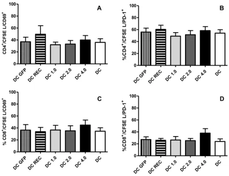
Fig. 4 – Mixed leukocyte reaction was carried out for 5 days, in a 1:10 DC:lymphocyte ratio.
Proliferation rates of T lymphocytes were expressed as percentage of proliferating T
lymphocytes (CFSE low events) within the (A) CD4+ population and (B) CD8+ population.
Data are presented as mean ± SEM of five independent assays (C) viability of lymphocytes
cultured with DC-4.0 in comparison with DC; **p=0.0015.
DC GF P
DC R EC
DC 1. 0
DC 2. 0
DC 4. 0 DC 0 10 20 30 40 50 % C D 4
+ p
ro lif er at in g ce lls A B
DC GF
P
DC R
EC
DC 1.
0
DC 2.
0
DC 4.
0 DC 0 10 20 30 40 50 % CD 8
+ p
ro lif er at in g ce lls