Evaluation of cytokines in type 1 diabetes patients
with and without retinopathy
Avaliação de citocinas em pacientes diabéticos tipo 1 com e sem retinopatia
Jéssica F. Cardoso1; Karina B. Gomes2; Ana Paula Fernandes2; Caroline P. Domingueti1
1. Universidade Federal de São João del-Rei (UFSJ), Minas Gerais, Brazil. 2. Universidade Federal de Minas Gerais (UFMG), Minas Gerais, Brazil.
First submission on 13/09/16; last submission on 16/12/16; accepted for publication on 28/12/16; published on 20/02/17
ABSTRACT
Introduction: Diabetes mellitus is one of the greatest public health problems in the world; the numerous complications of this disease are
the focus of great concern, as they are related to high morbidity and mortality. Among the complications of diabetes, diabetic retinopathy is the most common microvascular complication. Objectives: Evaluate the association of plasma levels of inlammatory cytokines interleukin-6 (IL-6), tumor necrosis factor-alpha (TNF-α), and interferon-gamma (IFN-γ), and anti-inlammatory cytokine interleukin-10 (IL-10) with diabetic retinopathy and the correlation of plasma levels of these cytokines together. Material and method: 125 patients with type 1 diabetes mellitus were included in this study – 55 with retinopathy and 70 without retinopathy. Cytokines IL-6, TNF-α, IFN-γ and IL-10 were determined by low cytometry. Results and discussion: A signiicant association of the levels of cytokines IL-6, TNF-α, IFN-γ and IL-10 with retinopathy was not observed, possibly because measurements were performed on plasma samples, but not on vitreous humor. However, a positive correlation of cytokines among themselves (p < 0.001) was observed. Conclusion: Cytokines IL-6, TNF-α, IFN-γ and IL-10 presented a positive correlation among themselves, what suggests that they may act together in the development and progression of vascular complications in diabetes patients.
Key words: diabetes mellitus type 1; diabetic retinopathy; cytokines; inlammation.
INTRODUCTION
According to the International Diabetes Federation (IDF), the prevalence of diabetes mellitus (DM) in 2014 was 387 million, and an increase of around 205 million new cases is estimated to occur by 2035(1). Its prevalence in Brazil can be compared
with that in developed countries, where DM is considered one of the greatest public health problems(2). The high prevalence is a
cause of great concern because of the numerous complications
resulting from this disease(3). In our country, DM is one of the
10 main causes of mortality. Besides, its morbidity is associated with elevated health care costs, impaired working capacity, and reduced life expectancy of individuals affected by this disease, what characterizes a huge socioeconomic impact(4, 5).
DM type 1 (DM1) represents approximately 5%-10% of cases. In spite of the shorter frequency, it is the main form to affect children and adolescents. This type of DM is associated
with deiciency in insulin production due to destruction of pancreatic beta cells, what can be measured by the action of autoantibodies or can be attributed to unknown causes, in which there is no evidence of autoimmune action(6).
Uncontrolled hyperglycemia is considered the major precipitating factor of severe chronic complications, with DM being one of the leading causes of cardiovascular diseases, blindness, kidney failure and lower-limb amputation. In addition, DM patients are predisposed to the development of certain infections(3). Among the chronic complications,
diabetic retinopathy (DR) is the most common microvascular complication. Approximately 90% of DM1 patients are estimated to present some degree of DR 20 years after the onset of the disease; among these, 60% will develop the most severe form of DR, called proliferative diabetic retinopathy (PDR)(7, 8). DR
is considered the main cause of blindness in individuals aged 20-74 years; around 12% of new cases of reduced visual acuity that impairs the conduction of routine activities are attributed
to DR. However, when diagnosis and treatment are carried out in a timely manner, that is, before irreversible alterations occur, the risk of blindness due to DR is reduced to nearly 5%(9).
Studies show that alterations in four distinct biochemical pathways are involved in the pathogenesis of DR. As a group, these alterations are responsible for causing oxidative stress that triggers the inlammatory process, which is intimately related to the pathophysiology of DR(10). Some studies have shown that
inlammation, and more speciically inlammatory cytokines, are important factors in the development of microvascular complications of DM, including retinopathy, neuropathy and nephropathy(11).
Cytokines are extracellular proteins of low molecular weight that serve as mediators of immune response. They act in highly complex pathways that regulate the inlammatory process, and they are necessary to conduct response to the lesion site. Low-grade chronic inlammation and activation of the innate immune system are known to be strongly related to the pathogenesis of DM(11).
Therefore, the study of the association of plasma levels of inlammatory cytokines with the presence of retinopathy in DM patients is important, as it can contribute to a better comprehension of the mechanisms responsible for the development of this microvascular complication, as well as to the discovery of novel therapeutic targets, what can be useful to ind new treatments to avoid the development of retinopathy and delay its progression.
OBJECTIVES
The present study was aimed at evaluating the association of plasma levels of inlammatory cytokines interferon-gamma (IFN-γ), interleukin-6 (IL-6), and tumor necrosis factor-alpha (TNF-α), and anti-inlammatory cytokine interleukin-10 (IL-10) with the presence of DR in DM1 patients, besides correlating plasma levels of these cytokines among themselves.
MATERIAL AND METHOD
This study was given an assent by the ethics research committee of Universidade Federal de Minas Gerais (UFMG) (CAAE – 0392.0.203.000-11) and by the ethics research committee of Santa Casa de Misericórdia de Belo Horizonte (006/2012). All participants were informed about the purposes
of the project, completed the baseline questionnaire, and signed the free informed consent.
One hundred twenty-ive adult individuals aged 18-60 years with clinical and laboratory diagnosis of DM1 participated in the study. Those who presented any of the following conditions were excluded: liver disease, alcohol abuse, coagulopathy, cancer, ongoing infectious process, hemodialysis, history of kidney transplant, and pregnancy.
Participants were classiied in two groups according to the presence or absence of retinopathy, which was evaluated by means of funduscopy – this piece of information was obtained from the analysis of patients’ records.
The clinical and biochemical parameters were obtained from the analysis of patients’ clinical charts, which were completed based on information given by patients and conirmed and/or complemented by analysis of medical records. Clinical and biochemical data were compiled from a period of 1-6 months prior to collection date of blood samples for cytokine measurements. Cytokines IFN-γ, IL-6, IL-10 and TNF-α were determined in plasma samples by low cytometry, using the Human Basic Kit Flow Cytomix (eBioscience®).
The produced results were analyzed with the statistical program SPSS 20.0. For the continuous variables, the normality test Shapiro-Wilk was used. For the variables that presented normal distribution, mean and standard deviation were calculated, and Student’s t test was employed for comparison between both groups. For the variables that did not present normal distribution, median and 25%-75% percentile were calculated, and Mann-Whitney U test was used for comparison between both groups. The categorical variables were presented as absolute and relative frequency, and the chi-square test was adopted for comparison among these variables. The Pearson method was used to investigate correlation among the assessed parameters. A value of p ≤ 0.05 was considered signiicant.
RESULTS
Table 1 describes the characteristics of 125 DM1 patients, grouped according to the presence or absence of DR. Patients with DR presented older age (p = 0.001) and longer duration
of diabetes (p < 0.001) than those without DR. Aside from that,
DR patients presented levels of urea (p < 0.001), creatinine
(p = 0.001), albuminuria (p < 0.001) and cystatin C (p < 0.001) higher than those without DR, besides glomerular iltration rate
neuropathy (p = 0.01) was observed in the group of DR patients. Regarding medication, there was higher frequency in the use of
statins (p = 0.001), anti-hypertensive agents (p < 0.001), and
acetylsalicylic acid (ASA) (p = 0.001) by DR patients. Statistically signiicant differences were not veriied for the variables sex, body mass index (BMI), glucose, glycated hemoglobin (HbA1c), aspartate aminotransferase (AST), alanine aminotransferase (ALT), albumin, family history of diabetes, tobacco smoking, physical activity and use of thyroxine.
The correlations of plasma levels of inlammatory cytokines among themselves in DM1 patients without retinopathy, with retinopathy, with neuropathy and with nephropathy are presented in Tables 2, 3, 4, and 5, respectively. In the patients without retinopathy, all cytokines presented signiicant correlation among themselves (p < 0.01). A similar result was observed in patients with nephropathy (p < 0.05 for the
correlation between IFN-γ and IL-6, and p < 0.01 for the other correlations). In the patients with retinopathy and those with neuropathy, all cytokines presented signiicant correlation
among themselves (p < 0.01), except the correlation between IFN-γ and IL-6, which was not signiicant.
Table 6 presents the plasma levels of cytokines IFN-γ, IL-6, IL-10 and TNF-α of patients with and without DR, but statistically signiicant differences were not found among these assessed parameters.
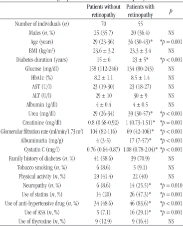
TABLE 1 − Characteristics of type 1 diabetes patients classiied according to presence or absence of retinopathy
Patients without retinopathy
Patients with retinopathy p
Number of individuals (n) 70 55 Males (n, %) 25 (35.7) 20 (36.4) NS
Age (years) 29 (23-36) 36 (30-43)* *p = 0.001 BMI (kg/m2) 23.6 ± 3.2 23.3 ± 3.4 NS
Diabetes duration (years) 15 ± 6 23 ± 5* *p < 0.001 Glucose (mg/dl) 158 (112-246) 134 (80-243) NS
HbA1c (%) 8.2 ± 1.1 8.5 ± 1.4 NS AST (U/l) 23 (19-30) 23 (18-27) NS ALT (U/l) 29 ± 10 30 ± 9 NS Albumin (g/dl) 4 ± 0.4 4 ± 0.5 NS
Urea (mg/dl) 29 (26-34) 39 (30-57)* *p < 0.001 Creatinine (mg/dl) 0.8 (0.68-0.92) 1 (0.73-1.51)* *p = 0.001 Glomerular iltration rate (ml/min/1.73 m2) 104 (82-116) 69 (42-106)* *p < 0.001
Albuminuria (mg/g) 4 (3-5) 17 (7-57)* *p < 0.001 Cystatin C (mg/l) 0.76 (0.64-0.87) 1.08 (0.78-2.04)* *p < 0.001 Family history of diabetes (n, %) 41 (58.6) 39 (70.9) NS
Tobacco smoking (n, %) 6 (8.6) 5 (9.1) NS Physical activity (n, %) 29 (41.4) 22 (40) NS
Neuropathy (n, %) 6 (8.6) 14 (25.5)* *p = 0.010 Use of statins (n, %) 14 (20) 26 (47.3)* *p = 0.001 Use of anti-hypertensive drug (n, %) 34 (48.6) 46 (83.6)* *p < 0.001 Use of ASA (n, %) 5 (7.1) 16 (29.1)* *p = 0.001 Use of thyroxine (n, %) 9 (12.9) 9 (16.4) NS
Variables presenting normal distribution were expressed as mean ± standard deviation, and compared using Student’s t test. Variables not presenting normal distribution were expressed as median (25%-75% percentile), and compared using Mann-Whitney U test. Categorical variables were expressed as absolute and relative frequencies n (%), and compared using the chi-square test.
*p < 0.05 for patients with retinopathy compared with patients without retinopathy. NS: not significant; BMI: body mass index; AST: aspartate aminotransferase; ALT: alanine transaminase; ASA: acetylsalicylic acid.
TABLE 2 − Correlation among inlammatory cytokines in type 1 diabetes patients without retinopathy
IFN-γ (pg/ml) IL-6 (pg/ml) IL-10 (pg/ml) TNF-α (pg/ml)
IFN-γ (pg/ml) - - -
-IL-6 (pg/ml) 0.757** - - -IL-10 (pg/ml) 0.826** 0.602** - -TNF-α (pg/ml) 0.54** 0.461** 0.686**
-Correlations (R2) were evaluated using the Pearson method.
IFN-γ: interferon-gamma; IL: interleukin; TNF-α: tumor necrosis factor-alpha; **p < 0.01.
TABLE 3 − Correlation among inlammatory cytokines in type 1 diabetes patients with retinopathy
IFN-γ(pg/ml) IL-6 (pg/ml) IL-10 (pg/ml) TNF-α(pg/ml)
IFN-γ (pg/ml) - - -
-IL-6 (pg/ml) 0.256 - -
-IL-10 (pg/ml) 0.956** 0.373** - -TNF-α (pg/ml) 0.393** 0.477** 0.51**
-Correlations (R2) were evaluated using the Pearson method.
IFN-γ: interferon-gamma; IL: interleukin; TNF-α: tumor necrosis factor-alpha; **p < 0.01.
TABLE 4 − Correlation among inlammatory cytokines in type 1 diabetes patients with neuropathy
IFN-γ (pg/ml) IL-6 (pg/ml) IL-10 (pg/ml) TNF-α (pg/ml)
IFN-γ (pg/ml) - - -
-IL-6 (pg/ml) 0.41 - -
-IL-10 (pg/ml) 0.811** 0.732** - -TNF-α (pg/ml) 0.744** 0.713** 0.885**
-Correlations (R2) were evaluated using the Pearson method.
IFN-γ: interferon-gamma; IL: interleukin; TNF-α: tumor necrosis factor-alpha; **p < 0.01.
TABLE 5 − Correlation among inlammatory cytokines in type 1diabetes patients with nephropathy
IFN-γ (pg/ml) IL-6 (pg/ml) IL-10 (pg/ml) TNF-α (pg/ml)
IFN-γ (pg/ml) - - -
-IL-6 (pg/ml) 0.312* - -
-IL-10 (pg/ml) 0.939** 0.406** - -TNF-α (pg/ml) 0.412** 0.492** 0.535**
-Nephropathy was defined by the presence of albuminuria ≥ 30 mg/g and/or glomerular filtration rate < 60 ml/min/1.73 m2. Correlations (R2) were evaluated using the Pearson method.
DISCUSSION
DR patients present older age (p = 0.001) and longer duration of DM (p < 0.001) than those without DR. According
to Esteves et al. (2008), duration of DM is an important
factor associated with DR. In DM1, whose onset may be easily identiied, it is estimated that after around 20 years of disease, almost all DM1 patients manifest some degree of DR(12, 13).
Moreover, DR patients present levels of urea (p < 0.001), creatinine (p = 0.001), albuminuria (p < 0.001), and cystatin C
(p < 0.001) higher than patients without DR, and a glomerular
iltration rate (p < 0.001) lower than patients without DR, what indicates association of DR with diabetic nephropathy, described in the literature. Jawa et al. (2004) observed that DM1 patients that presented nephropathy in most cases display other signs of microvascular complications (neuropathy and retinopathy), with the presence of advanced retinopathy almost always associated with histological alterations in renal glomeruli, and also with increased urinary protein excretion; increased albuminuria, urea, and plasma creatinine are strong indicators of the presence of retinopathy(14, 15).
The group of patients with DR presents higher frequency of neuropathy (p = 0.01), one of the microvascular complications of DM, intimately related to patient’s age, duration of diabetes, and glycemic control. Commonly, the presence of diabetic neuropathy is associated with other microvascular complications, such as retinopathy and nephropathy(16, 17).
Higher frequency of the use of statins (p = 0.001), anti-hypertensive medication (p < 0,001), and ASA (p = 0.001) by patients that present DR was observed. Statin is a medicine used to treat hypercholesterolemia, considered a risk factor for DR(18). Hypertension is also considered a risk factor for
the development of DM complications. Several studies have
demonstrated its strong association with the development and progression of DR(17, 19, 20). ASA is classiied as an antiplatelet
drug, often used clinically at the dose of 75 mg/day by patients that present high risk for cardiovascular disease (hypertension, diabetes, dyslipidemia, tobacco use, and family history of cardiovascular diseases) to reduce the risk of cardiovascular events(21).
Statistically signiicant associations were not observed between retinopathy and the levels of glucose, HbA1c, and plasma albumin. A possible reason for the divergent results found in the present study is the fact that assessment of HbA1c was not done over the years in the DM1 patients of this study: it is important to consider the possibility that patients with DR can have presented high levels of HbA1c in years preceding DR diagnosis. Concerning the plasma levels of albumin, a plausible explanation is that albuminuria in patients with DR is slightly to moderately increased. This picture can have a signiicant association with DR, but it was not enough to cause reduced plasma levels of albumin.
DM1 is deined as an inlammatory disease of pancreatic islets, in which beta cells are destroyed by autoantibodies, cytotoxic T cells, and inlammatory mediators(22). Histologically,
DM1 is represented by a lymphocytic mononuclear inlammatory iniltrate (insulitis) and by the absence of pancreatic beta cells. When migration of inlammatory cells characteristic of the insulitis process occurs, there is the important participation of several cytokines, especially the pro-inlammatory ones, such as TNF-α, IFN-γ and interleukin-1 beta (IL-1β),which favor the process of autoimmune and inlammatory response typical of DM1(23). As stated by Dogan et al. (2006)(24), it is possible to
detect high levels of inlammatory markers in newly diagnosed DM1 patients, what suggests that systemic inlammation is related to the onset of the disease. However, the levels of these markers are known to remain high during DM progression, and they can possibly contribute to the development of complications.
Reis et al. (2012)(25) analyzed plasma samples of 42 DM1
patients and 24 healthy patients as a control group, inding higher circulating levels of IL-6 in DM1 patients than in the control group. He et al. (2014)(26) carried out a study with
35 children with DM1 and 30 healthy individuals, in which levels of cytokines (IL-1, IL-6, IL-10, IL-12 and TNF-α) were investigated. The authors observed levels of cytokines higher in children with DM1 than in the control group.
IL-10 is an anti-inlammatory cytokine that seems to act trying to block DM evolution(27). Its increase in DM1 patients
TABLE 6 − Cytokines of type 1 diabetes patients classiied according to the presence or absence of retinopathy
Patients without retinopathy Patients with retinopathy p
IFN-γ (pg/ml) 98.4 ± 23.2 104.8 ± 29.8 NS IL-6 (pg/ml) 16.4 ± 2.6 16.3 ± 2 NS IL-10 (pg/ml) 1,166 ± 215.6 1,154.5 ± 214.5 NS TNF-α (pg/ml) 169.1 ± 101.9 175.8 ± 94.9 NS
Variables were expressed as mean ± standard deviation, and compared by means of Student’s t test.
can be resulting from a compensatory mechanism to the increase of pro-inlammatory cytokines. For this reason, the cytokines involved in the pathogenesis of DM1 are important for the development of the disease, acting both in the positive control (pro-inlammatory cytokines) and in the negative control (anti-inlammatory cytokines) of the inlammatory process, and they can act in a cascade or alone(25, 26, 28).
Thus, the results obtained in the present study are in agreement with the literature, which conirms the relationship of plasma levels of cytokines among themselves in DM1 patients without retinopathy, and also in those with DM complications, such as retinopathy, neuropathy, and nephropathy. Therefore, it is possible to suggest that cytokines can act in group, promoting a subclinical inlammatory state in DM1 patients, which can contribute to the development and the progression of DM complications.
There was no signiicant association of plasma levels of inlammatory cytokines IL-6, TNF-α, IFN-γ and IL-10 with DR in DM1 patients. Prolonged hyperglycemia is known to cause several biochemical alterations related to this disease, such as the alteration in the gene expression, non-enzymatic glycosylation of proteins, the increased oxidative stress that leads to an excess of oxidation end products, and, at last, iniltration of inlammatory cells followed by cytokine production in the diabetic retina(29). Accordingly, some authors characterize DR
as a low-level chronic inlammation, with the presence of inlammatory cytokines, and elevated numbers of circulating activated leukocytes(30, 31).
In the present study, cytokine levels did not show correlation with the presence of DR. A possible explanation for this fact is that cytokine levels can increase predominantly in retina, at a speciic site, presenting a systemic small increase in blood circulation in DR patients. This hypothesis can be conirmed by the result of a study conducted by Cheung et al.
(2012)(32), in which the authors assessed levels of different
cytokines in samples of vitreous/aqueous humor, and found association between levels of IL-6 and DR. Besides, some authors demonstrated that cytokine levels are higher in the aqueous humor than in the plasma of patients with DR(33, 34),
so justifying the results found in the present study.
Despite the relevant results, this work presented some limitations: its design is cross-sectional, what did not enable patients’ follow-up over the years; data collection based on completion of the baseline questionnaire by patients, which were conirmed by medical records, did not prove adequate, presenting some incomplete pieces of information, what
precluded the analysis between patients with non-proliferative DR (NPDR) and proliferative DR (PDR); and the fact that the used sample was plasma, not vitreous humor.
CONCLUSION
It has been suggested that plasma levels of inlammatory cytokines play a fundamental role in DR, contributing to the development of a low-grade chronic inlammatory state, which can promote the emergence and the aggravation of lesions in the retina. In the present work, association of cytokines IL-6, IL-10, IFN-γ and TNF-α with DR was not observed, possibly because the used samples were plasma, not aqueous/vitreous humor, what suggests the hypothesis that the increased levels of these inlammatory mediators occur predominantly at the local level, in the retina, during the development of this complication. However, positive correlations were observed of the assessed cytokines among themselves in patients without retinopathy and in those with DM complications, such as retinopathy, neuropathy, and nephropathy, suggesting that they can act as a group in the development and progression of microvascular and neuropathic complications of patients with diabetes. Also it was not possible to evaluate association of cytokines with the different DR stages, but it would be very interesting to verify if plasma measurement of cytokines can be useful for prognosis and monitoring of DR evolution. Therefore, it is necessary that further studies be conducted in this area, aimed at better comprehension of mechanisms involved in development and progression of DR, which can be useful for the discovery of new laboratory markers and the development of more effective treatments, to avoid that this complication develops or to retard its progression.
ACKNOWLEDGEMENTS
The authors thank Fundação de Amparo à Pesquisa do Estado de Minas Gerais (Fapemig), Conselho Nacional de Desenvolvimento Cientíico e Tecnológico (CNPq), and Comissão de Aperfeiçoamento de Pessoal do Nível Superior (Capes).
CONFLICT OF INTEREST
RESUMO
Introdução: O diabetes mellitus é considerado um dos maiores problemas de saúde pública no mundo; as numerosas complicações decorrentes dessa patologia são foco de grande preocupação, pois estão relacionadas com elevadas morbidade e mortalidade. Entre as complicações decorrentes do diabetes, a retinopatia diabética é considerada a complicação microvascular mais comum. Objetivos: Avaliar a associação dos níveis plasmáticos das citocinas inflamatórias interleucina 6 (IL-6), fator de necrose tumoral alfa (TNF-α) e interferon gama (IFN-γ) e da citocina anti-inflamatória interleucina 10 (IL-10) com a retinopatia diabética, bem como a correlação dos níveis plasmáticos dessas citocinas entre si. Material e método: Foram incluídos no estudo 125 pacientes diabéticos tipo 1, sendo 55 portadores de retinopatia e 70 sem retinopatia. As citocinas IL-6, TNF-α, IFN-γ e IL-10 foram determinadas pelo método de citometria de fluxo. Resultados e discussão: Não foi observada associação significativa dos níveis das citocinas IL-6, TNF-α, IFN-γ e IL-10 com a retinopatia, possivelmente devido às dosagens terem sido realizadas em amostras de plasma e não de humor vítreo/aquoso. Porém, foi observada correlação positiva das citocinas entre si (p < 0,001). Conclusão: As citocinas IL-6, TNF-α, IFN-γ e IL-10 apresentaram correlação positiva entre si, o que sugere que elas podem atuar em conjunto no desenvolvimento e na progressão das complicações vasculares nos pacientes diabéticos.
Unitermos: diabetes mellitus tipo 1; retinopatia diabética; citocinas; inflamação.
REFERENCES
1. International Diabetes Federation. IDF Diabetes Atlas. 7th edition. Brussels, Belgium: International Diabetes Federation; 2015.
2. Malerbi DA, Franco LJ. Multicenter study of the prevalence of diabetes mellitus and impaired glucose tolerance in the urban Brazilian population aged 30-69 yr. Diabetes Care. 1992; 15(11): 1509-15. 3. King GL. The role of inlammatory cytokines in diabetes and its complications. J Periondontal. 2008; 79(8): 1527-34.
4. Batista MCR, Priore SE, Rosado LEFPL, Tinôco ALA, Franceschini SCC. Avaliação dos resultados da atenção multiproissional sobre o controle glicêmico, peril lipídico e estado nutricional de diabéticos atendidos em nível primário. Rev Nutr. 2005; 18(2): 219-28.
5. Bosco A, Lerário AC, Soriano D, et al. Retinopatia diabética. Arq Bras Endocrinol Metab. 2005; 49(2): 217-27.
6. American Diabetes Association. Standards of medical care in diabetes. Diabetes Care. 2016; 39: S4-5.
7. Tarr MJ, Kaul K, Chopra M, Kohner EM, Chibber R. Pathophysiology of diabetic retinopathy. ISRN Ophthalmology. 2013; 2013: 13.
8. Fong DS, Aiello LP, Ferris FL, Klein R. Diabetic retinopathy. Diabetes Care. 2004; 27(10): 2540-53.
9. SBD. Sociedade Brasileira de Diabetes. Diretrizes da Sociedade Brasileira de Diabetes (2016-2015). São Paulo: A. Araújo Silva Farmacêutica; 2016.
10. Rangasamy S, McGuire PG, Das A. Diabetic retinopathy and inlammation: novel therapeutic targets. Middle East Afr J Ophthalmol. 2012; 19(1): 52-9.
11. Navarro-González FJ, Mora-Fernández C. The role of inlammatory cytokines in diabetic nephropathy. J Am Soc Nephrol. 2008; 19(3): 433-42.
12. Boelter MC, Azevedo MJ, Gross JL, Lavinsky J. Fatores de risco para retinopatia diabética. Arq Bras Oftalmol. 2003; 66(2): 239-47.
13. Esteves J, Laranjeira AF, Roggia FM, et al. Fatores de risco para a retinopatia diabética. Arq Bras Endocrinol Metab. 2008; 52(3): 431-41. 14. Jawa A, Kcomt J, Fonseca AV. Diabetic nephropathy and retinopathy. Med Clin N Am. 2004; 88(4): 1001-36.
15. Parving HH, Hommel E, Mathiesen E, et al. Prevalence of microalbuminuria, arterial hypertension, retinopathy and neuropathy in patients with insulin dependent diabetes. BMJ. 1988; 296(6616): 156-60. 16. Chen HT, Lin HD, Won JG, et al. Cardiovascular autonomic neuropathy, autonomic symptoms and diabetic complications in 674 type 2 diabetes. Diabetes Res Clin Pract. 2008; 82(2): 282-90.
17. Almeida FK, Esteves JF, Gross JL, Biavatti K, Rodrigues TC. Formas graves de retinopatia predizem aterosclerose subclínica em indivíduos com diabetes tipo 1. Arq Bras Cardiol. 2011; 97(4 S2): 346-9.
18. Alves C, Veiga S, Souza T. Dislipidemia e risco de doença cardiovascular em crianças e adolescentes com diabetes melito tipo 1. Rev Paul Pediatria. 2007; 25(1): 82-9.
19. Jannuzzi FF, Cintra FA, Rodrigues RCM, São-João TM, Gallani MCBJ. Adesão medicamentosa e qualidade de vida em idosos com retinopatia diabética. Rev Latino-Am Enfermagem. 2014; 22(6): 902-10.
20. Rodrigues TC, Pecis M, Canani LH, et al. Caracterização de paciente com diabetes mellitus tipo 1 do sul do Brasil: complicações crônicas e fatores associados. Rev Assoc Med Bras. 2010; 56(1): 67-73.
21. Rosito AG, Kunchenbecker R, Berwanger O, Barros E. Terapêutica cardiovascular: das evidências para a prática clínica. Porto Alegre: Artmed Editora; 2007. 524 p.
23. Sesterheim P, Saitovitch D, Staub HL. Diabetes mellitus tipo 1: multifatores que conferem suscetibilidade à patogenia autoimune. Scientia Medica. 2007; 17(4): 212-7.
24. Dogan Y, Akarsu S, Ustundag B, et al. Serum IL-1 beta, IL-2, and IL-6 in insulin-dependent diabetic children. Mediators Inlamm. 2006; 2006(1): 29206.
25. Reis JS, Amaral CAV, Valp CMO, et al. Oxidative stress and interleukin-6 secretion during the progression of type 1 diabetes. Arq Bras Endocrinol Metab. 2012; 56(7): 56-7.
26. He JS, Xie PS, Luo DS, Sun CJ, Zhang YG, Liu FX. Role of immune dysfunction in pathogenesis of type 1 diabetes mellitus in children. Asian Pac J Trop Med. 2014; 7(10): 823-6.
27. Balda CA, Silva AP. Aspectos imunológicos do diabetes melito tipo 1. Rev Assoc Med Bras. 1999; 45(2): 175-80.
28. Ozer G, Teker Z, Cetiner S, et al. Serum IL-1, IL-2, TNF alpha and INF gamma levels of patients with type 1 diabetes mellitus and their siblings. J Pediatr Endocrinol Metab. 2003; 16(2): 203-10.
29. Motta MMS, Coblentz J, Melo LGN. Aspectos atuais na isiopatologia do edema macular diabético. Rev Bras Oftalmol. 2008; 67(1): 45-9. 30. Kowluru RA, Zhong Q, Kanwar M. Metabolic memory and diabetic retinopathy: role of inlammatory mediators in retinal pericytes. Exp Eye Res. 2010; 90(5): 617-23.
31. Lutty GA. Effects of diabetes on the eye. Invest Ophthalmol Vis Sci. 2013; 54(14): ORSF81-7.
32. Cheung CMG, Vania M, Ang M, Chee SP, Li J. Comparison of aqueous humor cytokine and chemokine levels in diabetic patients with and without retinopathy. Mol Vis. 2012; 18: 830-7.
33. Funatsu H, Yamashita H, Shimizu E, Kojima R, Hori S. Relationship between vascular endothelial growth factor and interleukin-6 in diabetic retinopathy. Retina. 2001; 21(5): 469-77.
34. Ohara K, Funatsu H, Kitano S, Hori S, Yamashita H. The role of cytokines in the pathogenesis of diabetic retinopathy. Nihon Ganka Gakkai Zasshi. 2001; 105(4): 213-7.
CORRESPONDING AUTHOR
Caroline Pereira Domingueti