_____________________________
*)Corresponding author: [email protected]
doi:
10.2298/SOS1201103Z
UDK 541.124-16
Formation of Magnetic Microstructure of the Nanosized
NiFe
2O
4Synthesized Via Solid-State Reaction
T. Žák
1, V.
Ć
osovi
ć
2*), A.
Ć
osovi
ć
3, B. David
1, N. Talijan
2, D. Živkovi
ć
41
Institute of Physics of Materials AS CR, v.v.i., Žižkova 22, CZ-616 62 Brno, Czech
Republic
2
Institute of Chemistry, Technology and Metallurgy, University of Belgrade,
Njegoševa 12, 11000 Belgrade, Serbia
3
Institute for Technology of Nuclear and Other Mineral Raw Materials, Franse d’
Eperea 86, 11000 Belgrade, Serbia
4
Technical Faculty in Bor, University of Belgrade, Vojske Jugoslavije 12, 19210 Bor,
Serbia
Abstract:
Magnetic NiFe2O4 structure formation was studied through structural, compositional and magnetic characterization of obtained reaction products of a simple, high yielding and low-cost solid-state reaction. Initial annealing of the starting oxides mixture at 700oC did not allow us to observe formation of the desired magnetic phase. In contrast, subsequent thermomagnetic measurements up to 800oC indicated the considerable increase of the magnetic moment, which can be reasonably assigned to the changes in phase composition and formation of magnetic NiFe2O4 structure during the heating cycle of measurements. Nanosized NiFe2O4 phase formation has been confirmed by the following XRD and MS phase analyses and its nanocrystalline structure by XRD and SEM/TEM techniques. The obtained hysteresis loop taken after TM measurements suggest the increased volume of magnetically active material and thus additionally support the previous findings.
Keywords: NiFe2O4 nanoparticles, Solid-state reaction, Phase composition, Magnetic properties
1. Introduction
The soft or low coercivity ferrites are the most widely used group of magnetic materials utilized by electrical, electronic, information and home appliance industry, thus representing very important basic functional materials. Essentially, they are non-conductive ceramic materials based on iron oxides as well as nickel, zinc, and/or manganese compounds [1]. Due to low coercivity, their magnetization can be easily reversed without significant dissipation of energy. At the same time, their high electrical resistivity facilitates fairly low energy losses at high frequencies. Hence, they are extensively used in such applications where reduction of the different losses accompanying high-frequencies is more important than the static magnetic properties [2].
ferrites like nickel ferrite (NiFe2O4), a well-known ferrimagnetic material, have in fact an
inverse spinel structure. This structure is represented by the formula (Fe+3)A(Ni+2Fe+3)BO4 in
which half the atoms of iron occupy the tetrahedral (A) sites and the other half together with magnetic atoms occupy the octahedral (B) sites [3,4]. Mixed spinel structure with the certain degree of inversion is common as well.
The synthesis of nanosized NiFe2O4 has been studied by many investigators [5-8].
Accordingly, there are various, both wet and solid-state, synthesis techniques available e.g. co-precipitation, sono-chemical precipitation, hydrothermal synthesis, spray drying, freeze-drying, pulsed wire discharge and high energy ball milling, to name a few [7-10]. As each of them is being characterized by different disadvantages ranging from low yield, impurity formation, extensive agglomeration to complicated synthesis schemes, investigation of alternative processing routes is still burning topic [8,11,12].
It is widely recognized that the properties of nanocrystalline ferrites are size dependent and known to be very sensitive to the applied synthesis route [13-15]. Previous studies found in literature [16,17] point to difference between metal oxides obtained by grinding of solid metallic salts with sodium hydroxide and those obtained via hydroxides in solution. It is suggested that during solid-state reaction hydroxides decompose by a strong heat of reaction to produce oxides. However, the usual drawback of the conventional solid-state reactions for the preparation of Ni-ferrites is that at high temperatures, formation of a polycrystalline ferrite compound with a large crystallite size is favored. Consequently, this leads to loss of their peculiar structural and magnetic properties [7]. Nevertheless, the undesired grain growth during the solid-state reaction can be suppressed by addition of different inhibitors such as sodium chloride [16,18].
Given that the magnetic properties of NiFe2O4 ferrite magnetic materials are directly
related to their structure and phase composition, formation of magnetic microstructure of the nanosized nickel ferrite synthesized via solid-state reaction was analyzed and discussed through structural, compositional and magnetic characterization of obtained reaction products.
2. Experimental
The investigated nickel ferrite material was prepared by a simple high yielding and low-cost solid-state reaction route, using analytical grade NiSO4·6H2O, Fe(NO3)3·9H2O,
NaOH and NaCl as precursors. Adopted synthesis route is modified molten sodium chloride route proposed by S. L. Darshane et al. [16] where sodium chloride is used as grain growth inhibitor. Solid precursors are mixed in the molar ratio of 1:2:8:10, respectively and ground together in an agate mortar for about 60 min. During the mixing process, exothermal reaction takes place yielding in mixture of nickel and ferrite oxides. In order to synthesize NiFe2O4
this mixture is subjected to calcination at 700oC for 4 h. Pure polycrystalline NiFe2O4
nanoparticles are obtained with subsequent crushing and washing of produced powder, with deionized water, to remove present sodium chloride.
Additional advantage of the adopted synthesis route is that the grain growth during the solid-state reaction is inhibited by the sodium chloride, given that it facilitates formation of the coating around the nanoparticles that prevents the aggregation of nuclei to large particles [18].
done against an α-iron foil. For the spectra fitting and decomposition, the “CONFIT” software package was used [19]. The thermomagnetic curves were measured on the EG&G vibrating sample magnetometer in the field of 4 kAm-1 in vacuum. The heating and cooling rate was 4 °Cmin-1 with 30 min. delay at maximum of 800°C. Method of interpretation of comparable thermomagnetic measurement (TM) could be found in previous investigations [20]. Morphology and structure of the obtained powder samples were analyzed by means of scanning electron microscopy (SEM) and transmission electron microscopy (TEM). Magnetic properties were measured on the temperature of the ambient on Vibrating sample magnetometer (VSM) with magnetic field strength of 800 kAm-1.
3. Results and Discussion
In line with the mechanism of NiFe2O4 formation proposed by Darshane et al. [16]
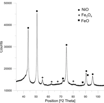
the mixture of nickel and ferrite oxides was firstly annealed at 700oC. The subsequent XRD analysis has revealed only presence of the oxides from the starting mixture confirming that nickel ferrite phase has not been formed (Fig.1). It could be that some of the processing conditions were not met and/or that the annealing time used in this study does not allow us to observe the formation of the desired magnetic phase.
Fig. 1. X -ray diffractogram of the sample annealed at 700oC
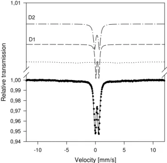
Further analysis of the phase composition using Mössbauer spectroscopy supported findings of the XRD analysis. Nevertheless, confirmation of the phase composition using MS is not so straight forward. Considering that the main characteristic of the 57Fe Mössbauer spectroscopy is that it can only “see” iron containing phases there was no possibility to gain any information about nickel oxide and metallic nickel in the analyzed sample. Consequently, the obtained MS spectrum (Fig. 2) contains only information regarding iron containing phases.
the spectrum are typical of Fe3O4 nanoparticles in the superparamagnetic state. Hence, the
doublet probably originated in the fast relaxation of the particle magnetic moment in the superparamagnetic state.
Velocity [mm/s]
-10 -5 0 5 10
Relati
ve transmission
0,94 0,95 0,96 0,97 0,98 0,99 1,00 1,01
D1 D2
Fig. 2. Mössbauer spectra of the powder sample annealed at 700oC
Furthermore, a quadrupole splitting QS = 0.84(2) mm/s and isomer shift IS = 0.29(1) mm/s for the second doublet (D2) in the spectrum are consistent with the parameters found in study of Saeed Kamali-M et al. [22] for superparamagnetic iron oxide nanoparticles. It is further stated that this doublet emanates from ferric iron in a non-spherical local surrounding, possibly coming from the rim of the iron oxide core.
Generally speaking, it is clear that the role of surface atoms or atoms on some irregular position rises with diminishing size of particles. The influence of surface (or interface) atoms on Mössbauer spectra is very known as well [23]. The broad wings in the spectrum suggest that there is some superposition of weak sextets near blocking temperature where both superparamagnetic and ferromagnetic interaction are present.
Taking into consideration reaction conditions used in similar solid state reaction [8] that suggests formation of NiFe2O4 phase at slightly higher annealing temperature,
themomagnetic measurements up to 800oC were utilized to observe desired phase formation. The temperature dependence of the total magnetic moment of the investigated powder sample at an applied field of 4 kAm-1 is presented on Fig. 3.
In regard to the determined phase composition of the studied starting material and that it is essentially nonmagnetized, during heating in the temperature range ambient temperature to 800 oC, no significant decrease of total magnetic moment can be observed. In contrast, cooling curve displays rapid and considerable increase of total magnetic moment with a decrease of temperature. This behaviour can be reasonably considered as a result of the formation of the optimal phase composition and formation of magnetic microstructure during heating cycle which essentially represents thermal treatment. Additionally, the observed increase of total magnetic moment during the subsequent cooling cycle can be attributed to “field cooling process” as well as overall decrease in thermal energy. Given that, starting from the magnetically disordered state, during cooling, magnetic moments tend to minimize energy against the external magnetic field and the final state results in an anisotropic moment distribution, appearing outside as an enlargement of a bulk magnetic moment.
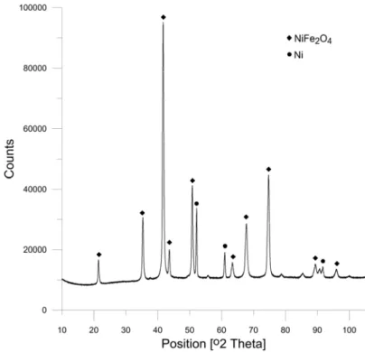
In order to gain insight into newly formed phase composition of the studied sample after TM, XRD analysis was used. The obtained X-ray diffractogram is given in Fig.4.
Fig. 4. X-ray diffractogram of the investigated Ni-ferrite powder after TM
According to obtained XRD results for the sample after TM measurements, it consists of 89 wt% NiFe2O4 phase and 11 wt% Ni, with their crystallite sizes being estimated to 25
and 45 nm, respectively. The presence of the Fe3O4 phase in the nickel ferrite sample was
that are specific only for Fe3O4 phase are in positions where only very weak signal or almost
none was measured. Hence, the presence of the Fe3O4 phase, if any, is practically negligible.
Phase composition of the obtained Ni-ferrite powder after TM was further analyzed using Mössbauer spectroscopy and the corresponding spectrum is presented in Fig.5. The obtained results indicate that about 60% of the iron atoms belong to the complete regular nickel ferrite structure. The rest is either on some “redundant” tetrahedral position or on position that is the not the full-value octahedral one as such atoms do not have the supposed number of nearest iron neighbors. Such result can be explained by taking into account the results of XRD analysis i.e. determined phase composition and corresponding crystallite sizes.
Velocity [mm/s]
-10 -5 0 5 10
Relative tr
ansmiss
ion
0,94 0,95 0,96 0,97 0,98 0,99 1,00 1,01
Fig. 5. Mössbauer spectra of the investigated Ni-ferrite magnetic material after TM
Studies found in literature suggest that the ratio of the tetrahedral (A) and octahedral (B) sites in the ultrafine samples may be somewhat different from that in the bulk materials [7]. It was found that the small particle size promotes a mixed spinel structure whereas bulk form is an inverse spinel [8]. Furthermore, in a similar study by Morrish and Hanada [24] of ultrafine NiFe2O4 particles with a crystallite size of ca. 25 nm, authors also pointed out the
possibility that the sample was either non-stoichiometric or not completely inverse, which can also be the case with the studied sample, given that presence of metallic nickel was determined by XRD. Limited Ni2+ substitution for Fe2+ as an outcome of the used solid-state reaction should be considered as well [7]. In line with the XRD results, MS phase analysis did not find presence of Fe3O4 phase.
Morphology and structure of the Ni-ferrite powder obtained after TM measurements up to 800oC was initially studied using SEM. The presented SEM image (Fig.6) illustrates presence of nanoscale NiFe2O4 particles that form much larger agglomerates which
Fig. 6. SEM image of the obtained Ni-ferrite powder after TM
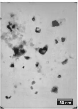
In order to gain better insight into the morphology, TEM analysis was utilized. TEM micrograph of the investigated magnetic powder (Fig.7) clearly shows individual nanoparticles of the nanocrystalline NiFe2O4. Moreover, the average particle size estimated
from the TEM image is consistent with the crystallite size determined by XRD.
Fig. 7. TEM micrograph of the obtained Ni-ferrite powder after TM
a) b)
Fig. 8. Hysteresis loops of the investigated powder samples a) annealed at 700oC and b) after thermomagnetic measurements up to 800oC
The presented hysteresis loops (Fig. 8) demonstrate substantial difference in the magnetic quality. As revealed by the structural analyses, the sample annealed at 700oC (Fig. 8a) shows superparamagnetic behavior with considerably small magnetization and no coercivity. Seeing that during the TM the structure changes into NiFe2O4, which is
ferrimagnetic, net magnetization and overall magnetic quality increase significantly (Fig. 8b) due to the increased volume of magnetically active material.
4. Conclusions
Nanosized NiFe2O4 magnetic material was prepared by a simple high yielding and
low-cost solid-state reaction route. Formation of magnetic microstructure was studied and discussed through structural, compositional and magnetic characterization of obtained reaction products. Initial annealing of the starting oxides mixture at 700oC did not allow us to observe formation of the desired magnetic phase. This could probably be because some of the processing conditions were not met and/or that the annealing time used in this study was not adequate. In contrast, the considerable increase of the magnetic moment with a decrease of temperature was observed on the obtained thermomagetic curve. It can be reasonably assigned to the changes in phase composition and formation of magnetic NiFe2O4 structure
during the heating cycle of measurements up to 800oC as well as to “field cooling process” and overall decrease in thermal energy during the following cooling cycle. NiFe2O4 phase
formation has been confirmed by the subsequent XRD and MS phase analyses and its nanocrystalline structure by XRD and SEM/TEM techniques. Increase of the net magnetization and overall magnetic quality illustrated by the obtained hysteresis loop taken after TM measurements suggest the increased volume of magnetically active material and thus additionally support the previous findings.
Acknowledgement
the Grant Agency of the Czech Republic. The presented work is carried out through joint scientific cooperation of the Serbian Academy of Sciences and Arts and the Academy of Sciences of the Czech Republic under project: Advanced Multicomponent Metal Systems and Nanostructured Materials with Diverse Functional Properties.
References
1. C.B.Carter, M.G Norton, Ceramic materials: science and engineering, Springer, New York, 2007.
2. K.H.J. Buschow, F.R. de Boer, Physics of magnetism and magnetic materials, Kluwer Academic/Plenum Press, New York, 2003.
3. G.A. Sawatzky, F. van der Woude, A.H. Morrish, J. Appl. Phys., 39 (1968) 1204. 4. D. Kedem, T. Rothem, Phys. Rev. Lett., 18 (1967) 165.
5. S. Komarneni, E. Fregeau, E. Breval, R. Roy, J. Am. Ceram. Soc., 71(1) (1988) C26. 6. T. Pannaprayil, R. Marande, S. Komarneni, S.G. Sankar, J. Appl. Phys., 64 (1988)
5641.
7. T. Kodama, Y. Wada, T. Yamamoto, M. Tsuji, Y. Tamamura, J. Mater. Chem., 5(9) (1995) 1413.
8. .A. Ceylan, S. Ozcan, C. Ni, S.I. Shah, J. Magn. Magn. Mater., 320 (2008) 857. 9. K.V.P.M. Shafi, Y. Koltypin, A. Gedanken, R. Prozorov, J. Balogh, J. Lendvai, I.
Felner, J. Phys. Chem. B, 101 (1997) 6409.
10. Y. Kinemuchi, K. Ishizaka, H. Suematsu, W. Jing, K. Yatsui, Thin Solid Films, 407 (2002) 109.
11. .A.S. Albuquerque, J.D. Ardisson, W.A.A. Macedo, J.L. López, R. Paniago, A.I.C. Persiano, J. Magn. Magn. Mater. 226-230 (2001) 1379.
12. V. Sepelak, D. Baabe, D. Mienert, D. Schultze, F. Krumeich, F.J. Litterstb, K.D. Becker, J. Magn. Magn. Mater. 257 (2003) 377.
13. S.N. Dolia, R. Sharma, M.P. Sharma, N.S. Saxena, Indian J. Pure Ap. Phy., 44 (2006) 774.
14. .A. Verma, T.C. Goel, R.C. Mediratta, Mater. Sci. Technol. 16 (2000) 712. 15. R.K. Tiwary, S.P. Narayan, O.P. Pandey, J. Min. Metall. B, 44B (2008) 91.
16. S.L. Darshane, S.S. Suryavanshi, I.S. Mulla, Ceramics International, 35 (2009) 1793. 17. X.R. Ye, D.Z. Jia, J.Q. Yu, X.Q. Xin, Z.L. Xue, Adv. Mater., 11 (1999) 941.
18. J.B.Wiley, R.B. Kaner, Science 255 (1992) 1093.
19. T. Žák, Y. Jirásková, Surf. Interface Anal., 38 (2006) 710.
20. N. Talijan, V. Ćosović, T. Žák, A. Grujić, J. Stajić-Trošić, J. Min. Metall. B,45B (1) (2009) 111.
21. G.F. Goya, Solid State Communications, 130 (2004) 783.
22. S. Kamali-M, T. Ericsson, R. Wäppling, Thin Solid Films, 515 (2006) 721.
23. J.-M. Grenèche and M. Miglierini, in Mössbauer Spectroscopy in Materials Science, edited by M. Miglierini and D. Petridis, Kluwer, Dordrecht, 1999.
24. .A.H. Morrish, K. Hanada, J. Appl. Phys., 52 (1981) 2496.
Садржај: И NiFe2O4
, .
И . П
700oC
. 800oC
.
NiFe2O4 ђ
(XRD) 57Fe Mössbauer
( Ѕ). NiFe2O4 ђ
XRD, SEM TEM .
, NiFe2O4
.