the Diagnosis of Alzheimer’s Disease
Carol Man Gao1., Alice Y. Yam1., Xuemei Wang1
, Erika Magdangal1, Cleo Salisbury1, David Peretz1, Ronald N. Zuckermann1¤, Michael D. Connolly1¤, Oskar Hansson3, Lennart Minthon3, Henrik Zetterberg2, Kaj Blennow2, Joseph P. Fedynyshyn1*, Sophie Allauzen1
1Research and Development, Novartis Vaccines and Diagnostics, Emeryville, California, United States of America,2Clinical Neurochemistry Laboratory, Department of Neuroscience and Physiology, Sahlgrenska University Hospital, Mo¨lndal, Sweden,3Clinical Memory Research Unit, Department of Clinical Sciences Malmo¨, Lund University, Malmo¨, Sweden
Abstract
Alzheimer’s Disease (AD) is the most prevalent form of dementia worldwide, yet the development of therapeutics has been hampered by the absence of suitable biomarkers to diagnose the disease in its early stages prior to the formation of amyloid plaques and the occurrence of irreversible neuronal damage. Since oligomeric Abspecies have been implicated in the pathophysiology of AD, we reasoned that they may correlate with the onset of disease. As such, we have developed a novel misfolded protein assay for the detection of soluble oligomers composed of Abx-40 and x-42 peptide (hereafter Ab40 and Ab42) from cerebrospinal fluid (CSF). Preliminary validation of this assay with 36 clinical samples demonstrated the presence of aggregated Ab40 in the CSF of AD patients. Together with measurements of total Ab42, diagnostic sensitivity and specificity greater than 95% and 90%, respectively, were achieved. Although larger sample populations will be needed to confirm this diagnostic sensitivity, our studies demonstrate a sensitive method of detecting circulating Ab40 oligomers from AD CSF and suggest that these oligomers could be a powerful new biomarker for the early detection of AD.
Citation:Gao CM, Yam AY, Wang X, Magdangal E, Salisbury C, et al. (2010) Ab40 Oligomers Identified as a Potential Biomarker for the Diagnosis of Alzheimer’s Disease. PLoS ONE 5(12): e15725. doi:10.1371/journal.pone.0015725
Editor:Sergio T. Ferreira, Federal University of Rio de Janeiro, Brazil
ReceivedAugust 2, 2010;AcceptedNovember 21, 2010;PublishedDecember 30, 2010
Copyright:ß2010 Gao et al. This is an open-access article distributed under the terms of the Creative Commons Attribution License, which permits unrestricted use, distribution, and reproduction in any medium, provided the original author and source are credited.
Funding:This work was funded by Novartis Vaccines and Diagnostics. Several of the authors are or were employed by the funder and, as such, the funders played a role in the study design, data collection and analysis, decision to publish, and preparation of the manuscript.
Competing Interests:Several of the authors are employed by Novartis Vaccines and Diagnostics and thus may have a conflict of interest. In addition, the submitted work is included in a pending patent application on amyloid-beta aggregates as biomarkers for Alzheimer’s Disease (patent#61/265,340, submitted by Novartis AG on Nov 30, 2009). This does not alter the authors’ adherence to all the PLoS ONE policies on sharing data and materials.
* E-mail: joseph.fedynyshyn@novartis.com
¤ Current address: Lawrence Berkeley National Laboratory, Molecular Foundry, Berkeley, California, United States of America
.These authors contributed equally to this work.
Introduction
Alzheimer’s Disease (AD) is a neurodegenerative disorder characterized by progressive memory loss and cognitive dysfunc-tion. It is the most prevalent form of dementia, estimated to affect 13 million people worldwide [1]. While the precise mechanism underlying the disease is not fully understood, the aggregation of amyloid beta (Ab) appears to play an important role [2–4]. Ab peptides of various lengths (typically 1–40 and 1–42) are cleavage products of the amyloid precursor protein that aggregate and form insoluble plaques in AD brains. Post mortem identification of these plaques together with neurofibrillary tangles and neuronal loss is currently the definitive and only fully accepted diagnostic confirmation of AD [5,6]. However, recent reports suggest that smaller, soluble Aboligomers are more likely to be the pathogenic agents of disease [3,4,7–10].
A growing number of in vitro generated oligomers of varied size and structure have been implicated in AD [4]. However, the actual identity of the oligomer participating in AD pathogenesis remains elusive. Its chemical composition is also poorly defined, although several lines of evidence suggest that AD-associated oligomers are primarily composed of Ab42 [3]. For instance, one
unifying feature of AD is the presence of Ab42-containing plaques in the brain parenchyma [11,12]. This suggests that any soluble oligomers would also be composed of Ab42. In addition, Ab42 appears to be more amyloidogenic than Ab40 and is found more frequently in plaques despite existing at much lower physiological concentrations [13]. Lastly, several presenilin mutations linked to familial forms of AD are known to increase production of Ab42 cleavage products [14], further implicating this Ab peptide in pathogenesis. Consequently, it is generally assumed that cytotoxic oligomers mediating AD are composed of Ab42 peptides.
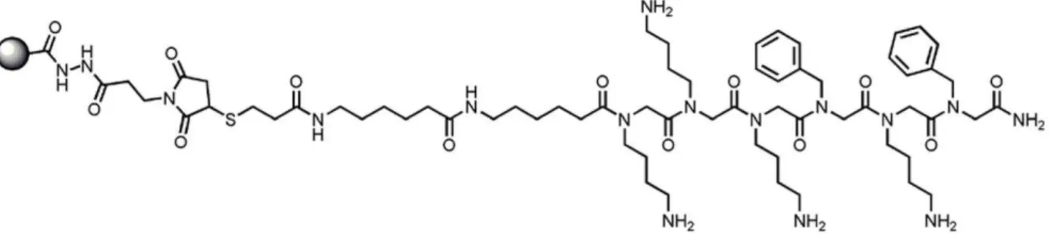
30 mg/mL) via maleimide chemistry (Figure 1). Control beads consisted of similarly conjugated glutathione molecules.
Ab42 aggregates from AD brain homogenate (ADBH) were captured with 3ml ASR1 beads for 1 hour at 37uC in 100ml 80%
plasma by spiking with 75 nl of 10% brain homogenate (Fig. 2C) or the indicated concentrations of Ab aggregates (Fig. 2D) in capture buffer (50 mM Tris, 150 mM NaCl, 1% Tween-20, 1% Triton X-100 pH 7.5). 10% ADBH was estimated to contain ,1 pg/nL Ab42 aggregates (data not shown). The beads were then washed with TBST (50 mM Tris, 150 mM NaCl, 0.05% Tween-20 pH 7.5), and bound proteins were eluted with 0.1 M NaOH at 80uC for 30 minutes and neutralized with 0.12 M NaH2PO4, 0.4% Tween-20. The eluted Ab was subsequently detected by an Ab42-specific ELISA, utilizing an Abx-42 specific capture antibody (12F4, Covance, Princeton, NJ) and HRP-conjugated 4G8 antibody (Covance) for detection. To test the conformational nature of ASR1 binding, ADBH was pretreated with 5.4 M guanidine thiocyanate for 30 minutes at room temperature prior to dilution into 80% plasma and MPA detection. AD and control brain samples were obtained from the tissue bank of the Swiss National Reference Centre of Prion Diseases (Zu¨rich, Switzerland).
In vitro Ab42 oligomers spiked into normal CSF and in vivo oligomers from clinical AD CSF were similarly detected by the MPA with a few modifications. 250ml 80% human CSF in
capture buffer was incubated with 30ml ASR1 beads. ASR1 beads
were subsequently washed with TBST and 1% Zwittergent 3-14 before Ab42 and Ab40 were eluted and detected in a multiplex format by MSD immunoassay (Meso Scale Discovery, Gaithers-burg, Maryland) according to the manufacturer’s instructions. In vitro Ab42 oligomers are estimated to be 12mers and were prepared as described [23]. Normal pooled CSF for oligomer spiking experiments was purchased from Analytical Biological Services Inc. (Wilmington, DE).
ELISA
ELISA plates were coated with 2mg/mL 12F4 antibody in
coating buffer (0.1 M NaH2PO4, 1% NaCl, pH 6), washed with TBST, and blocked with 1% BSA, 3% sucrose in TBS for 1 hour at 37uC. 0.2mg/mL HRP-conjugated 4G8 antibody in conjugate
diluent (0.1% BSA, 0.01% casein in PBS) was diluted 1:1 with the sample and applied to the 12F4-coated plates for 1 hour at room temperature before detection by a chemiluminescent substrate (Pierce Supersignal ELISA Femto Substrate, Thermo Fisher Scientific, Rockford, IL).
Patients and CSF sampling
The AD group consisted of 26 patients consulting the memory disorder clinic at Malmo¨ University Hospital, Sweden, mean age 71.867.3 years. All patients underwent physical, neurological and psychiatric examination, cognitive tests, careful clinical history and functional assessment. Patients diagnosed with AD had to meet the DSM-IIIR criteria of dementia [24] and the criteria of probable AD defined by NINCDS-ADRDA [25]. The AD patients were followed over time with repeated clinical evaluations, which increases the clinical diagnostic accuracy. The control group, in total 10 cases, mean age 69.469.7 years, was defined based on absence of memory complaints or any other cognitive symptoms, and no signs of active neurological or psychiatric disease. All patients and controls gave informed consent to participate in the study. The study was conducted according to the provisions of the Helsinki Declaration and was approved by the ethics committee of Lund University, Sweden. CSF was collected in polypropylene tubes, centrifuged, aliquoted, and stored at280uC pending analyses.
Statistical analysis
Statistical comparison of two populations was performed using two-tailed t-test using GraphPad Prism for Windows, v 5.01 (GraphPad Software, San Diego, CA). Receiver operating
characteristic curves (ROC) were generated using R (R Founda-tion for Statistical Computing, Vienna, Austria).
Results
An aggregate-specific reagent captures Abaggregates from AD brain homogenate in a conformation-dependent manner
We have developed an Aggregate Specific Reagent (ASR1, Figure 1) that preferentially binds aggregated proteins over monomeric proteins. ASR1 is a peptoid, a peptidomimetic containing N-substituted glycines [26], which have been shown to be resistant to proteolytic digestion from enzymes commonly found in body fluids [27]. The ASR1 sequence is derived from a PrP peptide that has a strong ability to capture aggregated PrP in solution [19]. ASR1 can capture picomolar amounts of insoluble aggregated PrP from a solution containing an excess of normally folded PrP (Figure S1 and [19]).
As amyloid aggregates are characterized by similar cross-beta sheet structure, they share conformational epitopes that are recognized by amyloid-binding molecules such as Congo Red and thioflavin T [15,16] as well as conformation-specific antibodies [17,18]. We hypothesized that ASR1 might also
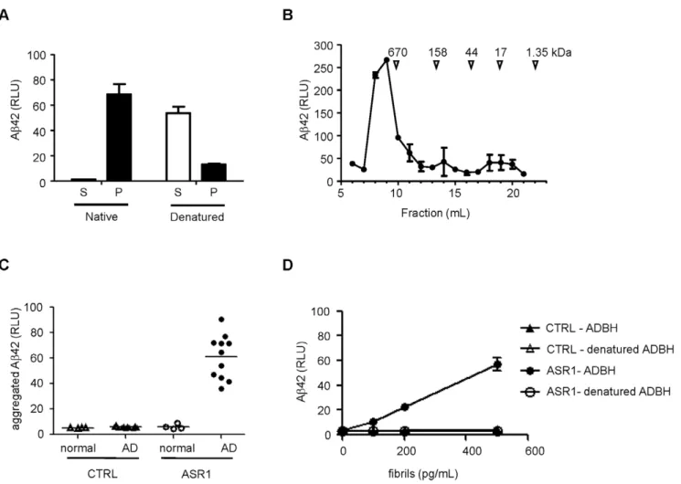
Figure 2. MPA detection of a conformational epitope in Abaggregates from AD brain homogenates (ADBH).A.ADBH was centrifuged at 134,000gfor 1 hr, and denatured Ab42 in the supernatant (S) and pellet (P) fractions was detected by ELISA.B.ADBH was fractionated by size exclusion chromatography and Ab42 was detected by ELISA.C.75 nl of normal (open symbols) or AD (closed symbols) brain homogenate was subjected to the MPA using control (triangles) or ASR1-coated (circles) beads.D.Abaggregates from an ADBH were examined by the MPA with (open symbols) or without (filled symbols) a pretreatment with 5.4 M guanidine thiocyanate using control (triangles) or ASR1-coated (circles) beads. Error bars represent the standard deviation of triplicate reactions.
doi:10.1371/journal.pone.0015725.g002
the aggregates with a chemical denaturant prior to the MPA abolished all binding (Figure 2D).
oligomers spiked into normal CSF, we asked whether the MPA could detect endogenous oligomers reported to be present in AD
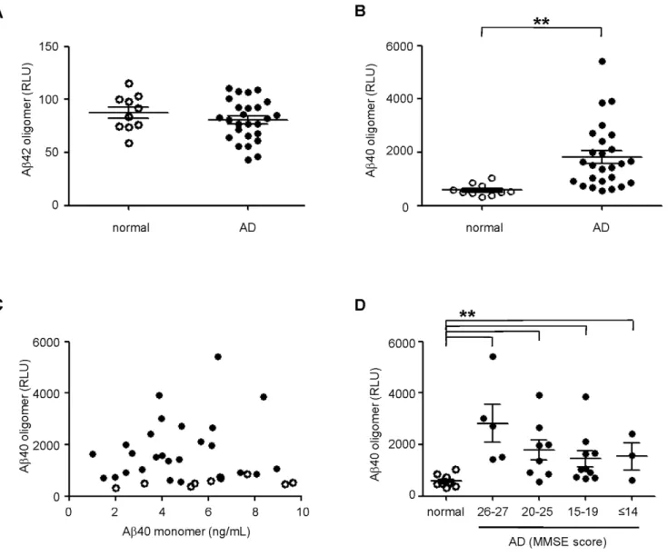
Figure 4. MPA detection of Ab40 oligomers from AD CSF.Normal (open circles) and AD (closed circles) CSF were analyzed by the MPA. Oligomers containing Ab42(A)and Ab40(B)were captured by ASR1 beads followed by detection using a multiplex immunoassay. Significant differences between normal and AD population were calculated by t-test (Ab42,p= 0.31; Ab40, **p,0.01).C.Lack of correlation between MPA-detected Ab40 signal and the concentration of Ab40 monomers in AD CSF (R2= 0.037).D.The distribution of Ab40 oligomers by disease severity was examined by categorization of AD patients according to MMSE scores (**p,0.01 between normal and all AD groups). The mean and standard error of the mean (SEM) are shown for all groups.
CSF [20–22]. CSF from 26 clinically diagnosed AD patients at varying stages of disease and 10 aged-matched controls were examined by the MPA. As before, CSF was incubated with ASR1 beads and the captured Ab was detected by a multiplex immunoassay specific for Ab40 and Ab42. Upon examination of captured Ab42, we did not detect any difference between AD and control populations (Figure 4A). However, we observed surpris-ingly clear and significant differences in the Ab40 signal between the two groups (Figure 4B). Importantly, Ab40 signals from the MPA did not correlate with the concentration of total Ab40 in the CSF (presumably this immunoassay format would bias the detection of total Ab toward monomeric species, so they are indicated as such henceforth). This suggests that the Ab40 signals were associated with specific MPA oligomeric capture and not nonspecific binding of Ab40 monomers to the capture beads (Figure 4C). When we further categorized the AD samples into groups of increasing disease severity based on clinical Mini-Mental State Examination (MMSE) scores, we found that the Ab40 oligomers were not only found in individuals with late-stage AD and low MMSE scores but also in patients with early stage AD and higher MMSE scores (Figure 4D). Therefore, using the MPA, we have identified Ab40 oligomers as a potential biomarker that could diagnose AD in the early stages of disease.
Ab40 oligomers are a novel biomarker for the diagnosis of AD
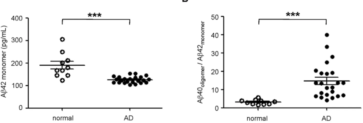
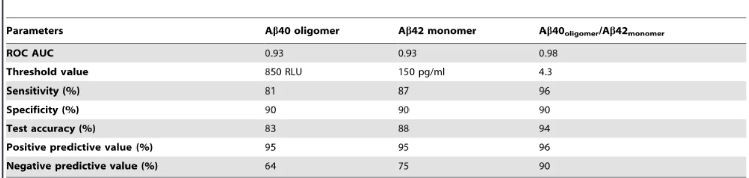
To validate our results against other investigated biomarkers for AD, we also measured the levels of monomeric Ab42 in this set of samples. As expected, Ab42 was significantly decreased in the CSF of AD patients relative to normal individuals (Figure 5A), consistent with previously published results [28]. Since low levels of monomeric Ab42 and high levels of oligomeric Ab40 are linked to AD, we combined the two biomarkers to strengthen their predictive value. The resulting ratio of oligomeric Ab40 to monomeric Ab42 indeed enhanced the differentiation of control and AD populations (Figure 5B). Additional ROC analysis demonstrated diagnostic sensitivity and specificity of 95% and 90%, respectively (Figure 6, Table 1). Positive and negative predictive value was estimated to be near 95% and 90%, respectively, whereas oligomeric Ab40 and monomeric Ab42 biomarkers alone had negative predictive values that were equal to or less than 75% (Table 1).
Discussion
AD is a growing epidemic that impacts nearly 50% of our elderly population greater than 85 years old [29]. However, the development of therapeutics to treat the disease has been hampered by the absence of suitable biomarkers to diagnose the disease. Currently, a clinical diagnosis of AD can only be confirmed with the identification of Ab plaques in postmortem brain tissue [5,6]. Premortem clinical diagnosis is also problematic as it relies heavily on subjective reporting and observed cognitive decline that can occur over a period of years. Since the first clinical signs of cognitive dysfunction may appear after significant neuronal loss has occurred, there is a compelling need for biomarkers that can diagnose early AD.
Because Aboligomers are suggested to play a key role in AD pathogenesis [3,4,7–10] and because they have been detected in
Figure 5. Synergistic combination of Ab40 oligomer signal with monomeric Ab42 concentration.A.The concentration of monomeric Ab42 was measured in normal (open circles) and AD (closed circles) CSF (***p,0.001).B.The ratio of Ab40 oligomers (RLU) and Ab42 concentration (pg/mL) was calculated and plotted for both normal (open circles) and AD (closed circles) populations (***p,0.001). Three AD samples were not included in this analysis because of insufficient sample to measure Ab42 concentrations. The mean and SEM are shown for all groups.
doi:10.1371/journal.pone.0015725.g005
Figure 6. Receiver operating characteristic (ROC) curve. ROC analysis was performed to compare the diagnostic value of 3 biomarkers: oligomeric Ab40 (– –), monomeric Ab42 (- - -), and the ratio Ab40oligomer/Ab42monomer(––).
diagnostically relevant body fluids [10,20–22], we reasoned that Ab oligomers could be a powerful AD biomarker predicting disease progression. However, the concentration of oligomers in CSF remains poorly defined and only a few studies estimate that they may exist at very low levels [21,22]. Furthermore, these studies did not distinguish between Ab40 and Ab42-containing oligomers since they utilized either antibodies recognizing oligomeric structure or oligomer-specific ELISAs that employed capture and detection antibodies recognizing the same amino-terminal epitopes. In this study, we report the development of a novel assay that specifically detected low concentrations of aggregated Abin a small population of AD CSF. Because AD is largely correlated with the accumulation of Ab42 in the brain, we expected to identify oligomers composed of Ab42. To our surprise, we found only an enrichment of Ab40-containing oligomers in AD CSF. These oligomers were observed at multiple stages of AD, suggesting that they could be a biomarker for early diagnosis of AD.
Because Ab42 oligomers are widely implicated in AD pathology, the correlation we observed of AD with Ab40 but not Ab42 oligomers was surprising. One explanation is that we detected oligomers associated with the vascular Ab40 deposits that are seen in patients with cerebral amyloid angiopathy (CAA). CAA is a pathological feature of AD characterized by the cerebrovas-cular deposition of Abpeptides with the major species being Ab40 [30,31] and has been estimated to impact greater than 90% of AD cases [32,33]. Therefore, our findings might reflect an increase of oligomeric Ab40 associated with these Abdeposits.
Another possibility is that Ab40 oligomers may be more soluble and reach CSF more readily than Ab42 oligomers which may rapidly aggregate and deposit in the brain parenchyma. Consequently, high levels of Ab40 oligomers in the CSF may be a surrogate marker of an amyloidogenic cascade in the brain.
A third explanation for our inability to identify Ab42 oligomers may be the sensitivity of our detection system. While both Ab40 and Ab42 containing oligomers may be present in CSF and captured by ASR1 beads, the subsequent detection of the constituent monomers may be limited by the sensitivity of the immunoassay (Mesoscale detection limits for Ab40 and Ab42 are 5 and 8 pg/mL, respectively). Furthermore, the concentration of Ab42 in CSF is approximately 10-fold lower than that of Ab40, suggesting that any Ab42-containing oligomers may exist at concentrations below our detection limit.
Although Ab40 aggregates have been documented in AD, the biological relevance of Ab40 oligomers in AD pathogenesis remains unclear. While nearly all cases of AD report Ab 42-containing plaques in the brain, approximately two thirds of them
also report Ab40 deposits [11], and significant levels of Ab40 have also been found in AD cortical brain tissue [34]. One interpretation of these results is that aggregation of the less soluble Ab42 would precede and recruit subsequent aggregation of Ab40, as has been previously suggested [12,35]. In support of this idea, purification of oligomers from AD brain tissues have isolated both Ab40, Ab42, and Ab40/42 heterodimers [8,36]. The impact of Ab40 oligomers on AD pathogenesis is unclear although some in vitro models suggest that Ab42 oligomers are significantly more cytotoxic [37]. Nevertheless, preparations of soluble Ab40 are sufficiently toxic to impact long term potentiation in cell culture systems and cognitive function when injected into mice [38].
Additional studies are clearly needed to understand the role of Ab40 oligomers in AD pathogenesis. Since oligomeric Ab is suggested to be a direct causative agent of AD, we believe they may be an ideal predictor of disease progression. Indeed, although the results do not reach significance, our data suggest that there could be an inverse correlation between oligomer concentration and disease severity (Figure 4D) similar to the decline that is observed in the Ab42 levels of AD patients. Future studies will include larger patient populations and prospective CSF sampling to screen for incipient AD. Additionally, if Ab oligomers are to become a relevant biomarker for AD, its predictive value must be measured in patients with mild cognitive impairment. Neverthe-less, our results showing the detection of Ab40 oligomers in CSF from patients in the initial stages of AD suggest that the MPA could be a sensitive assay for the early diagnosis of AD.
Supporting Information
Figure S1 Prion Protein (PrP) captured from plasma spiked with vCJD or normal brain homogenates.vCJD (closed circle) and normal (open circle) brain homogenates were spiked into normal human plasma at the indicated concentrations and subjected to the MPA. Captured prion protein was eluted and detected by a prion-specific ELISA. Materials and Methods: vCJD and normal 10% brain homogenates (w/v) (‘‘Blue’’ and ‘‘Clear’’ samples, respectively, from National Institute for Biological Standards and Control, United Kingdom) were spiked into normal human plasma (SeraCare Life Sciences, West Bridgewater, MA), after which 200ml of the solution was incubated with 50ml of 5x capture buffer and 9ml ASR1 beads for 1 hour at 37uC. The beads were washed and captured prion protein was subsequently eluted, denatured, and detected by sandwich ELISA [19]. The vCJD brain homogenate had an estimated 4mg/mL of aggregated PrP.
Acknowledgments
We thank Ruixiao Lu and Ping Shi for helpful discussions on statistical analysis and assistance in generation of the ROC curve, and Dr. Adriano Aguzzi for generously providing samples.
Author Contributions
Conceived and designed the experiments: CMG AYY SA JPF DP. Performed the experiments: CMG EM AYY. Analyzed the data: CMG EM AYY JPF. Contributed reagents/materials/analysis tools: XW CS RNZ MDC OH LM HZ KB. Wrote the paper: AYY.
References
1. Ferri CP, Prince M, Brayne C, Brodaty H, Fratiglioni L, et al. (2005) Global prevalence of dementia: a Delphi consensus study. Lancet 366: 2112–2117. 2. Stefani M, Dobson CM (2003) Protein aggregation and aggregate toxicity: new
insights into protein folding, misfolding diseases and biological evolution. J Mol Med 81: 678–699.
3. Haass C, Selkoe DJ (2007) Soluble protein oligomers in neurodegeneration: lessons from the Alzheimer’s amyloid beta-peptide. Nat Rev Mol Cell Biol 8: 101–112.
4. Lansbury PT, Lashuel HA (2006) A century-old debate on protein aggregation and neurodegeneration enters the clinic. Nature 443: 774–779.
5. Caroli A, Frisoni GB (2009) Quantitative evaluation of Alzheimer’s disease. Expert Rev Med Devices 6: 569–588.
6. Urbanelli L, Magini A, Ciccarone V, Trivelli F, Polidoro M, et al. (2009) New perspectives for the diagnosis of Alzheimer’s disease. Recent Pat CNS Drug Discov 4: 160–181.
7. Lesne S, Koh MT, Kotilinek L, Kayed R, Glabe CG, et al. (2006) A specific amyloid-beta protein assembly in the brain impairs memory. Nature 440: 352–357.
8. Shankar GM, Li S, Mehta TH, Garcia-Munoz A, Shepardson NE, et al. (2008) Amyloid-beta protein dimers isolated directly from Alzheimer’s brains impair synaptic plasticity and memory. Nat Med 14: 837–842.
9. Lambert MP, Viola KL, Chromy BA, Chang L, Morgan TE, et al. (2001) Vaccination with soluble Abeta oligomers generates toxicity-neutralizing antibodies. J Neurochem 79: 595–605.
10. Klyubin I, Betts V, Welzel AT, Blennow K, Zetterberg H, et al. (2008) Amyloid beta protein dimer-containing human CSF disrupts synaptic plasticity: prevention by systemic passive immunization. J Neurosci 28: 4231–4237. 11. Gravina SA, Ho L, Eckman CB, Long KE, Otvos L, Jr., et al. (1995) Amyloid
beta protein (A beta) in Alzheimer’s disease brain. Biochemical and immunocytochemical analysis with antibodies specific for forms ending at A beta 40 or A beta 42(43). J Biol Chem 270: 7013–7016.
12. Iwatsubo T, Odaka A, Suzuki N, Mizusawa H, Nukina N, et al. (1994) Visualization of A beta 42(43) and A beta 40 in senile plaques with end-specific A beta monoclonals: evidence that an initially deposited species is A beta 42(43). Neuron 13: 45–53.
13. Walsh DM, Selkoe DJ (2007) A beta oligomers - a decade of discovery. J Neurochem 101: 1172–1184.
14. Citron M, Westaway D, Xia W, Carlson G, Diehl T, et al. (1997) Mutant presenilins of Alzheimer’s disease increase production of 42-residue amyloid beta-protein in both transfected cells and transgenic mice. Nat Med 3: 67–72. 15. Sawaya MR, Sambashivan S, Nelson R, Ivanova MI, Sievers SA, et al. (2007)
Atomic structures of amyloid cross-beta spines reveal varied steric zippers. Nature 447: 453–457.
16. Leliveld SR, Korth C (2007) The use of conformation-specific ligands and assays to dissect the molecular mechanisms of neurodegenerative diseases. J Neurosci Res 85: 2285–2297.
17. Kayed R, Head E, Thompson JL, McIntire TM, Milton SC, et al. (2003) Common structure of soluble amyloid oligomers implies common mechanism of pathogenesis. Science 300: 486–489.
18. Kayed R, Head E, Sarsoza F, Saing T, Cotman CW, et al. (2007) Fibril specific, conformation dependent antibodies recognize a generic epitope common to amyloid fibrils and fibrillar oligomers that is absent in prefibrillar oligomers. Mol Neurodegener 2: 18.
19. Lau AL, Yam AY, Michelitsch MM, Wang X, Gao C, et al. (2007) Characterization of prion protein (PrP)-derived peptides that discriminate full-length PrPSc from PrPC. Proc Natl Acad Sci U S A 104: 11551–11556.
20. Pitschke M, Prior R, Haupt M, Riesner D (1998) Detection of single amyloid beta-protein aggregates in the cerebrospinal fluid of Alzheimer’s patients by fluorescence correlation spectroscopy. Nat Med 4: 832–834.
21. Georganopoulou DG, Chang L, Nam JM, Thaxton CS, Mufson EJ, et al. (2005) Nanoparticle-based detection in cerebral spinal fluid of a soluble pathogenic biomarker for Alzheimer’s disease. Proc Natl Acad Sci U S A 102: 2273–2276. 22. Fukumoto H, Tokuda T, Kasai T, Ishigami N, Hidaka H, et al. (2010) High-molecular-weight {beta}-amyloid oligomers are elevated in cerebrospinal fluid of Alzheimer patients. Faseb J.
23. Barghorn S, Nimmrich V, Striebinger A, Krantz C, Keller P, et al. (2005) Globular amyloid beta-peptide oligomer - a homogenous and stable neuro-pathological protein in Alzheimer’s disease. J Neurochem 95: 834–847. 24. American Psychiatric Association Work Group to Revise DSM-III (1987)
Diagnostic and statistical manual of mental disorders: DSM-III-R. Washington, DC: American Psychiatric Association. xxix, 567.
25. McKhann G, Drachman D, Folstein M, Katzman R, Price D, et al. (1984) Clinical diagnosis of Alzheimer’s disease: report of the NINCDS-ADRDA Work Group under the auspices of Department of Health and Human Services Task Force on Alzheimer’s Disease. Neurology 34: 939–944.
26. Nguyen JT, Porter M, Amoui M, Miller WT, Zuckermann RN, et al. (2000) Improving SH3 domain ligand selectivity using a non-natural scaffold. Chem Biol 7: 463–473.
27. Simon RJ, Kania RS, Zuckermann RN, Huebner VD, Jewell DA, et al. (1992) Peptoids: a modular approach to drug discovery. Proc Natl Acad Sci U S A 89: 9367–9371.
28. Blennow K, Hampel H, Weiner M, Zetterberg H (2010) Cerebrospinal fluid and plasma biomarkers in Alzheimer disease. Nat Rev Neurol 6: 131–144. 29. Hebert LE, Scherr PA, Bienias JL, Bennett DA, Evans DA (2003) Alzheimer
disease in the US population: prevalence estimates using the 2000 census. Arch Neurol 60: 1119–1122.
30. Weller RO, Nicoll JA (2003) Cerebral amyloid angiopathy: pathogenesis and effects on the ageing and Alzheimer brain. Neurol Res 25: 611–616. 31. Haglund M, Kalaria R, Slade JY, Englund E (2006) Differential deposition of
amyloid beta peptides in cerebral amyloid angiopathy associated with Alzheimer’s disease and vascular dementia. Acta Neuropathol 111: 430–435. 32. Jellinger KA (2010) Prevalence and impact of cerebrovascular lesions in
Alzheimer and lewy body diseases. Neurodegener Dis 7: 112–115.
33. Jellinger KA, Attems J (2005) Prevalence and pathogenic role of cerebrovascular lesions in Alzheimer disease. J Neurol Sci 229–230: 37–41.
34. Portelius E, Bogdanovic N, Gustavsson MK, Volkmann I, Brinkmalm G, et al. (2010) Mass spectrometric characterization of brain amyloid beta isoform signatures in familial and sporadic Alzheimer’s disease. Acta Neuropathol. 35. Jarrett JT, Berger EP, Lansbury PT, Jr. (1993) The carboxy terminus of the beta
amyloid protein is critical for the seeding of amyloid formation: implications for the pathogenesis of Alzheimer’s disease. Biochemistry 32: 4693–4697. 36. Noguchi A, Matsumura S, Dezawa M, Tada M, Yanazawa M, et al. (2009)
Isolation and characterization of patient-derived, toxic, high mass amyloid beta-protein (Abeta) assembly from Alzheimer disease brains. J Biol Chem 284: 32895–32905.
37. Hoshi M, Sato M, Matsumoto S, Noguchi A, Yasutake K, et al. (2003) Spherical aggregates of beta-amyloid (amylospheroid) show high neurotoxicity and activate tau protein kinase I/glycogen synthase kinase-3beta. Proc Natl Acad Sci U S A 100: 6370–6375.