Jemds.com
Original Article
J. Evolution Med. Dent. Sci./ eISSN- 2278-4802, pISSN- 2278-4748/ Vol. 5/ Issue 36/ May 05, 2016 Page 2159
FUNDUS CHANGES IN PREECLAMPSIA
Sujatha Krishnakumar1, Priyanka Chatterjee2
1Assistant Professor, Department of Ophthalmology, Shridevi Institute of Medical Sciences and Research Hospital, Tumkur.
2Assistant Professor, Department of Obstetrics and Gynaecology, Shridevi Institute of Medical Sciences and Research Hospital, Tumkur.
ABSTRACT
AIM
To study the occurrence of fundus changes in preeclampsia and association between the severity of the disease, blood pressure and proteinuria with the fundus changes.
METHODS
This study included the patients who were admitted with the diagnosis of preeclampsia. Parameters such as age, gravida, gestation period, blood pressure and proteinuria were noted from the case sheets. Patients were asked for history of any ophthalmic complaints. The pupils were dilated and fundus examination was done with direct ophthalmoscope at bedside. All the findings were noted on a data sheet and were analysed using SPSS program.
RESULTS
Among the 46 patients diagnosed with preeclampsia, the mean age of patients was 27 years (range 19-35 years); the gestation period ranged from 25 weeks to 41 weeks; 24 (52.16%) were primigravida, 18 (39.13%) patients had mild preeclampsia, 27 (58.69%) had severe preeclampsia and 1 (2.18%) had eclampsia. Retinal changes (hypertensive retinopathy) were noted in 27 (58.69%) patients - Grade I in 24 (52.1%) and Grade II in 3 (6.5%). Haemorrhages or exudates or retinal detachment were not seen in any patient. There was statistically significant positive association of retinal changes and blood pressure (P=0.0139) and severity of the preeclampsia (P =0.0114). However, there was no significant association between the fundus changes and proteinuria.
CONCLUSION
59% of patients with preeclampsia had fundus changes (Grade I and II hypertensive retinopathy). This was significantly associated with blood pressure and severity of the disease. This study shows that fundus examination is an important tool in assessing the severity of preeclampsia.
KEYWORDS
Fundus Changes Preeclampsia.
HOW TO CITE THIS ARTICLE: Krishnakumar S, Chatterjee P. Fundus changes in preeclampsia. J. Evolution Med. Dent. Sci. 2016;5(36):2159-2162, DOI: 10.14260/jemds/2016/503
INTRODUCTION
Preeclampsia is a disorder in pregnancy where there is
elevated blood pressure that is ≥ 4 /9 mmHg or a rise of 30
mmHg of systolic pressure or a rise of 15 mmHg of diastolic pressure, taken on two occasions after rest, in combination with generalized oedema and/or proteinuria. The pathological changes of this disease appear to be related to vascular endothelial dysfunction and its consequences (Generalized vasospasm and capillary leak). The retinal vascular changes generally, but not always, correlate with the severity of systemic hypertension. Vasospastic manifestations are reversible and the retinal vessels rapidly return to normal after delivery.(1)
SUBJECTS AND METHODS
This cross sectional, observational study was conducted over a period of ten months (April 2015 - January 2016). All the patients who fulfilled the diagnostic criteria of preeclampsia (>24 weeks of pregnancy, high arterial blood pressure and proteinuria) admitted in obstetric ward, Shridevi Institute of
Financial or Other, Competing Interest: None. Submission 21-03-2016, Peer Review 15-04-2016, Acceptance 21-04-2016, Published 05-05-2016. Corresponding Author:
Dr. Sujatha Krishnakumar,
Jyothishri, 7C 28/1140,
Sreekanteshwaram, Trivandrum-695023. E-mail: [email protected] DOI: 10.14260/jemds/2016/503
Medical Sciences and Research Hospital, Tumakuru, were included in this study. Patients who had pre-existing diabetes or hypertension or renal disease or hazy media, which did not permit fundus visualization were excluded from the study. Patients were asked for history of any ophthalmic symptoms and anterior segment was examined with torch light at the bedside.
Both pupils were dilated with 1% tropicamide eye drops
and fundus examination was done with direct
ophthalmoscope in a semi dark room attached to the ward. Hypertensive retinopathy changes seen in right or left or both eyes were taken as positive findings in that patient. Age, para, gravida, blood pressure, proteinuria were noted from the case records. The preeclampsia was graded as mild, severe and eclampsia. All the findings were noted on a data sheet.
The retinal changes (Hypertensive retinopathy) were graded according to Keith Wagener classification into: Grade I
– mild generalized arterial attenuation, particularly of small branches; Grade II–more severe Grade I + focal arteriolar attenuation; Grade III–Grade II + haemorrhages, hard exudates, cotton wool spots; Grade IV – Grade III = optic disc swelling (Papilloedema).(2)
Jemds.com
Original Article
J. Evolution Med. Dent. Sci./ eISSN- 2278-4802, pISSN- 2278-4748/ Vol. 5/ Issue 36/ May 05, 2016 Page 2160
and increased serum creatinine; Eclampsia---severe
preeclampsia + convulsions. Proteinuria was tested using dipstix method and was graded as + = 0.3 gm/L, ++ = 1 gm/L, and +++ = 3 gm/L.(3)
The results were analysed using SPSS program. Chi-square test was used to determine the association between the retinal changes and blood pressure, proteinuria and severity of preeclampsia. A P value <0.05 was taken as significant. This research project was approved by Ethical Committee of the Institution.
RESULTS
A total of 46 patients were examined. The mean age of patients was 27 years (Range 19-35 years). The gestation period ranged between 25 and 41 weeks. Twenty-four (52.16%) were primigravida (First time pregnant), 15 (32.6%) were multigravida (2-4 pregnancies) and 10 (21.7%) were grand multis (5 or more pregnancies). Seventeen (36.9%) had mild preeclampsia, 27 (58.7%) had severe preeclampsia and 1 (2.1%) had eclampsia.
Blurring of vision was present in two patients of severe preeclampsia; visual acuity was 6/9 in both eyes in both patients) and in one patient of eclampsia (Visual acuity was 6/12 in both eyes). The visual acuity was normal (6/6 in both eyes) in 43 patients. Retinal changes (Hypertensive retinopathy) were noted in 27 (58.7%) patients.
(n=46)
Retinal Changes (Hypertensive Retinopathy) in Preeclampsia
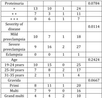
The association between retinal changes and different parameters is shown in Table 2. There was statistically significant positive association between the presence of retinal changes and blood pressure (P=0.0139) and severity of PIH (P=0.0114). However, age (P=0.2424) and gravida (P=0.3667) and proteinuria (P=0.0784) were not associated with occurrence of retinopathy in our study.
Parameter
Retinal Changes
Total P
Nil n=19
Gr I n= 24
Gr II n= 3
Blood
pressure 0.0139
<150 mmHg systolic
12 6 0 18
<100 mmHg diastolic >150 mmHg
systolic
7 18 3 28
>100 mmHg diastolic
Proteinuria 0.0784
+ 13 10 1 24
+ + 7 3 1 11
+ + + 0 6 1 7
Severity of
disease 0.0114
Mild
preeclampsia 10 7 1 18
Severe
preeclampsia 9 16 2 27
Eclampsia 0 0 1 1
Age 0.2424
19-24 years 10 15 0 25
25-30 years 7 8 2 17
31-35 years 2 1 1 4
Gravida 0.0667
Primi 8 11 1 20
Multi 7 9 0 16
Grand multi 4 4 2 10
Table 2: Showing the association of Retinopathy with Different Variables in Patients with Preeclampsia
(n=46)
DISCUSSION
In the present study, hypertensive retinopathy changes (Grade I and II) were seen in 59% of patients with preeclampsia. Haemorrhages, exudates and retinal detachment were not seen in any of the patients in this study. Since the antenatal check-up of pregnant ladies has improved very much in Tumakuru, hypertension was detected early during the antenatal visits and treatment was started immediately. This could be the probable reason for the presence of only Grade I and Grade II hypertensive retinopathy changes in our study.
Preeclampsia is responsible for maternal deaths, especially in the developing countries. During the period 1997-2000, eclampsia was the cause of death in 7.8% and preeclampsia in 4.1% cases in India. The eyes may be affected in 30% to 100% of patients with preeclampsia; the most common abnormality seen in the fundus is narrowing of retinal arterioles.(1) Various changes in the fundus and visual
problems reported in patients of preeclampsia and eclampsia from different countries include spasm and focal/generalized narrowing of retinal arterioles, haemorrhages, exudates, peripapillary or focal retinal oedema, serous retinal detachment.(4), isolated cases of acute ischemic optic
neuropathy.(5), transient blindness.(6),(2), cortical
blindness.(7,8,5), bilateral retinal detachment.(5), exudative
retinal detachment in one eye and severe macular oedema in the other eye.(3), retinal pigment epithelial lesions.(9),
temporary decrease in vision secondary to severe retinal arteriolar spasm and retinal oedema.(10), permanent blindness
secondary to central retinal artery occlusion and optic atrophy.(11) Although transient blindness has been reported in
1% to 3% of patients with eclampsia.(12) with current methods
of treatment, the present incidence is probably much lower. Optic atrophy secondary to retinal vascular involvement is unusual, but may cause visual impairment.(10),(13)
In a study of 275 cases of preeclampsia and 125 cases of eclampsia, Reddy.(12) from India has reported retinal changes
in 53.4% preeclampsia and in 71.2% in eclampsia patients
Grades of Retinopathy
Number of Patients
with Changes Percentage
No changes 19 41.30%
Grade I 24 52.17%
Grade II 3 6.53%
Grade III 0 0%
Grade IV 0 0%
Table 1:Retinal Changes (Hypertensive
Jemds.com
Original Article
J. Evolution Med. Dent. Sci./ eISSN- 2278-4802, pISSN- 2278-4748/ Vol. 5/ Issue 36/ May 05, 2016 Page 2161 (overall 59%, 236 out of 400). The most common retinal
change noted was narrowing of arterioles (45.7%, 183 out of 400 cases). He found that retinal changes were significantly more in patients with severe hypertension.
Jaffe and Schatz.(13) from USA have reported significant
correlation between the reduction in arteriole to vein ratio, number of focal arteriolar constrictions and severity of preeclampsia. They did not find any haemorrhages, exudates, cotton wool spots or retinal detachment in their study of 17 mild preeclamptic and 14 severe preeclamptic patients.
Rasdi et al(3) from Malaysia studied a group of patients
with hypertensive disorders of pregnancy (gestational hypertension, chronic hypertension, preeclampsia/eclampsia,
chronic hypertension with superadded
preeclampsia/eclampsia). The retinal changes were seen in 21.5% (5 out of 28 patients) of preeclampsia/eclampsia. They found generalized arteriolar narrowing (5/28), cotton wool spot (1/28), haemorrhage (1/28) and serous retinal detachment (1/28). They noted the resolution of all the above retinal changes except narrowing of arteries during the puerperium period.
Tadin et al(14) from Croatia have reported 45% of retinal
changes in their study of 40 patients with preeclampsia. They found a statistical correlation between proteinuria, blood pressure and hypertensive retinopathy. The degree of retinopathy was directly proportional to severity of preeclampsia. They stated that hypertensive retinopathy is a valid and reliable prognostic factor in determining the severity of preeclampsia; examination of fundus is a valuable and necessary diagnostic procedure in pregnant women with preeclampsia.
Karki et al(15) from Nepal have reported 13.7% of fundus
changes in their study of 153 subjects with preeclampsia. They assessed the foetal outcome in these patients and concluded that retinal and optic nerve head changes were associated with low birth weight; choroidal and optic nerve head changes were associated with low Apgar score; and fundus evaluation in patients with preeclampsia is an important procedure to predict adverse foetal outcomes.
The prevalence of hypertensive retinopathy changes (59%) seen in our study is higher than 13.7%.(15) 21.5%.(3)
45%.(14), but similar to 59%.(16) reported in the literature. The
absence of haemorrhages and exudates observed in present study has been supported by Jaffe and Schatz.(13)
Exudative retinal detachment is seen rarely in preeclampsia patients. It is thought to be caused by choroidal ischemia.(17) Retinal pigment epithelial lesions called Elschnig
spots, may also be found in preeclamptic patient with choroidal infarcts. The prognosis in these cases is good with visual symptoms and retinal pigment epithelial changes resolve spontaneously within weeks of delivery.(9) Presence of
macular oedema or papilloedema or retinal detachment are the warning signs for termination of pregnancy to save the 13 vision of the mother.(18) The management of retinal
detachment is not surgery, but termination of pregnancy after controlling blood pressure so that vision can be saved in the affected eye.
Cortical blindness refers to reduced vision from bilateral damage to any portion of the visual pathways posterior to the lateral geniculate nucleus. Eye examination is typically normal including a normal pupillary light reflex. It can occur in antepartum and postpartum period, lasting for several hours
to several days in preeclampsia and eclampsia patients.(16)
Other presenting symptoms include headache, seizures and loss of consciousness. MRI shows hypertense signals on T2-weighted images and hypotense signals on T1-T2-weighted images in occipital cortex. These findings are consistent with transient ischemic events as a result of cerebral oedema.(8),(16)
Management includes magnesium sulphate for seizure prophylaxis, anti-hypertensives for severe hypertension, fluid restriction to avoid worsening of cerebral oedema, ophthalmologic and neurologic consultation as well as neuroimaging. Prompt delivery is curative with resolution of neuroimaging findings.(8)
The retinal changes were more often seen in patients with severe hypertension and severity of the disease in present study; they were significantly associated with these factors (Table 1). A similar association between hypertensive retinopathy and the above two parameters was reported in earlier studies.(12)-(15) We did not find any significant
association between proteinuria and retinal changes. We did not find any case of serous retinal detachment in present study, which is similar to the previously reported studies.(12)
However, Rasdi et al(3) reported a case of serous retinal
detachment from Malaysia.
In general, it is believed that the presence of changes in the retinal arterioles and retinal haemorrhages may indicate similar changes in the placenta. Since the well-being of the foetus depends on the placental circulation, ophthalmoscopic
examination of mother’s fundus may give a clue to similar
micro-circulation changes in the placenta and indirectly to the foetal well-being. Fundus examination in patients with preeclampsia is an important clinical evaluation to predict adverse foetal outcomes.
In conclusion, visual symptoms are few in patients with preeclampsia and often absent unless the macula is involved. Sudden onset of headache, which is resistant to routine therapy in these patients may be the warning symptom before the onset of first convulsion. The presence of papilloedema in the eyes may indicate raised intracranial tension and such patients may develop convulsions. In cases of toxaemia of pregnancy, the retinal changes usually regress with decrease in blood pressure and may disappear completely after delivery due to lack of placental toxins. Hence, regular fundus examinations help in assessing the severity of the disease and the response to the treatment given.
REFERENCES
1. Richard RO. Pregnancy induced hypertension
(preeclampsia-eclampsia) In: Schachat AP, Murphy RB, editors. Retina.. St Louis: Mosby 1994;2nd edn:1405–12.
2. Achanna S, Monga D, Sivagnanam. Transient blindness in pregnancy induced hypertension. Asia Oceania J Obstet Gynaecol 1994;20(1):49–52.
3. Rasdi AR, Nik-Ahmad-Zuki NL, Bakiah S, et al.
Hypertensive retinopathy and visual outcome in hypertensive disorders in pregnancy. Med J Malaysia 2011;66(1):42–7.
4. Kanski JJ. Clinical ophthalmology-a systematic approach. Oxford: Butterworth heinmann 1989;2nd edn:p 329.
Jemds.com
Original Article
J. Evolution Med. Dent. Sci./ eISSN- 2278-4802, pISSN- 2278-4748/ Vol. 5/ Issue 36/ May 05, 2016 Page 2162 6. Nalliah S, Thavarasha AS. Transient blindness in
pregnancy induced hypertension. Int J Gynaecol Obstet 1989;29(3):249–51.
7. Grimes DA, Ekbladh LE, McCartney WH. Cortical
blindness in preeclampsia. Int J Gynaecol Obstet 1980;17(6):601–3.
8. Apollon KM, Robinson JN, Schwartz RB, et al. Cortical
blindness in severe preeclampsia: computed
tomography, magnetic resonance imaging, and single-photon-emission tomography findings. Obstetrics & Gynaecology 2000;95(6 pt 2):1017-9.
9. Gandhi J, Ghosh S, Pillari VT. Blindness and retinal changes in preeclamptic toxaemia. N Y State J Med 1978;78(12):1930–2.
10. Somerville-Large LB. A case of permanent blindness due
to toxaemia of pregnancy. Br J Ophthalmol
1950;34(7):431–4.
11. Dieckmann WJ. St Louis: Mosby-Year book Inc: The toxaemias of pregnancy 1952;2nd edn:576–611.
12. Reddy SC. Ocular fundus changes in toxaemia of pregnancy. The Antiseptic 1989;86(7):367–72.
13. Jaffe G, Schatz H. Ocular manifestations of preeclampsia. Am J Ophthalmol 1987;103(3 pt1):309–15.
14. Tadin I, Bojić L, Mimica M, et al. Hypertensive
retinopathy and preeclampsia. Coll Antropol
2001;25(Suppl):77–81.
15. Karki P, Malla KP, Das H, et al. Association between pregnancy induced hypertensive fundus changes and fetal outcome. Nepal J Ophthalmol 2010;2(1):26-30.
16. Cunningham FG, Fernandez CO, Hernandez C. Blindness
associated with preeclampsia and eclampsia. Am J Obstet Gynaecol 1995;172(4 Pt1):1291-8.
17. Valluri S, Adelberg D, Curtis R, et al. Diagnostic indocyanine green angiography in preeclampsia. Am J Ophthalmol 1996;122(5):672–7.