ARTICLE
Higher mortality rate is associated with
advanced age and periodic lateralized
epileptiform discharges in patients with
refractory status epilepticus
Alta taxa de mortalidade está associada com idade avançada e descargas periódicas
lateralizadas em pacientes com estado de mal epiléptico refratário
Paulo Breno Noronha Liberalesso, Eliana Garzon, Elza M.T. Yacubian, Américo C. Sakamoto
Status epilepticus (SE) is considered the main emergen-cy in clinical neurology, requiring immediate diagnosis and treatment. Recent data suggest an incidence from 100,000 to 200,000 cases, per year1.
he SE deinitions proposed by the Committee of Classiication and Terminology of the International League Against Epilepsy (ILAE – 1964 and 1981)2,3 do not consider
the event duration factor. herefore, diferent investigators
have used diferent deinitions, impairing the uniformity of the concept. Many authors consider SE a single epileptic seizure or two or more recurrent epileptic seizures without full recovery of consciousness, lasting 30 minutes or more4.
his deinition has been widely used over the last 20 years, although several authors have suggested that such con-cept should include epileptic seizure lasting more than ive or ten minutes5,6. However, the increase in mortality and in
MD, PhD, Hospital São Paulo, Escola Paulista de Medicina, Federal University of São Paulo, São Paulo SP, Brazil.
Correspondence: Paulo Breno Noronha Liberalesso; Rua Benjamin Constant 90, apto. 73; 80060-020 Curitiba PR - Brasil; E-mail: [email protected]
Conflict of interest: There is no conlict of interest to declare. Received 29 September 2012; Accepted 05 October 2012.
ABSTRACT
Objective: To evaluate clinical data, electroencephalogram, etiology, classiication, treatment, morbidity, and mortality in acute refractory status epilepticus. Methods: Fifteen patients, mean age of 41.3 years-old, six males, with refractory status epilepticus, were retrospectively studied. All of them were followed by serial electroencephalogram or continuous electroencephalographic monitoring. Results: The most common comorbidity was hypertension. Seven (46.7%) patients were diagnosed with previous symptomatic focal epilepsy. More than one etiology was identiied in 40.0% of the cases. Status epilepticus partial complex was the most common (n=14, 93.3%), and discrete seizures were the most observed initial ictal pattern. Continuous intravenous midazolam was used in nine (60.0%) patients and continuous thiopental in three (20.0%). Nine (60.0%) participants died, one (6.6%) had neurological sequelae, and ive (33.3%) presented no neurological sequelae.
Conclusions: Higher mortality rate was associated with advanced age and periodic lateralized epileptiform discharges. Midazolam proved to be a safe drug. The refractory status epilepticus is related to high mortality.
Key words: status epilepticus, treatment, prognosis, age.
RESUMO
Objetivo: Avaliar os dados clínicos, o eletroencefalograma, a etiologia, a classiicação, o tratamento, a morbidade e a mortalidade do estado de mal epiléptico. Métodos: Quinze pacientes, idade média de 41,3 anos, seis masculinos, foram avaliados retrospectivamente. Todos eles foram acompanhados por eletroencefalogramas seriados ou monitoração eletrencefalográica contínua. Resultados: A comorbidade mais comum foi hipertensão arterial. Sete (46,7%) pacientes tinham epilepsia focal sintomática prévia. Mais de uma etiologia foi identiicada em 40,0% dos casos. O estado de mal epiléptico parcial complexo foi o mais frequente (n=14; 93,3%) e discrete seizures foram os padrões ictal inicial mais observados. Midazolam contínuo foi usado em nove (60,0%) pacientes e tiopental contínuo em três (20,0%). Nove (60,0%) partici-pantes morreram, um (6,6%) teve sequelas neurológicas e cinco (33,3%) não apresentaram sequelas. Conclusões: Alta taxa de mortalidade foi associada com idade avançada e com a presença de descargas periódicas epileptiformes lateralizadas. Midazolam provou ser uma droga segura. Estado de mal epiléptico refratário está associado à alta mortalidade.
a deinitive neurological lesion related to epileptic seizures with more than 30 minutes appears to justify the mainte-nance of the current deinition.
Epidemiological data regarding morbidity and mortal-ity vary considerably and are closely related to patient’s age, etiology and duration of the seizures, early diagno-sis, and therapeutic conduct adopted7,8. Studies on animal
models have suggested that continuous and prolonged epileptiform activity per se may be related to irreversible neuronal damage9.
he most serious form of SE is denoted refractory SE (RSE), which is understood as SE that cannot be clinically and/or electrographically controlled with irst and second line antiepileptic drugs (AED)10. Other authors consider SE to
be refractory after failure of treatment with a third line AED11.
Some of them reserve the term RSE for epileptic seizure, with more than one hour12,13, while others reserve the term for
sei-zures lasting more than two hours14.
Clinical practice demonstrates that a considerable pro-portion of patients with SE develop refractoriness to clinical treatment, although few studies are available with speciic emphasis on the epidemiological aspects of RSE.
In the present study, we analyzed patients with RSE diag-nosis in order to identify the etiology of the condition, to eval-uate its clinical and electrographic evolution, the treatment, the complications during the acute phase, and the possible risk factors that contributed to mortality.
METHODS
All patients with RSE diagnosis admitted between January 2001 and September 2004 were retrospectively identiied. All medical records were reviewed by the same investigator, who also illed out all previously elaborated protocols. All partici-pants were submitted to continuous electroencephalograph-ic monitoring or to serial electroencephalographies (EEGs) lasting at least 20 minutes by using 21-channel digital equip-ment (Nihon Koden® and NEUROTEC®), with the electrodes positioned according to the 10-20 International System. We evaluated demographic data; medical history, etiology and clinical classiication of SE; electrographic patterns; use of irst, second, and third line AED; treatment during RSE; and short-term prognosis.
For SE classiication, we used the classiication of epi-leptic seizures and SE of Gastaut4:generalized convulsive SE
(tonic-clonic, tonic, clonic, myoclonic); simple partial convul-sive SE; nonconvulconvul-sive generalized SE; complex partial non-convulsive SE; and simple partial nonnon-convulsive SE.
he following deinitions were adopted:
• SE: a single epileptic seizure or two or more recurrent ones without recovery of consciousness and lasting 30 minutes or more.
• RSE: SE for which no clinical and/or electrographic con -trol of the epileptic phenomenon was obtained after in-travenous administration of appropriate doses of irst and second line AED, such as benzodiazepines, phenyt-oin, and phenobarbital.
• Ictal patterns: ive distinct patterns were considered — discrete seizures (DS) merging seizures (MS), continuous ictal discharge (CID), CID with lat periods (CID-F), and periodic lateralized epileptic discharges (PLEDs).
• Control of SE: control was deined as the last clinical and/or electrographic seizure after which the patient did not sufer recurrence of an epileptic seizure for at least 48 hours.
• Failure of RSE treatment: permanence or recurrence within less than 48 hours after clinical and electrographic controls and the maximum tolerated dose of continuous-ly infused midazolam, thiopental, or propofol.
• Minimum tolerated dose: continuously infused dose of midazolam, thiopental or propofol related to the onset of severe side efects, which prevented the maintenance of the drug.
• Short-term prognosis: neurological status evaluated dur -ing the irst two weeks after RSE control.
he study was approved by the Hospital Ethics and Human Research Committee (1312/02).
RESULTS
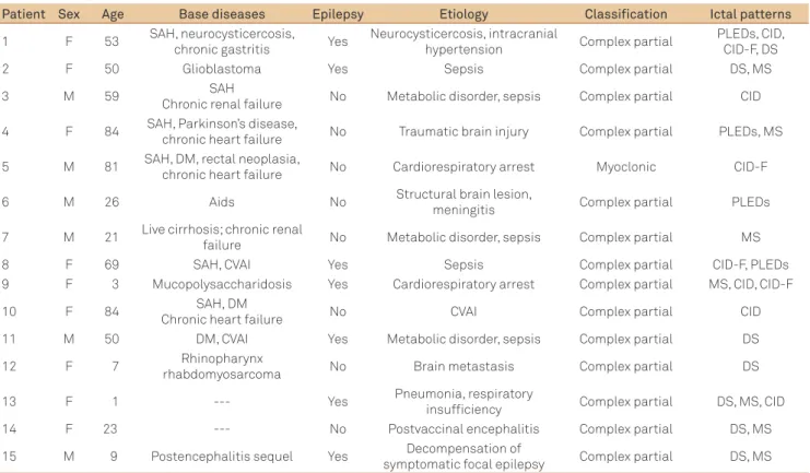
We clinically and electrographically evaluated 15 pa-tients with a RSE diagnosis, six (40.0%) of them males, ranging in age from 1 to 84 years-old (mean: 41.3 years-old; median: 50 years-old), six by continuous electroencephalo-graphic monitoring, and nine by serial EEGs (43 serial re-cordings). Fourteen patients presented comorbidities, the most prevalent being systemic arterial hypertension (n=6; 40.0%), chronic congestive heart failure (n=3; 20.0%), diabe-tes mellitus (n=3; 20.0%), chronic renal failure (n=2; 13.3%), and ischemic cerebrovascular accident (n=2; 13.3%). Seven patients (46.7%) had a previous diagnosis of symptomatic focal epilepsy (Table 1).
It was possible to identify the etiology of RSE in all cas-es, with more than one etiology being detected in six (40.0%) patients. Fourteen (93.3%) cases were classiied as complex partial SE and one (6.7%) as myoclonic SE. he most frequent initial ictal pattern was DS (n=6; 40%), with more than one ictal pattern being recorded in eight (53.3%) cases during the course of RSE. PLEDs occurred in three (20.0%) cases as the initial ictal pattern and during the course of RSE in one (6.7%) of them (Table 1).
Table 1. Demographic data, base diseases, epilepsy, classiication of status epilepticus, and ictal patterns.
Patient Sex Age Base diseases Epilepsy Etiology Classification Ictal patterns
1 F 53 SAH, neurocysticercosis,
chronic gastritis Yes
Neurocysticercosis, intracranial
hypertension Complex partial
PLEDs, CID, CID-F, DS
2 F 50 Glioblastoma Yes Sepsis Complex partial DS, MS
3 M 59 SAH
Chronic renal failure No Metabolic disorder, sepsis Complex partial CID
4 F 84 SAH, Parkinson’s disease,
chronic heart failure No Traumatic brain injury Complex partial PLEDs, MS
5 M 81 SAH, DM, rectal neoplasia,
chronic heart failure No Cardiorespiratory arrest Myoclonic CID-F
6 M 26 Aids No Structural brain lesion,
meningitis Complex partial PLEDs
7 M 21 Live cirrhosis; chronic renal
failure No Metabolic disorder, sepsis Complex partial MS
8 F 69 SAH, CVAI Yes Sepsis Complex partial CID-F, PLEDs
9 F 3 Mucopolysaccharidosis Yes Cardiorespiratory arrest Complex partial MS, CID, CID-F
10 F 84 SAH, DM
Chronic heart failure No CVAI Complex partial CID
11 M 50 DM, CVAI Yes Metabolic disorder, sepsis Complex partial DS
12 F 7 Rhinopharynx
rhabdomyosarcoma No Brain metastasis Complex partial DS
13 F 1 --- Yes Pneumonia, respiratory
insuficiency Complex partial DS, MS, CID
14 F 23 --- No Postvaccinal encephalitis Complex partial DS, MS
15 M 9 Postencephalitis sequel Yes Decompensation of
symptomatic focal epilepsy Complex partial DS, MS
F: female; M: male; SAH: systemic arterial hypertension; DM: diabetes mellitus: Aids: acquired immunodeiciency syndrome; CVAI: ischemic cerebrovascular accident; PLEDs: periodic lateralized epileptiform discharges; CID: continuous ictal discharges; CID-F: continuous ictal discharges with lat periods; MS: merging seizures; DS: discrete seizures.
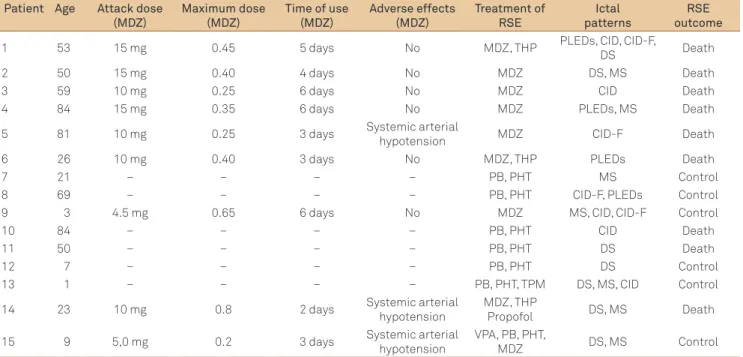
and phenobarbital as the third (n=7; 46.7%). After RSE diagnosis, continuous intravenous infusion of midazol-am was used for nine (60.0%) patients, followed by con-tinuous thiopental in three cases. In six (40.0%) patients, after the detection of RSE, the option was treatment by polytherapy without infusion of midazolam and/or thio-pental (Table 2).
In the group of patients treated with continuous midazol-am (n=7), the mean attack dose was 10.5 mg, and the mean maximum dose reached was 0.42 mg/kg/h, with a mean use time of 4.2 days. During continuous midazolam infusion, an adverse efect of the drug was observed in three (20.0%) pa-tients (arterial hypotension), with the administration of do-pamine being necessary in two cases (Table 2).
Three patients were treated with continuous thio-pental infusion, with a mean maximum dose reached of 3.9 mg/kg/h and a mean time of use of 3.5 days. The three patients presented arterial hypotension during infusion of the drug, with the administration of dopamine being nec-essary in two cases and discontinuation of the drug being required in only one.
Regarding the short-term course, nine (60.0%) patients died, five (33.3%) had no neurological sequels, and one (6.6%) had neurological sequels. In six (40.0%) patients, it was possible to obtain control of RSE within a mean time of 33.6 hours.
DISCUSSION
RSE physiopathology has peculiarities related to its du-ration and severity, with emphasis on hypoglycemia second-ary to excessive insulin release, faulty self-regulation of cere-bral blood low, excitotoxic neuronal damage, brain edema, increased body temperature related to muscle activity, and during a later phase multiple organ and system dysfunction followed by cardiovascular failure11,15. RSE has peculiar
char-acteristics compared to SE, with a higher risk of complica-tions and morbidity and mortality.
Age and sex
Although practically half the cases of SE afect children and adolescents, several studies have demonstrated that in-dividuals above the age of 60 years represent a population at risk15,16. In general, SE seems to present a bimodal age
distri-bution, with a irst incidence peak among children young-er than 12 months and anothyoung-er among eldyoung-erly patients2. In
the present series, most of the patients were adults (73.3%). Approximately two thirds of the patients were older than 50 years-old and one third was older than 80, suggesting that increasing age is accompanied by an increasing risk of refrac-toriness in cases of SE.
among males16, while others show predominance among
fe-males17. In the present study we observed a predominance
among females (n=9; 60.0%)
Comorbidities and etiology
Approximately one third of SE cases are deemed idio-pathic, occurring in neurologically healthy individuals and representing the irst manifestation of an epileptic picture18,19.
Age and presence of previous unprovoked epileptic sei-zures are important aspects for the evaluation of SE etiol-ogy. Among children and small infants, febrile/infectious situations, anoxic lesions, traumatic brain injury, meningo-encephalitis and chronic nonprogressive encephalopathy are responsible for a considerable portion of SE cases19,20;
whereas among adults and elderly individuals, cerebro-vascular disorders metabolic disorders and infection of the central nervous system are the most frequent16,17,21,22.
Among previously epileptic patients, changes in the treat-ment scheme or irregular use of AED is the factor triggering SE in most situations22. In a large study involving more than
200 patients, Barry and Hauser23 suggested that a
meticu-lous investigation of SE etiology should be performed even in previously epileptic patients, since half of these patients have a base disease or an acute brain injury as the factor triggering the current SE.
he etiology was classiied as acute symptomatic in 14 (93.3%) of our patients. SE was due to decompensation of previous symptomatic focal epilepsy in only one of our cases. Sepsis and metabolic disorders were conspicuous for their high frequency. In contrast to the literature, the
etiology related to cerebral ischemia was infrequent in our series, with only one case secondary to an ischemic cere-brovascular accident and two secondary to cardiorespira-tory arrest being observed.
Probably due to the predominance of elderly patients in our study, comorbidities were present in most cases (n=13; 86.7%), with emphasis on systemic arterial hypertension, dia-betes mellitus, and chronic congestive heart failure. he pres-ence of comorbidities/base diseases among elderly patients may impair the etiologic identiication of SE.
A previous history of symptomatic focal epilepsy was observed in practically half of our patients (n=7; 46.7%), al-though decompensation of previous refractory epilepsy was implicated as the etiology in only one case.
Classification of refractory status epilepticus According to Gastaut4, SE can be classiied as
gener-alized convulsive (tonic-clonic, tonic, clonic, myoclonic); simple partial convulsive (somatomotor, postural motor, continuous partial epilepsy); generalized nonconvulsive (typical and atypical absence); simple partial nonconvul-sive (somatosensory, sensory, autonomic, psychic or afec-tive, motor inhibitory), and complex partial nonconvulsive. Except for one of our patients diagnosed as having myo-clonic RSE, all others presented complex partial RSE. hese data agree with the current literature, which indicates that complex partial SE is the most frequent condition in criti-cally ill adult and elderly patients8.
Several studies have demonstrated that clinical evalua-tion may be insuicient for the diagnosis and classiicaevalua-tion
Table 2. Patients treated with continuous midazolam and short-term evolution of refractory status epilepticus. Patient Age Attack dose
(MDZ)
Maximum dose (MDZ)
Time of use (MDZ)
Adverse effects (MDZ)
Treatment of RSE
Ictal patterns
RSE outcome
1 53 15 mg 0.45 5 days No MDZ, THP PLEDs, CID, CID-F,
DS Death
2 50 15 mg 0.40 4 days No MDZ DS, MS Death
3 59 10 mg 0.25 6 days No MDZ CID Death
4 84 15 mg 0.35 6 days No MDZ PLEDs, MS Death
5 81 10 mg 0.25 3 days Systemic arterial
hypotension MDZ CID-F Death
6 26 10 mg 0.40 3 days No MDZ, THP PLEDs Death
7 21 – – – – PB, PHT MS Control
8 69 – – – – PB, PHT CID-F, PLEDs Control
9 3 4.5 mg 0.65 6 days No MDZ MS, CID, CID-F Control
10 84 – – – – PB, PHT CID Death
11 50 – – – – PB, PHT DS Death
12 7 – – – – PB, PHT DS Control
13 1 – – – – PB, PHT, TPM DS, MS, CID Control
14 23 10 mg 0.8 2 days Systemic arterial
hypotension
MDZ, THP
Propofol DS, MS Death
15 9 5,0 mg 0.2 3 days Systemic arterial
hypotension
VPA, PB, PHT,
MDZ DS, MS Control
of SE, with serial or continuous electroencephalographic re-cordings being required. According to DeLorenzo24, about 8%
of comatose patients with no motor manifestation sugges-tive of epileptic seizures are in nonconvulsive SE, and this di-agnosis can be conirmed exclusively by the EEG. he perma-nence of continuous epileptiform activity over a prolonged period is positively related to increased mortality and neu-rological morbidity, and diagnosis and treatment should be established as early as possible25. In our series, two patients
were unconscious and did not show any motor manifesta-tion that might suggest the presence of epileptic seizures, re-vealing the importance of the EEG for the investigation of pa-tients in a comatose state.
Ictal patterns
Treiman et al.26 suggested the existence of ive ictal
pat-terns that represent the natural history of the electrograph-ic course of SE: DS with interelectrograph-ictal slowing (this electrograph-ictal pattern was observed as the initial ictal pattern in six cases and dur-ing the course of RSE in one case); MS (this ictal pattern was observed in two cases as the initial ictal pattern and in ive cases during the course of RSE); continuous ictal discharges (this ictal pattern was observed in the initial recording in two of our cases and during the course of SE in three other cas-es); continuous ictal discharges with lat periods (this pattern was initially observed in two cases and during the course of SE in two other patients); and PLEDs (this pattern was ob-served in the initial recording in three patients and during the course of RSE in only one case).
PLEDs represent an electrographic inding of particularly diicult interpretation, with many investigators considering them an unequivocal ictal pattern27, although they may be
observed in situations not related to SE.
In contrast to the results reported by Treiman et al.26
re-garding patients with generalized convulsive SE, our data did not conirm the presence of a stereotyped sequence of ictal patterns during the course of complex partial SE.
Treatment
he objective of treating patients with SE is clinical and electrographic controls within the shortest possible time, since there is evidence suggesting that a longer duration of SE is accompanied by an increased refractoriness to treatment.
Although SE diagnosis is based on at least 30 minutes of continuous ictal activity, this theoretical deinition has no implications regarding the decision about the beginning of clinical treatment. Although there are variations in proto-cols, most authors consider benzodiazepines, phenytoin, and phenobarbital the AED of choice for the initial management of these patients. In a study in which 111 SE episodes were evaluated clinically and electrographically in 102 patients, SE could not be controlled with irst or second line AED in 11.7% of cases, which corresponded to RSE. Benzodiazepines
are potent AED that should be used only in the presence of epileptic seizures, always accompanied by another AED of longer-lasting action such as the phenytoin. Intravenous formulations of diazepam and lorazepam (the latter not available in Brazil) are the irst line drugs most commonly used, because of their rapid initiation of action due to their high liposolubility28,29.
Phenobarbital has been used in many cases as a second line drug in the treatment of SE, especially in newborn and young infants. Shaner et al.29 compared the eicacy of
phe-nobarbital and diazepam in combination with phenytoin for the initial treatment of SE, but they observed no signiicant diferences between treatments.
Patients with nonconvulsive SE, particularly those with a history of epilepsy, should not be treated in an aggressive manner such as by induction of barbituric coma, which in-creases the risk of neurological morbidity and mortality, of-setting the beneits of early control of SE.
In the present study, the first line AED most frequently used for the treatment of SE was diazepam, the second line drug was phenytoin and the third one was phenobar-bital. Once RSE was observed, continuous infusion of mid-azolam was the therapeutic conduct in most cases, with systemic arterial hypotension being detected as a side ef-fect in three patients and easily controlled with dopamine. The three patients treated with thiopental developed se-vere systemic arterial hypotension, which required dis-continuation of the drug in one case and treatment with dopamine in the other two.
Short-term prognosis
Several series have demonstrated that the prognosis of patients with SE is closely related to etiology and that both mortality and risk of neurological sequels are more frequent among adults/elderly subjects than among children30.
he morbidity-mortality rate varies considerably in dif-ferent studies depending on factors such as patient age, presence of base diseases and clinical complications, classi-ication of SE, and the therapeutic conduct adopted. Maytal et al.30, studying aspects related to prognosis in 193 children
and adolescents who had presented SE, observed a 3.6% mortality rate during the irst three months and a 9.1% of neurological sequels. hese values were signiicantly low-er than those detected by othlow-er investigators in a slow-eries of adults/elderly subjects with SE, in which mortality ranged from 9.3 to 88.0%16,17.
in RSE than in nonrefractory SE22. On the other hand, cases
related to discontinuation or alteration of AED among previ-ously epileptic patients and those related to alcoholism have lower morbidity-mortality. Other aspects such as sex and race do not seem to afect the prognosis of SE22.
Regarding the short-term prognosis, the present data agree with those reported in the literature, with more than half of our patients (n=9; 60%) dying, and approximately one quarter showing no additional neurological sequels during the irst two weeks after controlling RSE.
In conclusion, in the present series we observed a pre-dominance of RSE in adults/elderly individuals, with two thirds of our patients being over the age of 50. All patients older than more 80 years-old and more than half of the ones
older than 50 years-old died, conirming the high mortality rate occurring in elderly individuals.
All patients who presented PLEDs at some time during the course of RSE died, conirming previous literature reports that associate this ictal pattern with high mortality rate. he presence of previous symptomatic focal epilepsy did not af-fect the prognosis of RSE.
Treatment of RSE with continuous midazolam infusion proved to be safe, with side efects being observed and easily controlled in three cases. In contrast, the administration of thiopental was accompanied by severe hypotension, requir-ing discontinuation of the drug in one patient.
Despite adequate treatment, RSE is related to a high rate of neurological mortality and morbidity.
1. DeLorenzo RJ, Hauser WA, Towne AR, et al. A prospective population-based epidemiologic study of status epilepticus in Richmond, Virginia. Neurology 1996;46:1029-1035.
2. Commission on Classiication and Terminology of the International League Against Epilepsy. A proposed international classiication of epileptic seizures. Epilepsia 1964;5:297-306.
3. Commission on Classiication and Terminology of the International League Against Epilepsy. Proposal for revised clinical and electroencephalographic classiication of epileptic seizures. Epilepsia 1981;22:489-501.
4. Gastaut H. Classiication of status epilepticus. In: Delgado-Escueta AV, Wasterlain CG, Treiman DM, Porter RJ (ed). Status epilepticus. Mechanisms of brain damage and treatment. New York: Raven Press; 1983. pp. 15-35.
5. Lowenstein DH, Bleck T, Macdonal RL. It’s time to revise the deinition of status epilepticus. Epilepsia 1999;40:120-122.
6. Lowenstein DH, Alldredge BK. Status epilepticus. N Engl J Med 1998;338:970-976.
7. Aminoff MJ, Simon RP. Status epilepticus: causes, clinical features and consequences in 98 patients. Am J Med 1980;69:657-666. 8. DeLorenzo RJ, Towne AR, Pellock JM, Ko D. Status epilepticus
in children, adults and the elderly. Epilepsia 1992;33(Suppl.4): S15-S25.
9. Meldrum BS, Brierley JB. Prolonged epileptic seizures in primates: ischemic cell change and its relationship to ictal physiological events. Arch Neurol 1973;28:10-17.
10. Jagoda A, Riggio S. Refractory status epilepticus in adults. Ann Emerg Med 1993;22:1337-1348.
11. Bleck TP. Advances in the management of refractory status epilepticus. Crit Care Med 1993;21:955-957.
12. Hanley DF, Kross JF. Use of midazolam in the treatment of refractory status epilepticus. Clin Ther 1998;20:1093-10105.
13. Mayer SA, Claassen J, Lokin J, Mendelsohn F, Dennis LJ, Fitzsimmons BF. Refractory status epilepticus. Frequency, risk factors, and impact on outcome. Arch Neurol 2002;59:205-210.
14. Prasad A, Worrall BB, Bertram EH, Bleck TP. Propofol and midazolam in the treatment of refractory status epilepticus. Epilepsia 2001;42:380-386.
15. Meldrum BS, Horton RW. Physiology of status epilepticus in primates. Arch Neurol 1973;28:1-9.
16. Knake S, Rosenow F, Vescovi M, et al. Incidence of status epilepticus in adults in Germany: A prospective, population-based study. Epilepsia 2001;42:714-8.
17. Vignatelli L, Tonon C, D’Alessandro R. Incidence and short-term prognosis of status epilepticus in adults in Bologna, Italy. Epilepsia 2003;44:964-968.
18. Aicardi J, Chevrie JJ. Convulsive status epilepticus in infants and children: a study of 239 cases. Epilepsia 1970;11:187-197.
19. Dunn DW. Status epilepticus in children: etiology, clinical features, and outcome. J Child Neurol 1988;3:167-173.
20. Oliveira D, Oliveira MJ, Alves V, Temudo T. Estado de mal epiléptico en niño. Revisión de siete años. Rev Neurol 2000;30:414-418.
21. Hauser WA, Anderson VE, Loewenson RB. Seizure recurrence after a irst unprovoked seizure. N Engl J Med 1982;307:522-528.
22. Towne AR, Pellock JM, Ko D, DeLorenzo RJ. Determinants of mortality in status epilepticus. Epilepsia 1994;35:27-34.
23. Barry E, Hauser WA. Status epilepticus: the interaction of epilepsy and acute brain disease. Neurology 1993;43:1473-1478.
24. DeLorenzo RJ. Prevalence of nonconvulsive status epilepticus in comatose patients. Neurology 2000;54:340-350.
25. Lowenstein DH, Aminoff MJ. Clinical and EEG features of status epilepticus in comatose patients. Neurology 1992;42:100-104. 26. Treiman DM, Walton NY, Kendrick C. A progressive sequence of
electroencephalographic changes during generalized convulsive status epilepticus. Epilepsy Res 1990;5:49-60.
27. Garzon E, Fernandes RMF, Sakamoto AC. Serial EEG during human status epilepticus. Evidence for PLED as an ictal pattern. Neurology 2001;57:1175-1183.
28. Leppik IE, Derivan AT, Homan RW, et al. Double-blind study of lorazepam and midazolam in status epilepticus. JAMA 1983;249:1452-1454. 29. Shaner DM, McCurdy SA, Herring MO, et al. Treatment of status
epilepticus: a prospective comparison of diazepam and phenytoin versus phenobarbital and optional phenytoin. Neurology 1988;38:202-207.
30. Maytal J, Shinnar S, Moshé SL, Alvarez LA. Low morbidity and mortality of status epilepticus in children. Pedriatrics 1989;83:323-331.