O R I G I N A L A R T I C L E UDC: 616.832-004 DOI: 10.2298/VSP121105024R
Limb apraxia in multiple sclerosis
Apraksija udova kod multiple skleroze
Dragan Rapaiü*, Veselin Medenica†, Ružica Kozomara‡§, Lidija Ivanoviü*
*Faculty for Special Education and Rehabilitation, University of Belgrade, Belgrade, Serbia; †Medical College of Professional Studies “Milutin Milankoviü”, Belgrade, Serbia; ‡Military Medical Academy, Belgrade, Serbia; §Faculty of Medicine of the
Military Medical Academy, University of Defence, Belgrade, Serbia
Abstract
Background/Aim. There are almost no studies on apraxia in people with multiple sclerosis. Although the white matter is damaged in MS, it is not the only location in which the pathological changes are present. Demyelinated lesions in the cortex have recently been recognized as important com-ponents of multiple sclerosis pathology. The aim of this study was to determine whether apraxia is present among people with MS, and the importance of demographic char-acteristics and impairment of functional systems at con-ceptualization and execution of movements. Methods. The experimental group consisted of 30 patients, mean age 51.34 ± 7.70 years. The patients in the experimental group were diagnosed with MS according to the McDonald criteria. The control group consisted of 30 healthy subjects, mean age 50.30 ± 10.47 years. For research purposes, we used the following instruments: Questionnaire for Collecting Demo-graphic Data, Kurtzke Functional Systems Scores, Water-loo-Sunnybrook Apraxia Battery (WatAB). Execution of motion tasks that are a part of the WatAB were incorpo-rated in the System for the Observation and Analysis of Motor Behavior. Results. Our study showed that limb apraxia was common in people with MS. Apraxia was pres-ent during pantomime in 26.70% of the patipres-ents, and during the imitation of movements in 44.80% of the patients. Gender, age, education level, duration of disease and a form of MS did not determine the quality of conceptualization and execution of movements. The time elapsed from the last exacerbation was a determinant of quality of executed movements. Impairments of functional systems predicted impairments of movement execution. The expanded dis-ability scale score correlated with the severity of apraxia.
Conclusion. Our study confirm the presence of apraxia in MS. It is necessary to carry out further studies using func-tional magnetic resonance imaging, as well as the conduct longitudinal studies to determine the precise structure of motor behavior in people with MS.
Key words:
apraxias; diagnosis; multiple sclerosis; questionnaires; severity of illness index.
Apstrakt
Uvod/Cilj. Istraživanja apraksije kod osoba sa multiplom sklerozom (MS) gotovo da nema. Iako je ošteýenje bele mase prisutno, ono ne predstvlja jedinu lokaciju u CNS-u na kojoj su prisutne patološke promene kod MS. U skorije vreme smatra se da je kora velikog mozga veoma važna lokacija na kojoj dolazi do patoloških promena kod osoba koje boluju od MS. Cilj ovog istraživanja bio je da se utvrdi da li meĀu oso-bama sa MS ima onih kod kojih je prisutna apraksija, kao i znaÿaj demografskih karakteristika i ošteýenja funkcionalnih sistema za konceptualizaciju i izvoĀenje pokreta. Metode. Ek-sperimentalnu grupu ÿinilo je 30 uÿesnika starosti 51,34 ± 7,70 godina. Uÿesnicima eksperimentalne grupe dijagnostikovana je MS prema Mekdonaldovom dijagnostiÿkom kriterijumu. Kontrolnu grupu saÿinjavalo je 30 zdravih osoba starosti 50,30 ± 10,47 godina. Za potrebe istraživanja korišýeni su podaci dobijeni Upitnikom za prikupljanje demografskih podataka i osnovnih podataka o bolesti, Kurzke-ovim skorovima funkci-onalnih sistema i Adaptiranom Vaterlo baterijom za procenu apraksije (ova baterija korišýena je u kombinaciji sa Sistemom za opservaciju i analizu motornog ponašanja). Rezultati. Naše istraživanje je pokazalo da apraksija udova predstavlja ÿestu pojavu od osoba sa MS. Apraksija na zadacima izvoĀenja pantomime bila je prisutna kod 26,70% bolesnika, a na zada-cima izvoĀenja imitacije pokreta kod 44,80% bolesnika. Pol, godine života, stepen obrazovanja, dužina trajanja bolesti i ob-lik bolesti nisu determinisali kvalitet konceptualizacije i
izvo-Āenja pokreta kod osoba sa MS. Vreme proteklo od poslednje egzacerbacije predstavljalo je determinantu kvaliteta izvedenih pokreta. Ošteýenja funkcionalnih sistema kod osoba sa MS predviĀala su pristustvo ošteýenja izvršenja pokreta. Proširena skala funkcionalne onesposobljenosti bila je u korelaciji sa te-žinom apraksije. Zakljuÿak. Naše istraživanje otkrilo je prisu-stvo apraksije kod MS. Potrebno je izvršiti dalja istaživanja uz korišýenje funkcionalne magnetne rezonance, kao i sprovoĀ e-nje longitudinalnih studija kako bi se preciznije utvrdila struk-tura motornog ponašanja kod osoba sa MS.
Kljuÿne reÿi:
Introduction
Apraxia is defined as a disorder of learned movements, which is not caused by muscle and/or neurological factors (e.g. weakness, akinesis, aphasia, cognitive resources de-cline, vision problems, etc.) 1–3. Neuropsychology was mostly engaged in studying apraxia especially in persons with brain injury, Alzheimer's disease, Parkinson's disease, corticobasal degeneration, and other. The most investigated relationship was the one between the localized impairment and apraxia occurrence.
Multiple sclerosis (MS) is usually regarded as a disease of the white matter 4. Lesions of the white matter that include demyelination and neuronal damage are very visible on magnetic resonance imaging (MRI) and macroscopically during autopsy 5, 6. Detecting white matter lesions with MRI is an essential signal for the presence of MS. Cortex has re-cently been recognized as an important location in which pathological changes occur in patients suffering from MS 7, 8. MS is a chronic inflammatory disease of the central nervous system characterized by multifocal demyelination and axonal damage, which affects both white and gray matter of the cerebral cortex, deep gray matter nuclei and the spinal cord. The appearance of apraxia is expected due to lesions or de-generation of certain areas of cortex and/or by damaging the parietofrontal pathways 9.
There are almost no scholarly papers on apraxia in per-sons with MS. The only research that addresses the relation-ship of MS and apraxia is the one conducted by Kamm et al. 9.
For that reasonsthe aim of this study was to determine the frequency of aproxia in patients with MS, and its relation to demographic characteristics, severity of illness, type of illness, disease duration, and time elapsed since the last ex-acerbation.
Methods
This study included participants of both sexes, 18–65 years of age, divided into control and experimental groups of similar size. The sample was unrepresentative and con-vinient, depending on the avalilability of the participants.
The experimental group included patients with MS di-agnosed by the McDonalds diagnostic criteria 10. One of the criteria for inclusion in the sample was the score achieved on the Expanded Disability Status Scale (EDSS) that was greater than or equal to 1. All the patients of the experimen-tal group were the members of the Multiple Sclerosis Society of Serbia. Also, the inclusive criteria for the experimental group meant that the subject was able to independently read and understand the information from the form which con-firmed the consent to participate in the research. All the pa-tients of the experimental group read, understood and signed a form confirming the consent for participation in the re-search. The patients of the experimental group did not have the history of nor are currently subjected to alcohol and/or psychoactive substance use, in the last two years pregnant women, persons with the history of neurological damage which cannot be treated as a result of MS, persons with
de-mentia, persons suffering from psychiatric disorders, persons with significant motor disorders (such as tremor, bradykine-sia, dyskinesia), persons with peripheral conditions (e.g., ar-thritis) that may compromise motor function, individuals with developmental disabilities, persons that cannot understand the assessment procedure due to some cognitive deficit.
The control group formation criterion was to be healthy, and as for the experimental group, to read, understand and sign the form, confirming their consent to participate in re-search. Exclusive criteria for the control group were the same as for the experimental one.
The control group was introduced to demographic char-acteristics of the experimental group.
The System for the Observation and Analysis of Motor Behavior (SOAMB) consists of hardware and software com-ponents. Required central processing unit (CPU) and mem-ory of the system were provided by the computer Dell Inspi-ron PP29L. An additional monitor for displaying tasks was used, 20-inch diagonal display with 1,280 × 1,024 resolution and the image refresh rate of 75 Hz. For the audio material reproduction loudspeakers Genius were used. For the pur-pose of recording movements a web camera Logitech Webcam C905 was used, which could record in high defini-tion, 1,600 × 1,200 resoludefini-tion, 30 frames per sec.
The computer program for acquisition, analysis and partial data processing was created in Java programming language. The program allows creation of the profile for each participant. The task creation which participants should per-form, using sound, pictures or video files, was enabled.
In this study all the tasks of WatAB were developed in video format and uploaded into the program. In this way we avoided the possibility for the examiner to issue tasks at dif-ferent times and in difdif-ferent manners. For example, the pos-sibility that some of the examiner perform movement imita-tion tasks in different ways for different participants in the study was avoided, providing a greater degree of objectivity in the use of WatAB 11.
The research was conducted in the facilities of the Soci-ety of MS Serbia, on the territory of Belgrade and Aranÿ elo-vac.
For data analysis and processing, software packages Microsoft Excel and SPSS were used. From the statistical techniques we used descriptive, correlation, regression, and discriminate techniques. The results are presented in tables and figures.
Results
The demographic characteristics of the control and the ex-perimental group are presented inTable 1. Health status char-acteristics in the patients with MS are presented in Table 2.
The results of the participants of the control and experimental groups in the WatAB
statistically significant difference between the control and the experimental group on the conceptual scale, p < 0.05. The difference between the mean values of group character-istics was large [eta squared (n2) = 0.10)]. There was a sta-tistically significant difference in the recognition subscale (p < 0.05, n2 = 0.10).
Also, a statistically significant difference between the two groups on productive scale and almost all of its sub-scales was found.
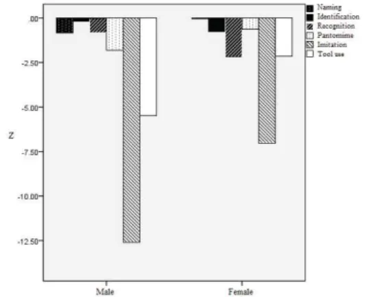
Figure 1 shows deviations of those with MS, compared to a typical population. Deviations are represented by Z scores.
The presence of apraxia in the patients with MS
In order to determine the number of the study patients with from MS and the presence of apraxia we used the Roy`s research group approach. We analyzed the subscale of pan-tomime, imitation as well as the whole production scale. The
patietns with a deviation up to 1 SD were classified as those with no present apraxia. Those with discrepancy in scores greater than 1 SD, and less than 2 SD were classified as bor-derline conditions, while those with a variation in
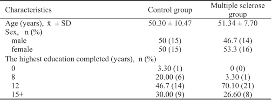
perform-Table 1 Demographic characteristics of the study participlants
Characteristics Control group Multiple sclerose
group Age (years), ʉ ± SD 50.30 ± 10.47 51.34 ± 7.70 Sex, n (%)
male 50 (15) 46.7 (14)
female 50 (15) 53.3 (16)
The highest education completed (years), n (%)
0 3.30 (1) 0 (0)
8 20.00 (6) 3.30 (1)
12 46.7 (14) 70.10 (21)
15+ 30.00 (9) 26.60 (8)
Table 2 Characteristics of the patients with multiple sclerosis (MS)
Distribution Age (year) Age at time of diagnosis (year)
Last remission
period (years) EDSS (points) Type of MS
n (%) ʉ ± SD ʉ ± SD ʉ ± SD ʉ ± SD
RRMS 10 (33.3) 49.41 ± 7.61 32.20 ± 10.28 5.14 ± 3.34 4.20 ± 2.26
PPMS 10 (33.3) 53.48 ± 7.38 35.50 ± 9.58 8.00 ± 0.23 4.00 ± 1.93
SPMS 2 (6.7) 58.92 ± 0.11 34.50 ± 7.78 3.00 ± 0.12 5.50 ± 3.53
N/A* 8 (26.6) 48.96 ± 8.19 35.42 ± 6.24 2.85 ± 5.43 4.94 ± 2.81
Total 30 (100) / / / /
Average / 51.34 ± 7.70 34.28 ± 8.74 4.49 ± 3.91 4.42 ± 2.30
*New available date is not present in documentation; EDSS – Expanded Disability Status Scale; RRMS – relapsed-remitting MS; PPMS – primary progressive MS; SPMS – secondary progressive MS.
Table 3 The scores of the control and the multiple sclerose (MS) group on the
Adapted Waterloo-Sunnybrook Apraxia Battery Types of tasks Control group
(ʉ ± SD)
MS group (ʉ ± SD) Conceptual scale* 97.17 ± 4.43 92.61 ± 9.81
naming 97.04 ± 4.99 94.25 ± 8.20
identification 97.83 ± 5.27 95.29 ± 8.27
recognition* 95.97 ± 5.92 85.60 ± 21.91
Production scale* 97.16 ± 2.90 84.84 ± 23.34
pantomime* 93.98 ± 6.08 85.07 ± 15.83
concurrent imitation* 99.20 ± 1.08 83.83 ± 27.60 delayed imitation* 98.09 ± 2.54 78.86 ± 33.84
Tool use* 98.39 ± 3.29 85.83 ± 24.34
*p < 0.05 (statistically significant difference).
ance in these subscales and scales greater than 2 SD be-longed to the group with the presence of apraxia. In this way, the obtained results showed that the performance on the sub-scale pantomime among those with apraxia can be classified in 8 (26.70%) participants. At the subscale movement imita-tion in the group with MS and the presence of apraxia, was classified in 13 (44.80%) participants. On the overall pro-duction scale apraxia was present in 12 (42.90%) of the pa-tients. The overall results of this procedure are shown in Ta-ble 4.
The WatAB results concerning gender, age and the level of education
The results of the patients in the subscales of the Wa-tAB in relation to gender were compared by t-test for inde-pendent samples, while the analysis of variance was used to compare these results in relation to age and the level of edu-cation. The results were compared on the subscales of nam-ing, identification, recognition, pantomime, imitation and use of tools/objects.
There was no statistically significant difference in rela-tion to the results of these subscales regarding gender (Figure 2), as well as age (group 1: < 45.42 years; the group 2: from 45.53 to 55.00 years; and the group 3: more than 55.01 years) and the degree of education (group 1: 0 years of edu-cation; the group 2: 8 years of eduedu-cation; the group 3: 12 years of education; group 4: 15+ years of education)].
Fig. 2 – Z-scores at the Adapted Waterloo-Sunnybrook Apraxia Battery in relation to gender.
The WatAB results concerning type, duration, degree of functional impairment of the system and the time elapsed from the last exacerbation
The results on the subscales of naming, identification, recognition, pantomime, imitation and use of tools/objects were compared in relation to type, duration, degree of
func-tional impairment of the system and the time from the last exacerbation. Comparisons were performed by analysis of variance.
There was no statistically significant difference in the results of these subscales in relation to the form of the dis-ease (the group 1: relapsing-remitting, the group 2: primary progressive, the group 3: secondary progressive).
There was no statistically significant difference in the re-sults, nor in the duration of MS (the group 1: up to 12.33; the group 2: from 12.34 to 18.83, and the group 3: over 18.84).
The results on subscales identification, recognition, pantomime, imitation and use of tools/objects in relation to the time from the last exacerbations were compared by the t -test of independent samples. The group 1 consisted of par-ticipants with the last exacerbation 4 years ago or less, while the group 2 consisted of participants with the last exacerba-tion 4 years or more before. We found only one statistically significant difference between the group 1 (= -1.57 ± 2.03) and the group 2 (= -0.65 ± 0.39) in the pantomime subscale, t (11) = -2.62, p = 0.024 (two-sided). The difference between the mean values o f the characteristics of the groups (mean difference = -2.22, 95% Cl: -4.09 to -0.35) was very large (n = 0.38).
In order to additionally explain the obtained results, we used the Pearson's correlation to establish whether there was a relationship between the time from the last exacerbation and the results of the subscales WatAB. The assumptions of the normality, linearity and homogeneity were satisfied. There was a correlation between achievements on the subscale of recognition and the time elapsed from the last exacerbation (r = 0.59, p < 0.05), as well as correlations between scores on the subscale of pantomime and the time from the last exacerbation (r = 0.58, p < 0.05). Shorter time elapsed from the last exacer-bation was accompanied by lower Z scores on the subscales of recognition and pantomime people with MS.
The possibility for the degree of functional systems impairments to predict the results in the subscales pantomime, imitation and use of tools of the WatAB
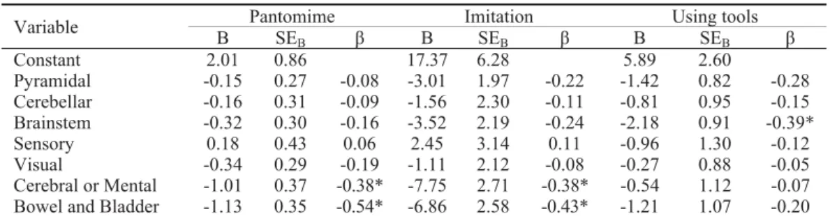
The possibility of the degree of damage measured by FSS to predict results in the WatAB subscales was estimated in a sample of patients with MS. We used the standard mul-tiple regression, for each of WatAB subscales.
For predicting the results on the subscale of pantomime, a preliminary analysis showed that the assumptions of nor-mality, linearity, multicollinearity and homogeneity of vari-ance were not violated. The model as a whole explained 50.1% of the total variance, F (7, 22) = 5.15, p < 0.05, so it can be said that scores on the FSS significantly predict the results
Table 4 The presence of apraxia in the patients with multiple sclerosis
Pantomime Imitation Production scale The presence of apraxia
n (%) n (%) n (%)
Yes 8 (26.70) 13 (44.80) 12 (42.90)
Borderline 6 (20.00) 4 (13.80) 2 (7.10)
on the subscale of pantomime in the WatAB. The regression coefficients and standard errors are shown in Table 5.
For predicting the results in the motion imitation subscale preliminary analysis showed that the assumptions of normal-ity, linearnormal-ity, multicollinearity and homogeneity of variance were not violated. The model as a whole explained 54.8% of the total variance, F (7, 21) = 5.85, p < 0.05, so it can be said that scores on the FSS significantly predict the results on the subscale of motion imitation on WatAB. The regression coef-ficients and standard errors are shown in Table 5.
For predicting results in the use of tools subscale pre-liminary analysis showed that the assumptions of normality, linearity, multicollinearity and homogeneity of variance were not violated. The model as a whole explained 46.8% of the total variance, F (7, 20) = 4.39, p < 0.05, so it can be said that scores on the FSS significantly predict the results on the subscale of using tools in WatAB. The regression coeffi-cients and standard errors are shown in Table 5.
The results in WatAB associated with EDSS scores
We used the Pearson's correlation to establish whether there was any relationship between the EDSS and achieve-ments of the WatAB subscales. The assumptions of the nor-mality, linearity and homogeneity were satisfied. There were strong correlations between the results on the subscales of pantomime (r = -0.72, p < 0.01), imitation (r = -0.76, p < 0.01), tool use (r = -0.75, p < 0.01) and the EDSS. A higher EDSS was accompanied by lower Z scores on the subscales of pantomime, imitation and tool use in people with MS.
Discussion
The obtained results indicate that limb apraxia very often occurs in population with MS. In this population apraxia is present more often in imitation than in pantomime tasks. The obtained results consist of the results of a few other studies in terms of the general presence of apraxia in persons with MS. According to Staff et al. 12 the percentage of apraxia presence in the population of persons with MS is much lower – only 13%. On the other hand, Kamm et al. 9 have found that limb apraxia is present in 26.3% of participants with MS.
Praxic abilities of women are better than in men throughout their entire development 13 and healthy women
perform voluntary movement with fewer errors than men 14. In patients with brain lesions praxis system is much more
dependent on the anterior region of the left hemisphere in women than in men 15–17, which may suggest that more fo-cused representation allows greater precision of voluntary movements control in women 15. Women have a better abil-ity to implement complex motion and pre-planning move-ment than men during the execution and control 18. Pozzilli et al. 19 in their study using MRI show that there are differences in lesion characteristics between men and women with MS. The authors conclude that lesions in men are less inflamma-tory, but more destructive than in lesions in women. There-fore, we considered the possibility that the general tendency for women to have less apraxic errors in the execution of movements than men does not have to be present in persons with MS. We showed the parallel display of the results of men and women on the WatAB subscales. It is obvious that women in the subscales of the pantomime, imitation and use of tools have minor deviations from the control group in comparison to men. However, the difference between men and women did not reach statistical significance.
The participants in the study did not have different re-sults in the WatAB in relation to age and disease duration. We believe that this result is quite expected considering the nature of progression of the disease, which is indicated by the existence of its subtypes according to the mode of pro-gression (relapsing-remitting, primary progressive, secon-dary progressive, etc). This means that the disease can quickly progress to some younger people than in older or vice versa, and that there is no rule or correlation between age and disease progression. Kamm et al. 9 designed a linear regression model, for which the result on the EDSS is the best single predictor, while among other predictors are the duration of MS and the age of the participants. Participants in their study had lower EDSS compared to participants in our study. That could be the reason for results diversity. Also, the authors, did not consider the individual effects of these variables within the model. Dimeck et al. 20 conducted a study in which the sample consisted of healthy participants who performed concurrent and delayed imitation of move-ment. A statistically significant difference between older and younger participants had been obtained. Changes in condi-tions of imitation also gave a statistically significant
differ-Table 5 Regression coefficients and standard errors (SE) in predicting the quality the performance
of pantomime, imitation and using tools
Pantomime Imitation Using tools
Variable
B SEB ȕ B SEB ȕ B SEB ȕ
Constant 2.01 0.86 17.37 6.28 5.89 2.60
Pyramidal -0.15 0.27 -0.08 -3.01 1.97 -0.22 -1.42 0.82 -0.28
Cerebellar -0.16 0.31 -0.09 -1.56 2.30 -0.11 -0.81 0.95 -0.15
Brainstem -0.32 0.30 -0.16 -3.52 2.19 -0.24 -2.18 0.91 -0.39*
Sensory 0.18 0.43 0.06 2.45 3.14 0.11 -0.96 1.30 -0.12
Visual -0.34 0.29 -0.19 -1.11 2.12 -0.08 -0.27 0.88 -0.05
ence in achievements. If the age and duration of disease play a role in the results of participants with MS, for more accu-rate determination of their impact a longitudinal study on a number of people, with occasional reevaluation of praxic abilities should be carried out. Otherwise, the influence of age and disease duration on the results of the tasks of apraxia assessment is not visible due heterogeneous illness progres-sion of the participants in the study.
It was expected that this claim would be supported by the results of the participants regarding MS type. However, there was no statistically significant difference between the groups, while conflicting results have been obtained by Kamm et al. 9. Participants in their study with the relapsing-remitting form of MS had significantly higher scores on the praxis assessment compared to the participants with primary progressive and secondary progressive forms of MS. There were no statistically significant differences between the groups of participants with primary progressive and secon-dary progressive type of MS. It remains to be further ex-plored.
The time from the last exacerbation has great effect only on the results on the subscale of pantomime. These re-sults contribute to the statement that the time after exacer-bations in people with MS leads to recovery of the func-tion, and that this recovery implies the features of concep-tualization and execution of movement. We will refrain from further interpretation of the obtained results because they are limited in the sense that they represent only the condition in one timely moment for each individual, that is, they do not have a longitudinal character. Stamenova et al. 21
investigated the long-term recovery of limb apraxia after brain injury. Participants in the study (with acute and chronic conditions) on all tasks showed signs of recovery except on the tasks of identifying actions. Faster recovery showed acute and subacute patients on the tasks of panto-mime. The study of Stamenova et al. 21 has similarity to the results from our study.
The degree of functional system impairments in patients with MS predicted the success of the performance of panto-mime imitation and use of tools. Patients with apraxia often do not have problems in using real tools. Our study confirms that significant predictors of the success in performing pan-tomime and imitation differ from significant predictors of the success in the use of tools. When performing pantomime and imitation, requirements for use of cognitive functions could be increased. In the case of pantomime performance it is necessary to create visual representations of the tools used or representations of social situations in which persons use some form of representative nontransitive pantomime. In case of movement imitation it is essential to receive infor-mation about the action performed by someone else, to de-code information about what is seen, to form ideas about the
movement that is to be executed and to properly activate ef-fectors for movement performance. On the other hand, when using the real tools, through the sensory system the informa-tion about the tool (shape, weight, etc.) is obtained. In this sense, action performance with real tools requires a higher degree of activation of other systems in the brain (especially learned actions), as well as greater load of the musculo-skeletal system.
The role of the system for bladder and bowel control in the prediction of apraxia may seem surprising at first glance, but it should be emphasized that from the neurological point of view some elements of this system overlap with other functional elements of other systems. The bladder and bowel control is partly influenced by our own will. Fowler et al. 22 suggest that clinical observation studies and observation studies using MRI suggest that the frontal lobe plays an im-portant role in determining the appropriate moment for mic-tion. Some studies 23, 24 show that the right inferior frontal gyrus, which is a part of the prefrontal cortex was active when the bladder was full in patients. It is believed that the prefrontal cortex is involved in planning complex cognitive behaviors, personality characteristics reflects, plays a role in the expression of appropriate social behavior as well as the functions of attention and response selection 25. This system damage in our research emerges as a predictor of quality of pantomime and imitation together with impairment of mental function.
Finally, higher EDSS were associated with poorer per-formance on tasks of pantomime, imitation and tool use. EDSS and apraxia connection exists solely to the production of movement tasks, but not the conceptualization of move-ment (identification, recognition and naming). EDSS is based on FSS and other motor skills, which is why this cor-relation is understandable. Unlike Kamm et al. 9, who showed that more MS patients having apraxia have higher EDSS, our research in this regard did not address the fre-quency of apraxia in relation to EDSS. Our research con-firms that if EDDS is higher, there will be present a more se-vere form of apraxia as shown by the results in all the pro-duction subscales.
Conclusion
Limb apraxia is frequent in persons with multiple scle-rosis, and its occurrence is different when it comes to forming pantomime and movement imitation. Praxic per-formance depends on the time elapsed from the last exacer-bation and the Expended Disability Scale score. Functional systems impairments in patients with MS may predict the quality of movement execution. A connection between sex, age, type of disease and apraxia in people with multiple scle-rosis should be the subject of further research.
R E F E R E N C E S
1. Poeck K. The clinical examination for motor apraxia.
Psychol-ogy: Neuropsychological studies of apraxia and related disorders. Amsterdam: Elsevier Science Publishers; 1985. p. 111î61. 3. Roy EA, Square PA. Neuropsychology of movement
sequenc-ing disorders and apraxia. In: Zaidel D, editor. Handbook of Perception and Cognition: Neuropsychology. New York: Erl-baum; 1994. p. 185î218.
4. Noseworthy JH, Lucchinetti C, Rodriguez M, Weinshenker BG. Mul-tiple sclerosis. N Engl J Med 2000; 343(13): 938î52.
5. Runge VM, Price AC, Kirshner HS, Allen JH, Partain CL, James AE. The evaluation of multiple sclerosis by magnetic reso-nance imaging. Radiographics 1986; 6(2): 203î12.
6. Nagara H, Inoue T, Koga T, Kitaguchi T, Tateishi J, Goto I. Forma-lin fixed brains are useful for magnetic resonance imaging (MRI) study. J Neurol Sci 1987; 81(1): 67î77.
7. Walker CA, Huttner AJ, O'connor KC. Cortical injury in multiple sclerosis; the role of the immune system. BMC Neurology 2011; 11(1): 152.
8. Brownell B, Hughes JT. The distribution of plaques in the cere-brum in multiple sclerosis. J Neurol Neurosurg Psychiatr 1962; 25: 315î20.
9. Kamm CP, Heldner MR, Vanbellingen T, Mattle HP, Müri R, Bohl-halter S. Limb apraxia in multiple sclerosis: prevalence and im-pact on manual dexterity and activities of daily living. Arch Phys Med Rehabil 2012; 93(6): 1081î5.
10.McDonald WI, Compston A, Edan G, Goodkin D, Hartung HP, Lublin FD, et al. Recommended diagnostic criteria for multiple sclerosis: guidelines from the International Panel on the diag-nosis of multiple sclerosis. Ann Neurol 2001; 50(1): 121î7. 11.Medenica V, Rapaic D, Nedovic G, Ivanovic L, Trgovcevic S, Potic S,
et al. Contemporary models and preservation assessment pos-sibilities in conceptual-production system of voluntary motor action. Healthmed 2012; 6(9): 3194î201.
12.Staff NP, Lucchinetti CF, Keegan MB. Multiple sclerosis with pre-dominant, severe cognitive impairment. Arch Neurol 2009; 66(9): 1139î43.
13.Chipman K, Hampson E. A female advantage in the imitation of gestures by preschool children. Dev Neuropsychol 2007; 31(2): 137î58.
14.Chipman K, Hampson E. A female advantage in the serial pro-duction of non-representational learned gestures. Neuropsy-chologia 2006; 44(12): 2315î29.
15.Kimura D. Sex differences in cerebral organization for speech and praxic functions. Can J Psychol 1983; 37(1): 19î35. 16.Kimura D. Neuromotor mechanisms in human
communica-tion. NewYork: Oxford University Press; 1993.
17.Kimura D, Hampson E. Neural and hormonal mechanisms mediating sex differences in cognition. In: Vernon PA, edi-tor. Biological approaches to the study of human intelli-gence. Norwood, NJ: Ablex Publishing Corp; 1993. p. 375î97.
18.Cohen NR, Pomplun M, Gold BJ, Sekuler R. Sex differences in the acquisition of complex skilled movements. Exp Brain Res 2010; 205(2): 183î93.
19.Pozzilli C, Tomassini V, Marinelli F, Paolillo A, Gasperini C, Bas-tianello S. 'Gender gap' in multiple sclerosis: magnetic reso-nance imaging evidence. Eur J Neurol 2003; 10(1): 95î7. 20.Dimeck PT, Roy EA, Hall CR. Aging and working memory in
gesture imitation. Brain Cognit 1998; 37(1): 124î6.
21.Stamenova V, Black SE, Roy EA. A model-based approach to long-term recovery of limb apraxia after stroke. J Clin Exp Neuropsychol 2011; 33(9): 954î71.
22.Fowler CJ, Griffiths D, Groat WC. The neural control of micturi-tion. Nat Rev Neurosci 2008; 9(6): 453î66.
23.DasGupta R, Kavia RB, Fowler CJ. Cerebral mechanisms and voiding function. BJU Int 2007; 99(4): 731î4.
24.Kavia RB, Dasgupta R, Fowler CJ. Functional imaging and the central control of the bladder. J Comp Neurol 2005; 493(1): 27î32.
25.Pardo JV, Fox PT, Raichle ME. Localization of a human system for sustained attention by positron emission tomography. Nature 1991; 349(6304): 61î4.