INTRODUCTION
Glaucoma is the leading cause of irreversible blindness worldwide(1,2).
This disease is associated with chronic and progressive optic neuro-pathy(1,3) characterized by loss of retinal ganglion cells(3), which leads
to visual ield deterioration(1,3,4). Moreover, glaucoma is associated
with traic accidents, restricted mobility, and falls, thus afecting qua
-lity of life(1). An important risk factor for glaucoma is intraocular
pressure (IOP), and its decrease is the mainstay of treatment(3).
IOP measurement has been a matter of debate for many years. In 1950, Goldmann introduced an approach to measure IOP, which is called applanation tonometry, and this approach is currently the gold standard(3-5). This approach is related to the elasticity of the cornea,
which indicates that it depends on corneal thickness and hysteresis(4).
Goldmann assumed that the average central corneal thickness (CCT)
would be approximately 500 µm(4-7), and excessively thin and thick
corneas would cause underestimations and overestimations of the IOP, respectively(4,7,8). With the advent of more sophisticated devices
capable of measuring CCT, it has become clear that CCT is much
more variable than predicted by Goldmann(5-7). More recently, some
studies, such as the Ocular Hypertension Treatment Study (OHTS), stated that CCT is an important confounder of Goldmann applanation
tonometer (GAT) measurements(5,6,8). In addition, factors, such as
astigmatism, examiner’s competence, gaze direction, tear thickness, corneal hydration, connective tis sue composition, bioelasticity, corneal curvature, and other corneal biomechanical properties, are
important sources of error in GAT measurements(2-4,8). Currently, an
accepted formula to correct IOP is not available(4,6,7).
The ocular response analyzer (ORA) was introduced in 2005, and
it was classiied as a non-contact tonometer(2,3,5,9). This tonometer
allows the measurement and evaluation of corneal biomechanical properties, namely corneal hysteresis (CH), corneal resistance factor
(CRF), and corneal compensated intraocular pressure (IOPcc)(3,5), as
well as CCT and Goldmann correlated intraocular pressure (IOPg)(3,5).
Briely, the ORA produces a rapid air pulse that deforms the corneal curvature(2,3,5,9) and records corneal deformation(2,9). When the cornea
moves inward, it reaches the irst applanation state (P1)(2,3,9). After a
slightly concave state(2,3,9), the air pulse pressure decreases and the
cornea moves outward, passing through the second applanation state (P2)(2,3,9). The average of P1 and P2 is IOPg, which is analogous
Corneal properties and glaucoma: a review of the literature and meta‑analysis
Propriedades da córnea e glaucoma: revisão da literatura e meta-análise
RicaRdo GaspaR1, Luís abeGão pinto2,3, david coRdeiRo sousa2,3
Submitted for publication: May 26, 2017 Accepted for publication: July 6, 2017
1 Faculdade de Medicina, Universidade de Lisboa, 1649-028 - Lisboa, Portugal.
2 Vision Sciences Study Center, CECV, Faculdade de Medicina, Universidade de Lisboa, 1649-028
Lisboa, Portugal.
3 Department of Ophthalmology, Hospital de Santa Maria, 1649-035, Lisboa, Portugal.
Funding: No specific financial support was available for this study.
Disclosure of potential conflicts of interest: None of the authors have any potential conflict of interest to disclose.
Corresponding author: Luís Abegão Pinto. Hospital de Santa Maria - Oftalmologia. Av. Prof. Egas Moniz - Lisboa - 1649-035 - Portugal - E-mail: [email protected]
ABSTRACT
Purpose: Studies have suggested that corneal biomechanical properties inluence intraocular pressure (IOP) measurements, namely central corneal thickness (CCT ) and corneal hysteresis (CH). The present study aimed to investigate the associations of CH and CCT with glaucoma development.
Methods: We performed a review of the literature and meta-analysis of obser-vational studies (2006-2016) that included both adult glaucoma patients and controls and reported CCT and CH as outcomes. Nineteen studies were conside red eligible, and the mean difference (MD) between groups (patient and control) for both variables was used for statistical analyses.
Results: A total of 1,213 glaucoma and 1,055 healthy eyes were studied. Quan-titative analysis suggested that CH was signiicantly lower in the glaucoma group than in the control group (MD=-1.54 mmHg, 95% CI [-1.68, -1.41], P<0.0001). Additionally, CCT was signiicantly lower in the glaucoma group than in the control group (MD=-8.49 µm, 95% CI [-11.36, -5.62], P<0.001).
Conclusion: Corneal properties appear to difer between glaucoma patients and healthy controls. Our results emphasize the importance of corneal biomechanical properties in IOP interpretation and should support further studies on the inluence of CH and CCT in glaucoma screening and diagnosis.
Keywords: Glaucoma; Corneal pachymetry; Cornea/physiopathology; Meta-analysis as topic
RESUMO
Objetivo: A literatura sugere que as propriedades biomecânicas da córnea, nomea-damente a espessura central da córnea (ECC) e a histerese corneana (HC), influenciam a medição da pressão intraocular (PIO). Este estudo teve como objetivo investigar a associação entre a ECC e a HC e o desenvolvimento de glaucoma.
Métodos: Revisão da literatura e meta-análise. Foram incluídos estudos observacio-nais, publicados entre 2006 e 2016, que integrassem um grupo controle e um grupo de pacientes com glaucoma em que estes dois grupos apresentassem, igualmente, a ECC e a HC como parâmetros. Dezenove estudos foram considerados elegíveis e a diferença média (MD) daqueles parâmetros nos dois grupos foi utilizada para análise estatística.
Resultados: Estudaram-se um total de 1.213 olhos com glaucoma e 1.055 olhos saudáveis. A análise quantitativa revelou que a HC é significativamente mais baixa no grupo de doentes com glaucoma quando comparada com o grupo controle (MD=-1,54 mmHg, intervalo de confiança de 95% [-1,68-1,41], P<0,00001). A ECC foi, também, significativamente mais baixa no grupo glaucoma quando comparada com os indivíduos saudáveis MD=-8,49 µm, intervalo de confiança de 95% [-11,36, -5,62], P<0,001).
Conclusão: Os pacientes com glaucoma parecem possuir propriedades corneanas diferentes das que apresentam os indivíduos saudáveis. Os resultados enfatizam a importância das propriedades biomecânicas da córnea na interpretação da PIO e devem contribuir para novos estudos sobre a influência da HC e da ECC no rastreio e diagnóstico do glaucoma.
to the IOP measured by GAT(2,5,9), being the diference between these
two values (P2 - P1) the value of CH(2,3,5).
The OHTS revealed that CCT is an important and independent risk factor for the development of glaucoma(4-6,10). These results were
validated in the European Glaucoma Prevention Study (EGPS)(4,5). In
fact, a two-fold increased risk of progression to glaucoma over 5
years was found for each 40-mm thinning of the central cornea(4),
indicating that a patient with a thin cornea has a high risk of
glauco-ma progression(4,6). However, this was not noted in other studies. For
instance, in the Early Manifest Glaucoma Trial (EMGT), with 5 years of follow-up, CCT was not a signiicant predictive factor for glaucoma
progression(4). The value of CCT as a signiicant predictive factor for
the progression of glaucoma was only noted in patients with high baseline IOP and not in those with low baseline IOP after 11 years
of follow-up(4). Furthermore, other studies such as the Barbados Eye
Study and the studies by Chauhan et al. and Congdon et al., did not
ind any association between CCT and glaucoma(2,4).
Interestingly, Congdon et al. showed that CH is associated with
glaucoma progression risk(2,5,9). This inding suggests that low CH is
associated with glaucomatous visual ield damage and optic nerve
defects(2,9). In fact, CH may be more strongly associated with
glau-coma diagnosis, risk of progression, and efectiveness of glauglau-coma treatment than CCT itself(2,9).
Nevertheless, the biological link between the biomechanical pro perties of the eye and glaucoma development and progression remains to be understood(4-6).
The present study aimed to investigate the associations of CH and CCT with glaucoma development.
METHODS
Our study is the irst review of the literature and meta-analysis to collect CCT and CH data from adults with glaucoma and heathy controls in order to discuss differences in these two outcomes in both groups. This study was conducted in July 2016.
E
LIGIBILITYCRITERIAIn this study, we only considered observational studies that in-cluded adult patients with a diagnosis of open-angle glaucoma and controls and that reported CCT and CH as outcomes.
Studies with any other ophthalmologic diagnosis that could afect IOP, those not written in English, those with an interventional design, those with a non-healthy control group, those with pediatric patients and volunteers (age <18 years), and those that did not pro-vide outcome values for each group separately were excluded.
In order to minimize confounding factors, only studies with primary open-angle glaucoma (POAG) were analyzed. Moreover, only non-interventional studies were considered to reduce the possible interference of procedures and medications with the pri-mary outcome.
I
NFORMATIONSOURCESANDSEARCHPROCEDUREMEDLINE was used as the information source, and the search terms used were “hysteresis,” “glaucoma,” and “corneal thickness” between January 2006 and July 2016. This limited search period was because of the introduction of the ORA, which allows the measurement of
CH(3), only in 2005(2,9). As CH and CCT were our primary outcomes,
we used the above-mentioned search terms in order to obtain access to a non-restrictive group of studies on this topic for further consideration.
S
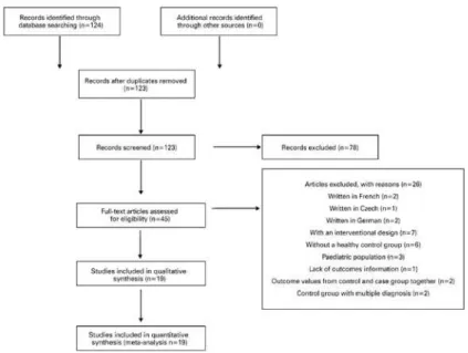
TUDYSELECTIONA total of 124 articles were identiied with the search criteria. The abstract from each article was used for screening, and one of the abstracts was found to be duplicated. After screening, we found
45 studies, and of these, 2 were written in French, 2 were written in German, 1 was written in Czech, 3 included pediatric populations, 1 had no outcome information, 2 had a case group with a glaucoma diagnosis and other diagnoses, 2 provided data from the control and case groups together, 6 had a non-healthy control group, and 7 were interventional studies.
Therefore, for comparative and quantitative purposes, 19 studies
between 2008 and 2016 were considered (Table 1)(11-29). The study
selection information is presented in igure 1, according to Preferred Reporting Items for Systematic Reviews and Meta-Analyses (PRISMA) guidelines(30).
D
ATACOLLECTIONPROCESSANDSTATISTICALANALYSISThe selected full texts were collected and assessed for demographic data and reported outcomes. For statistical analysis, we used the mean diference (MD) between groups (patient and control) for CH and CCT.
RESULTS
From a total of 124 studies screened, only 19 complied with our eligibility criteria, as shown in igure 1. Table 1 and igures 2 and 3 summarize the mean and standard deviation (SDs) of both CH and CCT for the control and case groups of each study(11-29).
S
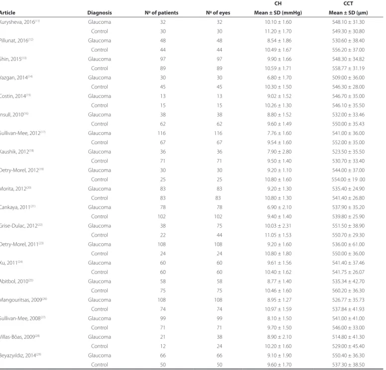
YNTHESISOFRESULTSA total of 1,213 glaucoma eyes from 1,159 glaucoma patients and 1,055 healthy eyes from 1,021 healthy subjects were considered in our study. Table 1 shows the baseline characteristics of these participants and their eye-related parameters.
Quantitative analysis showed that CH was signiicantly lower in the glaucoma group than in the control group (MD=-1.54 mmHg, 95% CI [-1.68, -1.41], p<0.00001; Figure 2). Additionally, CCT was signiicantly lower in the glaucoma group than in the control group (MD=-8.49 µm, 95% CI [-11.36 to -5.62], p<0.001; Figure 3).
DISCUSSION
The latest evidence regarding the true value of CCT as a risk factor for glaucoma is still unclear. While some studies have con-sidered CCT as an important risk factor for the development of
glau coma(4-6,10), others such as the EMGT, Barbados Eye Study, and
studies by Chauhan et al. and Congdon et al., did not find a simple
and linear relationship between CCT and glaucoma(2,4). According
to our study, there was a signiicantly lower CCT value among glau-coma patients than among controls (MD=-8.49 µm, 95% CI [-11.36 to -5.62], p<0.001).
The ORA device provides several biomechanical properties that are assumed to be less inluenced by CCT when compared with GAT factors, namely CH, which is a biomechanical property related to the viscoelasticity of the cornea. According to our results, there was a sig-niicantly lower CH among glaucoma patients than among controls (MD=-1.54 mmHg, 95% CI [-1.68, -1.41], p<0.00001), and this inding is consistent with previous results from other studies(2,9). As the ORA is a
non-contact tonometer(2,3,5,9), parameters measured using this device
may be more reliable than those measured using GAT(2,3).
Table 1. Baseline characteristics
Article Diagnosis No of patients No of eyes
CH CCT
Mean ± SD (mmHg) Mean ± SD (µm)
Kurysheva, 2016(11) Glaucoma 032 032 10.10 ± 1.60 548.10 ± 31.30
Control 030 030 11.20 ± 1.70 549.30 ± 30.80 Pillunat,2016(12) Glaucoma 048 048 08.54 ± 1.86 530.60 ± 38.40
Control 044 044 10.49 ± 1.67 556.20 ± 37.00 Shin,2015(13) Glaucoma 097 097 09.90 ± 1.66 548.30 ± 34.82
Control 089 089 10.59 ± 1.71 558.77 ± 31.19 Yazgan,2014(14) Glaucoma 030 030 06.80 ± 1.70 509.00 ± 36.00
Control 045 045 10.30 ± 1.50 546.30 ± 28.00 Costin,2014(15) Glaucoma 013 013 09.02 ± 1.52 546.70 ± 35.00
Control 015 015 10.26 ± 1.30 546.10 ± 35.50 Insull,2010(16) Glaucoma 038 038 08.80 ± 1.52 532.00 ± 33.46
Control 062 062 09.60 ± 1.49 550.00 ± 35.43 Sullivan-Mee,2012(17) Glaucoma 116 116 07.76 ± 1.60 541.00 ± 36.00
Control 067 067 09.54 ± 1.60 552.00 ± 35.00 Kaushik,2012(18) Glaucoma 036 036 07.90 ± 2.80 523.50 ± 35.50
Control 071 071 09.50 ± 1.40 530.70 ± 33.40 Detry-Morel, 2012(19) Glaucoma 030 030 09.20 ± 1.10 544.00 ± 37.00
Control 025 025 10.80 ± 1.60 554.00 ± 19 .00 Morita,2012(20) Glaucoma 083 083 09.20 ± 1.30 535.40 ± 24.90
Control 083 83 10.80 ± 1.30 541.40 ± 26.80
Cankaya,2011(21) Glaucoma 078 078 06.90 ± 2.10 537.90 ± 35.20
Control 102 102 09.40 ± 1.40 539.80 ± 25.90
Grise-Dulac,2012(22) Glaucoma 038 075 10.03 ± 2.31 551.50 ± 38.90
Control 022 044 11.05 ± 1.53 550.70 ± 29.30 Detry-Morel,2011(23) Glaucoma 108 108 09.20 ± 1.60 536.00 ± 61.00
Control 024 024 10.80 ± 1.80 550.00 ± 36.00 Xu,2011(24) Glaucoma 060 060 09.61 ± 1.56 541.40 ± 37.46
Control 060 060 10.40 ± 1.62 541.75 ± 26.07 Abitbol,2010(25) Glaucoma 058 058 08.77 ± 1.40 535.34 ± 42.70
Control 075 075 10.46 ± 1.60 560.20 ± 36.30 Mangouritsas,2009(26) Glaucoma 108 108 08.95 ± 1.27 526.77 ± 35.73
Control 074 074 10.97 ± 1.59 537.84 ± 41.93 Sullivan-Mee,2008(27) Glaucoma 099 099 08.10 ± 1.50 541.00 ± 41.00
Control 071 071 09.70 ± 1.50 546.00 ± 33.00 Villas-Bôas,2009(28) Glaucoma 021 038 08.90 ± 2.10 514.80 ± 41.30
Control 012 024 10.20 ± 1.60 529.00 ± 45.40 Beyazyıldız,2014(29) Glaucoma 066 066 09.10 ± 1.90 550.40 ± 36.30
Control 050 050 09.60 ± 1.70 537.30 ± 38.50 CCT= central corneal thickness; CH= corneal hysteresis; No=number; SD= standard deviation.
population(5-7), is very important. Thus, it can become a valuable tool
in various assessments, such as assessment of the stratiication risk of glaucoma patients and even prognosis. However, the ORA is not commonly found in ophthalmology clinics worldwide, and this limits the knowledge of CH in glaucoma.
The results of our study provide strong evidence on the use of CCT and CH. Furthermore, it should be pointed out that this is the irst review of the literature and meta-analysis on the topic of CH in glau-coma. However, we recognize that our study has some limitations.
Figure 1. Study low diagram.
CI= conidence interval; SD= standard deviation. Figure 2. Corneal hysteresis - forest plot.
2005. Therefore, this study had a relatively short review period. Finally, we recognize some other limitations, including the fact that this was not a systematic review, risk of bias evaluation was not performed, and only observational studies were considered to eliminate the risk of bias from interventions.
In conclusion, this study reveals a signiicant diference in CH and CCT between glaucoma patients and healthy controls. These results indicate that there may be better assessments beyond CCT measure-ment alone. Therefore, it is important to keep searching for new and more sophisticated tools to measure corneal properties such as CH in order to deepen our knowledge of this subject.
REFERENCES
1. Garway-Heath DF, Crabb DP, Bunce C, Lascaratos G, Amalitano F, Anand N, et al. Lata-noprost for open-angle glaucoma (UKGTS): A randomised, multicentre, placebo-controlled trial. Lancet. 2015;385(9975):1295-304.
2. Deol M, Taylor DA, Radclife, NM. Corneal hysteresis and its relevance to glaucoma. Cur Opin Ophthalmol. 2015;26(2):96-102.
3. Yaoeda K, Fukushima A, Shirakashi M, Fukuchi T. Comparison of intraocular pressure ad-justed by central corneal thickness or corneal biomechanical properties as measured in glaucomatous eyes using noncontact tonometers and the Goldmann applanation tonometer. Clin Ophthalmol. 2016;10:829-34.
4. Iester M, Mete M, Figus M, Frezzotti P. Incorporating corneal pachymetry into the ma-nagement of glaucoma. J Cataract Refract Surg. 2009;35(9):1623-8.
5. Brandt J. Central corneal thickness, tonometry, and glaucoma risk-a guide for the per plexed. Can J Ophthalmol. 2007;42(4):562-6.
6. European glaucoma prevention study group. Central corneal thickness in the European glaucoma prevention study. Ophthalmology. 2007;114(3):454-9.
7. Brandt JD, Beiser JA, Kass MA, Gordon MO; Ocular Hypertension Treatment Study Group. Central corneal thickness in the ocular hypertension treatment study (OHTS). Ophthalmology. 2001;108(10):1779-88.
8. Hong Y, Shoji N, Morita T, Hirasawa K, Matsumura K, Kasahara M, et al. Comparison of corneal biomechanical properties in normal tension glaucoma patients with diferent visual ield progression speed. International J Ophthalmol. 2016;9(7):973-8. 9. Garcia-Porta N, Fernandes P, Queiros A, Salgado-Borges J, Paraita-Mato M, González-Méi jome JM. Corneal biomechanical properties in diferent ocular conditions and new measurement techniques. ISRN Ophthalmology. 2014;2014:724546. doi: 10.1155/2014/724546. eCollection 2014.
10. Brandt JD, Gordon MO, Beiser JA, Lin SC, Alexander MY, Kass MA; Ocular Hypertension Treatment Study Group. Changes in central corneal thickness over time. The ocular hypertension treatment study. Ophthalmology. 2008;115(9):1550-6.
11. Kurysheva NI, Parshunina OA, Shatalova EO, Kiseleva TN, Lagutin MB, Fomin AV. Value of structural and hemodynamic parameters for the early detection of primary open-angle glaucoma. Cur Eye Res. 2016;3683:1-7.
12. Pillunat KR, Hermann C, Spoerl E, Pillunat LE. Analyzing biomechanical parameters of the cornea with glaucoma severity in open-angle glaucoma. Graefe’s Arch Clin Exp Ophthalmol. 2016;254(7):1345-51.
13. Shin J, Lee JW, Kim EA, Caprioli J. The efect of corneal biomechanical properties on rebound tonometer in patients with normal-tension glaucoma. Am J Ophthalmol. 2015;159(1):144-54.
14. Yazgan S, Celik U, Alagöz N, Taş M. Corneal Biomechanical comparison of pseudoexfolia-tion syndrome, pseudoexfoliative glaucoma and healthy subjects. Cur Eye Res. 2015; 40(5):470-5.
15. Costin BR, Fleming GP, Weber PA, Mahmoud AM, Roberts CJ. Corneal biomechanical properties afect Goldmann applanation tonometry in primary open-angle glaucoma. J Glaucoma. 2014;23(2):69-74.
16. Insull E, Nicholas S, Ang GS, Poostchi A, Chan K, Wells A. Optic disc area and correla-tion with central corneal thickness, corneal hysteresis and ocular pulse amplitude in glaucoma patients and controls. Clin Exp Ophthalmol. 2010;38(9):839-44. 17. Sullivan-Mee M, Katiyar S, Pensyl D, Halverson KD, Qualls C. Relative importance of
factors afecting corneal hysteresis measurement. Optom Vis Sci. 2012;89(5):e803-11. 18. Kaushik S, Pandav SS, Banger A, Aggarwal K, Gupta A. Relationship between corneal
biomechanical properties, central corneal thickness, and intraocular pressure across the spectrum of glaucoma. Am J Ophthalmol. 2012;153(5):840-9.e2.
19. Detry-Morel M, Jamart J, Hautenauven F, Pourjavan S. Comparison of the corneal biomechanical properties with the Ocular Response Analyzer® (ORA) in African and Caucasian normal subjects and patients with glaucoma. Acta Ophthalmol. 2012;90(2): 118-24.
20. Morita T, Shoji N, Kamiya K, Fujimura F, Shimizu K. Corneal biomechanical properties in normal-tension glaucoma. Acta Ophthalmol. 2012;90(1):48-53.
21. Cankaya AB, Anayol A, Özcelik D, Demirdogen E, Yilmazbas P. Ocular response ana lyzer to assess corneal biomechanical properties in exfoliation syndrome and exfoliative glaucoma. Graefe’s Arch Clin Exp Ophthalmol. 2012;250(2):255-60. 22. Grise-Dulac A, Saad A, Abitbol O, Febbraro JL, Azan E, Moulin-Tyrode C, et al. Assessment
of corneal biomechanical properties in normal tension glaucoma and Comparison with open-angle glaucoma, ocular hypertension, and normal eyes. J Glaucoma. 2012; 21(7):486-9.
23. Detry-Morel M, Jamart J, Pourjavan S. Evaluation of corneal biomechanical properties with the reichert ocular response analyzer. Eur J Ophthalmol. 2011;21(2):138-48. 24. Xu G, Lam DS, Leung CK. Inluence of ocular pulse amplitude on ocular response
analyzer measurements. J Glaucoma. 2011;20(6):344-9.
25. Abitbol O, Bouden J, Doan S, Hoang-Xuan T, Gatinel D. Corneal hysteresis measured with the ocular response analyzer® in normal and glaucomatous eyes. Acta Ophthal-mol. 2010;88(1):116-9.
26. Mangouritsas G, Morphis G, Mourtzoukos S, Feretis E. Association between corneal hysteresis and central corneal thickness in glaucomatous and non-glaucomatous eyes. Acta Ophthalmol. 2009;87(8):901-5.
27. Sullivan-Mee M, Billingsley SC, Patel AD, Halverson KD, Alldredge BR, Qualls C. Ocular Response Analyzer in subjects with and without glaucoma. Optom Vis Sci. o2008; 85(6):463-70.
28. Villas-Bôas F, Doi L, Sousa A, Melo Jr L. Correlation between diurnal variation of intra-ocular pressure, intra-ocular pulse amplitude and corneal structural properties. Arq Bras Oftalmol. 2009;72(3):296-301.
29. Beyazyıldız E, Beyazyıldız Ö, Arifoğlu HB, Altıntaş AK, Köklü ŞG. Comparison of ocular response analyzer parameters in primary open angle glaucoma and exfoliative glau-coma patients. Ind J Ophthalmol. 2014;62(7):782-7.