Journal of Investigative Medicine High Impact Case Reports
July-September 2016: 1 –7 © 2016 American Federation for Medical Research
DOI: 10.1177/2324709616665409 hic.sagepub.com
Creative Commons CC-BY: This article is distributed under the terms of the Creative Commons Attribution 3.0 License (http://www.creativecommons.org/licenses/by/3.0/) which permits any use, reproduction and distribution of the work without further permission provided the original work is attributed as specified on the SAGE and Open Access pages (https://us.sagepub.com/en-us/nam/open-access-at-sage). Case Report
Introduction
Posterior cerebral artery (PCA) cortical branches supply blood to the occipital lobe, the inferomedial temporal lobe, and portions of the posterior inferior parietal lobe.1 Most adult humans have the classic vascular anatomy in which both left and right PCAs originate from the basilar artery and are part of the vertebrobasilar system or posterior circula-tion.2 An anatomic variant of the PCA, known as fetal-type or fetal PCA (FPCA), has been detected by anatomic3-6 and angiographic7-10 studies in 11% to 46% of adult humans, either unilaterally or bilaterally. Differences in detection method and definition may account for the variance in reported prevalence. In the definition proposed by van Raamt et al, an FPCA is called a full FPCA if the P1 segment is not visualized on computed tomography angiography (CTA), magnetic resonance angiography (MRA), or after injection of contrast into the vertebral artery; a partial FPCA if the P1 segment is smaller than the posterior communicating artery (PcomA); or an intermediate FPCA if the P1 segment is as large as the PcomA.11 When present, the FPCA supplies
blood to parts of the cerebrum, which, in the majority of humans, are perfused by distal PCA branches.7,8 Like the middle cerebral artery (MCA) and the anterior cerebral artery (ACA), the FPCA is a branch of the internal carotid artery (ICA) and is therefore a part of the carotid system or anterior circulation.
Although the FPCA is considered a normal anatomic variant, its presence may modify the distribution and sever-ity of cerebral injury from thromboembolic events. A few patients with acute ischemic stroke and simultaneous or
1
Louisiana State University School of Medicine, New Orleans, LA, USA 2
Louisiana State University Health Sciences Center, New Orleans, LA, USA
Received June 20, 2016. Revised July 22, 2016. Accepted July 30, 2016.
Corresponding Author:
Stephen L. Lambert, BS, Louisiana State University School of Medicine, 1901 Perdido Street, New Orleans, LA 70112, USA.
Email: [email protected]
Fetal-Type Variants of the Posterior
Cerebral Artery and Concurrent
Infarction in the Major Arterial
Territories of the Cerebral Hemisphere
Stephen L. Lambert, BS
1, Frank J. Williams, MD
2,
Zhora Z. Oganisyan, MD
2, Lionel A. Branch, MD
2,
and Edward C. Mader Jr, MD
2Abstract
Fetal-type or fetal posterior cerebral artery (FPCA) is a variant of cerebrovascular anatomy in which the distal posterior cerebral artery (PCA) territory is perfused by a branch of the internal carotid artery (ICA). In the presence of FPCA, thromboembolism in the anterior circulation may result in paradoxical PCA territory infarction with or without concomitant infarction in the territories of the middle (MCA) or the anterior (ACA) cerebral artery. We describe 2 cases of FPCA and concurrent acute infarction in the PCA and ICA territories—right PCA and MCA in Patient 1 and left PCA, MCA, and ACA in Patient 2. Noninvasive angiography detected a left FPCA in both patients. While FPCA was clearly the mechanism of paradoxical infarction in Patient 2, it turned out to be an incidental finding in Patient 1 when evidence of a classic right PCA was uncovered from an old computed tomography scan image. Differences in anatomical details of the FPCA in each patient suggest that the 2 FPCAs are developmentally different. The FPCA of Patient 1 appeared to be an extension of the embryonic left posterior communicating artery (PcomA). Patient 2 had 2 PCAs on the left (PCA duplication), classic bilateral PCAs, and PcomAs, and absent left anterior choroidal artery (AchoA), suggesting developmental AchoA-to-FPCA transformation on the left. These 2 cases underscore the variable anatomy, clinical significance, and embryological origins of FPCA variants.
Keywords
2 Journal of Investigative Medicine High Impact Case Reports
nearly simultaneous PCA-ICA territory infarction were found to have an FPCA ipsilateral to the paradoxical PCA occlusion.12-15 Concurrent infarction occurred in the PCA and MCA territories of one hemisphere,12-14 except in one case where infarction occurred in the PCA, MCA, and ACA territories of the right hemisphere.15 We report 2 cases of concurrent ICA-PCA territory infarction in the setting of a unilateral FPCA. An FPCA was clearly visible on the left in both patients. Infarction occurred in the right MCA and PCA territories in Patient 1 and in the left ACA, MCA, and PCA territories in Patient 2.
Case Presentation
Both patients are 82-year-old right-handed women, both had a large ischemic stroke involving the anterior and posterior circulation territories of one hemisphere, and both were found to have an FPCA. All of these coincidences can be confusing, so we must point out that the patients were not related and that one patient had a stroke several months before the other. In the emergency department, both patients had focal neurological deficits and head CT did not show hemorrhage. Neither patient qualified for tissue plasminogen activator therapy because of the extent of cerebral injury and the presence of at least one more contraindication: the absence of a clear time of symptom onset in Patient 1 and
ongoing warfarin therapy in Patient 2 (prothrombin time = 21.3, international normalized ratio = 1.9).
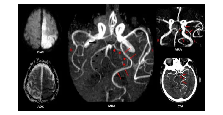
left vertebral artery (Figure 1: MRA). Because the FPCA was contralateral to the infarcted hemisphere, we hypothesized the presence of FPCAs bilaterally with the right FPCA invisible because it was occluded. However, an old CT image (taken 2½ years prior to admission and retrieved from her old records) revealed a classic right PCA (Figure 1: CT). Electrocardiography showed normal sinus rhythm, and echocardiography revealed moderate mitral and aortic regurgitation. Because of cerebral edema, her sensorium declined over the first few days and even-tually stabilized. Unfortunately, she did not regain full alertness and her deficits did not improve despite early physiotherapy in the acute care setting. After 3 weeks, she was transferred to a long-term care facility.
Patient 2 had 3 major stroke risk factors—hypertension, hyperlipidemia, and atrial fibrillation—for which she takes warfarin. Before the stroke, she lived in an assisted living facility and was able to drive a car. She was preparing a meal when another person found her slouched over a drawer. Emergency responders noted her to be somnolent, rousing occasionally to painful stimulation, but without speech output. On arrival to the emergency room, her vital signs were as fol-lows: BP = 167/82, HR = 85/min (irregular), RR = 20, and temperature = 36.7°C. Her neurological examination was remarkable for profound impairment of language function involving comprehension and expression, distal > proximal pattern of weakness of the right upper (2/5) and right lower
(1/5) extremities, and a right Babinski sign (NIHSS score = 31). It was impossible to assess her aphasia amid her depressed sensorium, the latter most likely due to cerebral edema and mass effect from the large stroke. Brain MRA on the second hospital day showed acute infarcts in the left ACA, MCA, and PCA territories (Figure 2: DWI and ADC). MRA and CTA did not show any flow-disrupting stenosis but an anatomic variant, known as PCA duplication, was present on the left—the dominant PCA was an FPCA and the smaller PCA was a classic PCA (Figure 2: MRA and CTA). Other findings include bilateral classic PCAs and posterior commu-nicating arteries (PcomAs) and an anterior choroidal artery (AchoA) on the right but not on the left (Figure2: middle MRA and CTA). Electrocardiography showed atrial fibrillation and echocardiography revealed biatrial enlargement. Her mental status declined over a few days due to worsening cerebral edema. An ictal contribution to the depressed sensorium was confirmed when electroencephalography showed intermittent epileptiform activity in the left temporal region. Intravenous levetiracetam was administered but it was the regression of cerebral edema that correlated with the resolution of cortical hyperexcitability. Because of the size of the infarct, warfarin was temporarily placed on hold. She became slightly more arousable but neither her language function nor her strength on the right showed signs of improvement. After 12 days, she was discharged to a skilled nursing facility.
4 Journal of Investigative Medicine High Impact Case Reports
Discussion
Both patients had acute ischemic strokes (high DWI signal intensity with corresponding low ADC) and concurrent infarc-tion (simultaneous occurrence of symptoms and similar DWI signal intensities) in the ICA and PCA territories of one hemi-sphere (Figures 1 and 2: DWI and ADC images). Noninvasive cerebral angiography detected a left FPCA in both patients (Figure 1: MRA images; Figure 2: MRA and CTA images). However, the clinical significance and developmental implica-tions of FPCA in each patient are quite different.
Emboli can move up the ICA, enter and occlude the FPCA or its branches, and result in a paradoxical PCA territory infarction—with12-15 or without16-20 attendant occlusion of other ICA branches. A left FPCA was clearly the reason for the left-sided paradoxical infarction in Patient 2. On the other hand, the reason for the right-sided paradoxical infarction in Patient 1 was not obvious. MRA showed high-grade stenosis of the right ICA petrous segment (see Figure 1 for complete MRA findings) indicating that right ICA thrombosis and embolism resulted in right MCA and PCA territory infarc-tion, the latter via a right FPCA. However, MRA showed a left FPCA and absent right PCA (except for a P1 remnant). As there was no concrete evidence of a right FPCA, we argued—as Eswaradass et al did in their case of concurrent ICA and PCA territory infarction15—that an FPCA was pres-ent ipsilateral to the infarct but was angiographically invisi-ble because it was occluded. This argument was proved wrong when we discovered evidence of a classic right PCA in the patient’s old CT scan files (Figure 1: CT). All things considered, the most likely mechanism of paradoxical PCA occlusion in Patient 1 is right ICA-to-PCA embolism through a patent PcomA. Although an FPCA has been implicated in nearly all cases of paradoxical PCA occlusion,12-20 the PcomA is a potential conduit for cross embolization from the ICA to the PCA P2 segment or its distal branches.12,21 The rarity of PcomA embolism may be due to the low ICA-PCA pressure gradient across the PcomA.12 Factors that increase the ICA-PCA pressure gradient, such as hemodynamic changes from stenotic lesions in the vertebrobasilar system or the site of ICA thrombosis relative to the PcomA orifice, may increase the risk of PcomA embolism.
Cardioembolism with concomitant PCA and MCA branch occlusion is a plausible mechanism of PCA-MCA territory infarction in Patient 1, especially since she had atrial fibrilla-tion in the past. However, she was in normal sinus rhythm throughout her hospital stay and echocardiography did not show any embolic source (albeit a transesophageal study was not performed). The study of Yang et al also argue against a cardioembolic mechanism in Patient 1.12 In this study, simulta-neous ipsilateral anterior and posterior circulation territory infarction was present in only 21 (1.5%) of 1388 acute stroke patients. Of the 21 patients, 16 had angiographic evidence of ICA stenosis ipsilateral to the infarct and only one suffered car-dioembolism. All but one patient with concurrent PCA-MCA infarction and an FPCA or a patent PcomA had significant ICA
stenosis ipsilateral to the infarct.12 In earlier reports of FPCA and concurrent PCA-ICA infarction, only the MCA (not the ACA) territory of the ICA was involved.12-14 Recently, Eswaradass et al reported the first case of simultaneous PCA, MCA, and ACA territory infarction in association with a pos-sible FPCA; MRA showed complete occlusion of the right ICA and a right FPCA was inferred based on the absence of the P1 segment on MRA.15 Patient 2 also had ACA, MCA, and PCA territory infarction but, unlike the case of Eswaradass et al, a definite FPCA was found (along with a classic PCA) ipsilateral to the stroke (Figure 2: MRA and CTA). In the absence of ICA stenosis and in the presence of atrial fibrillation, the 3-territory stroke in Patient 2 is most likely the result of cardioembolism to the right ACA, MCA, and FPCA (all 3 vessels are branches of the right ICA).
The label “fetal PCA” has been attached to a number of developmental variants of the adult cerebral arterial system where a significant portion of the distal PCA territory is per-fused through a branch of the ICA. However, experts do not always agree on whether or not a particular variant should be called “fetal PCA.”22 The new nomenclature of PCA variants proposed by Masoud et al avoids the term “fetal PCA.”23 Some authors emphasize the importance of distinguishing PCA variants that are “truly fetal” from those that are not.24 Different PCA variants can only be properly understood in the context of vascular neuroembryology.25 The reader whose field entails a deep understanding of vascular neuro-embryology can benefit immensely from the website http:// neuroangio.org/22,25 and from excellent publications on this subject, some of which are included in our bibliography.26-33 We also created highly simplified and idealized diagrams to help the reader acquire a basic idea of cerebrovascular embryology (Figure 3).
outcomes are possible but only these 3 patterns are relevant to our discussion. Figure 3B shows how the typical PCA pattern is achieved. The MCA and PCA take over the dominant roles of the AchoA and PchoA, respectively. Both choroidal arteries regress but usually do not disappear. Part of the PchoA is annexed by the evolving PCA. The cICA regresses becoming the PcomA. Figure 3C shows how the common FPCA variant develops. The cICA (PcomA) continues to be dominant and becomes the proximal FPCA segment that connects with the ICA. The P1 segment of the PCA regresses and may disappear (absent in diagram). This is the case in Patient 1. Figure 3D shows how the “true fetal” PCA variant develops. Two PCAs emerge (PCA duplication)—one PCA is derived from the AchoA (often dominant) and the other PCA is a classic PCA (often smaller). This is the case in Patient 2. Some authors refer to this anatomic variant, not as FPCA or “true fetal” PCA, but as hyperplastic AChA.24,32,33
With a prevalence rate in the 11% to 46% range3-10 and an established role in the pathogenesis of multi-territory or par-adoxical strokes,12-21 FPCA certainly deserves more clinical attention. FPCA should be suspected in patients with infarcts in the anterior and posterior circulation territories of one hemisphere. Currently, the fastest and safest method for detecting FPCA is MRA or CTA. High-grade carotid stenosis
and FPCA ipsilateral to the infarcted hemisphere may obvi-ate additional workup in search of a cardioembolic source. If the stroke cannot be explained by carotid disease, the likeli-hood of cardioembolism is high; transesophageal echocar-diography, mobile cardiac outpatient telemetry, or loop recorder should be considered if transthoracic echocardiog-raphy or Holter monitoring are unrevealing. By being aware that an FPCA exists, surgeons can plan their surgical approach or endovascular procedure to minimize the risk of perioperative cerebral hypoperfusion or embolization.34,35 Preoperative vascular imaging and assessment of collateral blood flow is important in planning carotid endarterectomy or stenting.36 FPCA ipsilateral to the stenotic ICA is a com-pelling reason to use embolic protection devices and to implement intraoperative monitoring to prevent cerebral hypoperfusion during carotid endarterectomy. Failure to rec-ognize an FPCA can also lead to errors in interpreting cere-bral perfusion studies. Wentland et al found that a unilateral FPCA can affect perfusion measurements and give rise to perfusion map asymmetries.37
6 Journal of Investigative Medicine High Impact Case Reports
reported a case of sequential infarction (first left PCA, then left MCA territory) in the setting of left FPCA and extracra-nial ICA stenosis; stabilization of cerebral perfusion was achieved by carotid endarterectomy.14 Albeit intuitively appealing, there is no solid evidence in favor of more aggres-sive antiplatelet or anticoagulant regimens in individuals with FPCA and major stroke risk factors. It is also not clear whether FPCA increases the overall risk of stroke indepen-dent of other risk factors. Yang et al found a “causative” FPCA ipsilateral to the infarct in 44.4% of acute stroke patients but only 18.5% of individuals in their control group have “incidental” FPCA.12 Two autopsy studies found more FPCAs in brains with infarcts than in infarct-free brains.38,39 According to van Raamt et al, a full FPCA carries a higher vascular insufficiency risk than a partial FPCA because lep-tomeningeal anastomoses do not form between the anterior and posterior circulation if a person has a full FPCA.11 After reviewing the literature, Brozici et al realized the wide range of individual variability in the size, distribution, and number of leptomeningeal anastomoses; the authors pointed out that studies addressing the range of variability or that link the variability to compensatory capacity are lacking.40 Clinical studies have had mixed results: increased risk of cerebral ischemia from FPCA,41 enhanced risk only with full FPCA,42 greater risk with partial FPCA,43 and no increase in stroke risk44 have all been observed. Whether FPCA increases the overall risk of stroke independent of other risk factors is still an open question.
Conclusion
These 2 cases of fetal PCA (FPCA) and concurrent PCA-ICA territory infarction illustrate the variability in anatomy, clinical significance, and embryological origin of PCA variants. FPCA is clearly the mechanism of paradoxical infarction in one patient. In the other patient, FPCA is only an incidental finding, with an old CT scan image (not an angiogram) serving as confirmatory evidence. Differences in angiographic anatomy indicate that the FPCAs of the 2 patients are developmentally distinct—the FPCA of one patient appears to be an extension of the embryonic left posterior communicating artery, while the FPCA of the other patient is most likely derived from an embryonic anterior choroidal artery.
FPCA can increase the extent and severity of anterior cir-culation strokes by allowing additional infarction in the PCA territory. The physician should therefore vigorously address the stroke risk factors of individuals with FPCA, such as ICA stenosis and atrial fibrillation. Whether FPCA increases the overall risk of stroke independent of other risk factors is not clear. The optimal stroke prevention regimen for individuals with FPCA and one or more stroke risk factors is also unclear. With increasing accessibility to noninvasive neurovascular imaging, it will not be long before clinical researchers find solutions to these problems.
Declaration of Conflicting Interests
The author(s) declared no potential conflicts of interest with respect to the research, authorship, and/or publication of this article.
Funding
The author(s) received no financial support for the research, author-ship, and/or publication of this article.
References
1. Chaves C, Caplan LR. Posterior cerebral artery. In: Caplan
LR, van Gijn J, eds. Stroke Syndromes. 3rd ed. Cambridge,
England: Cambridge University Press; 2012:405-418.
2. Sandhu JS, Wakhloo AK. Neuroangiographic anatomy and common cerebrovascular diseases. In: Bradley WG,
Daroff RB, Fenichel GM, Jankovic J, eds. Neurology in
Clinical Practice. Principles of Diagnosis and Management. 4th ed. Philadelphia, PA: Butterworth Heinemann; 2004: 625-643.
3. Alpers BJ, Berry RG, Paddison RM. Anatomical studies of the
circle of Willis in normal brain. AMA Arch Neurol Psychiatry.
1959;81:409-418.
4. Saeki N, Rhoton AL Jr. Microsurgical anatomy of the upper
basilar artery and the posterior circle of Willis. J Neurosurg.
1977;46:563-578.
5. Zeal AA, Rhoton AL Jr. Microsurgical anatomy of the
poste-rior cerebral artery. J Neurosurg. 1978;48:534-559.
6. Pedroza A, Dujovny M, Artero JC, et al. Microanatomy of
the posterior communicating artery. Neurosurgery. 1987;20:
228-235.
7. Jongen JC, Franke CL, Soeterboek AA, Versteege CW, Ramos LM, van Gijn J. Blood supply of the posterior cerebral artery
by the carotid system on angiograms. J Neurol. 2002;249:
455-460.
8. Bulsara KR, Zomorodi A, Provenzale JM: Anatomic
vari-ant of the posterior cerebral artery. AJR Am J Roentgenol.
2007;188:W395.
9. van der Lugt A, Buter TC, Govaere F, Siepman DA, Tanghe HL, Dippel DW. Accuracy of CT angiography in the
assess-ment of a fetal origin of the posterior cerebral artery. Eur
Radiol. 2004;14:1627-1633.
10. Kovač JD, Stanković A, Stanković D, Kovač B, Šaranović D. Intracranial arterial variations: a comprehensive evaluation
using CT angiography. Med Sci Monit. 2014;20:420-427.
11. van Raamt AF, Mali WP, van Laar PJ, van der Graaf Y. The fetal variant of the circle of Willis and its influence on the
cerebral collateral circulation. Cerebrovasc Dis. 2006;22:
217-224.
12. Yang JH, Choi HY, Nam HS, Kim SH, Han SW, Heo JH. Mechanism of infarction involving ipsilateral carotid and
pos-terior cerebral artery territories. Cerebrovasc Dis. 2007;24:
445-451.
13. Senol MG, Velioğlu M, Toğrol E, Ozdağ F, Saraçoğlu M. Simultaneous posterior and middle cerebral artery infarct.
Neurol India. 2009;57:673-674.
14. Kolukısa M, Gürsoy AE, Kocaman G, Dürüyen H, Toprak H, Asil T. Carotid endarterectomy in a patient with posterior cere-bral artery infarction: influence of fetal type PCA on atypical
15. Eswaradass PV, Ramasamy B, Ramadoss K, Gnanashanmugham G. Can internal carotid artery occlusion produce
simultane-ous anterior and posterior circulation stroke? Indian J Vasc
Endovasc Surg. 2015;2:130-132.
16. Hunter JM, Tehrani SK, Wood T, Geraghty R. Internal carotid artery stenosis presenting as ipsilateral posterior cerebral
artery ischaemic stroke: a lesson to be learnt. BMJ Case Rep.
2013;2013. doi:10.1136/bcr-2013-008848.
17. Cucchiara BL, Kasner SE. Carotid dissection causing occipital
lobe infarction. Neurology. 2005;65:1408.
18. Kuker W, Mull M, Block F, Thron A. Carotid artery dissec-tions presenting as isolated posterior cerebral artery
infarc-tions. J Neurol. 1997;244:324-327.
19. Hoque R, Gonzalez-Toledo E, Minagar A, Kelley RE. Circuitous embolic hemorrhagic stroke: carotid
pseudoaneu-rysm to fetal posterior cerebral artery conduit: a case report. J
Med Case Rep. 2008;2:61.
20. Gooneratne IK, Gamage R, Gunarathne KS. Internal carotid
artery dissection: an unusual cause of occipital infarction. Ann
Indian Acad Neurol. 2010;13:148-149.
21. Libman RB, Lustrin ES. Posterior cerebral artery infarction
associated with carotid dissection. J Stroke Cerebrovasc Dis.
1998;7:157-160.
22. Shapiro M. Posterior cerebral artery. http://neuroangio.org/ anatomy-and-variants/posterior-cerebral-artery. Accessed June 15, 2016.
23. Masoud H, Nguyen TN, Thatcher J, Barest G, Norbash AM. Duplication of the posterior cerebral artery and the “true fetal”
variant. Interv Neurol. 2015;4:64-67.
24. Uchino A, Saito N, Takahashi M, Okano N, Tanisaka M. Variations of the posterior cerebral artery diagnosed by MR
angiography at 3 tesla. Neuroradiology. 2016;58:141-146.
25. Shapiro M. Vascular neuroembryology. http://neuroangio. org/neurovascular-evolution/vascular-neuroembryology/. Accessed June 15, 2016.
26. Menshawi K, Mohr JP, Gutierrez J. A functional perspective
on the embryology and anatomy of the cerebral blood supply. J
Stroke. 2015;17:144-158.
27. Padget D. The circle of Willis: its embryology and anatomy. In:
Dandy WE, ed. Intracranial Arterial Aneurysms. Ithaca, NY:
Comstock Publishing; 1944:67-90.
28. Paget DH. The development of the cranial arteries in the human
embryo. Contrib Embryol. 1948;32:205-262.
29. Moffat DB: The development of the posterior cerebral artery. J
Anat. 1961;95:485-494.
30. Lasjaunias P, Berenstein A, TerBrugge KG. Surgical
Neuroangiography. Part 1. Clinical Vascular Anatomy and Variations. 2nd ed. Berlin, Germany: Springer-Verlag; 2001.
31. Luh GY, Dean BL, Tomsick TA, Wallace RC. The
persis-tent fetal carotid-vertebrobasilar anastomoses. AJR Am J
Roentgenol. 1999;172:1427-1432.
32. Takahashi S, Suga T, Kawata Y, Sakamoto K, Anterior cho-roidal artery: angiographic analysis of variants and anomalies.
AJNR Am J Neuroradiol. 1990;11:719-729.
33. Antonietti LC, Glastonbury CM, Adler F, Wintermark M. Hyperplastic anterior choroidal artery identified using magnetic
resonance angiography: a report of two cases. Cerebrovasc
Dis. 2006;22:450-452.
34. Zada G, Breault J, Liu CY, et al. Internal carotid artery aneu-rysms occurring at the origin of fetal variant posterior cerebral
arteries: surgical and endovascular experience. Neurosurgery.
2008;63(1 suppl 1):ONS55-61.
35. Chen Z, Niu Y, Tang J, et al. Endovascular treatment of pos-terior communicating artery aneurysms in the presence of the
fetal variant of posterior cerebral artery. Interv Neuroradiol.
2015;21:456-461.
36. Krishnaswamy A, Klein JP, Kapadia SR. Clinical
cerebrovas-cular anatomy. Catheter Cardiovasc Interv. 2010;75:530-539.
37. Wentland AL, Rowley HA, Vigen KK, Field AS. Fetal origin of the posterior cerebral artery produces left-right
asymmetry on perfusion imaging. AJNR Am J Neuroradiol.
2010;31:448-453.
38. Kameyama M, Okinaka SH. Collateral circulation of the brain with special reference to atherosclerosis of the major cervical
and cerebral arteries. Neurology. 1963;13:279-286.
39. Battacharji SK, Hutchinson EC, McCall AJ. The circle of Willis: the incidence of developmental abnormalities in normal
and infarcted brains. Brain. 1967;90:747-758.
40. Brozici M, van der Zwan A, Hillen B. Anatomy and
func-tionality of leptomeningeal anastomoses: a review. Stroke.
2003;34:2750-2762.
41. Bisaria KK. Anomalies of the posterior communicating
artery and their potential clinical significance. J Neurosurg.
1984;60:572-576.
42. Shaban A, Albright KC, Boehme AK, Martin-Schild S.
Circle of Willis variants: fetal PCA. Stroke Res Treat.
2013;2013:105937.
43. Arjal RK, Zhu T, Zhou Y. The study of fetal-type poste-rior cerebral circulation on multislice CT angiography and
its influence on cerebral ischemic strokes. Clin Imaging.
2014;38:221-225.
44. de Monyé C, Dippel DW, Siepman TA, Dijkshoorn ML, Tanghe HL, van der Lugt A. Is a fetal origin of the posterior cerebral artery a risk factor for TIA or ischemic stroke? A study with