Vol-7, Special Issue4-May, 2016, pp314-320 http://www.bipublication.com
Research Article
Studying the Effect of Hydro- alcoholic Extract of Valeriana officinalis on the
Number and Size of Raphe Magnus Neurons in Mature Rats
Nahid Sadeghi1, * Mohammadlatif Rastian2, and Sajjad Hatami Joni3
1MSc in Animal Physiology,
Teacher at Education Department of Yasuj, Iran 2
MSc in Medical-Surgical Nursing, Faculty of Nursing and Midwifery, Yasuj University of Medical Sciences, Yasuj, Iran
3General Phisician. Esfahan University of Medical Sciences, Esfahan, Iran
*Corresponding Author: Mohammadlatif Rastian. Email:[email protected]
ABSTRACT:
Introduction & Objective: Effective materials from Valerian officinalis L. have too much usage in the pharmacological industry. It is used as a sedative, anticonvulsion, and antidepressant drug. Serotonin has a widespread role in vital function such as sleep, awareness and calmness. In this study we evaluated the effect of hydrochloric extract of valerian on number and size of raphe magnus neurons in adult rat.
Materials & Methods:In this experimental study, which was conducted at Yasuj University of Medical Sciences in 2009, forty adult Wistar rats, each 170-250 gr, were divided randomly into four groups (one control group and three experimental groups). The animals were injected daily for one month with doses of 300, 400 and 600 mg/kg of the extract. The control group just received distilled water. After transcardial perfusion, the whole brain was separated, then 10 µm sections of the brain stem were prepared, and hematoxylin and eosin (H&E) staining were done. Number and size of raphe magna neurons were observed under light microscope. The gathered data were analyzed by the SPSS software using One-way ANOVA and LSD.
Results: The control group did not statistically show significant changes in number of raphe magna neurons. Comparison of the means of long and short diameter neurons showed significant increases in experimental groups with control group (P<0.05). In experimental groups the neuron nucleuses were more euchromatic than the control group.
Conclusion: Hydrochloric extract of valerian has no effect on raphe magnus neurons, but it is effective on neurons' size. It can be concluded that the extract increases both neurons activity and serotonin secretion.
Keywords:Valerian, Extraction, Raphemagnus, Reticular Formation.
INTRODUCTION
The reticular network takes place in the medulla oblongata, and pons, and midbrain as frills and branches. These branches form the raphe nuclei as
the interconnectedcell densities at the
midlines.The nucleustake place in a symmetrical
and regular state with tail head position in the
brainstem. The characteristic of reticular
(Rostral and Caudal) (1).The formations of these
neuronsinclude large multi-dimensional, the
spindle-shaped, and the small, pear-shaped (2 and 3). According to the previous studies, Raphe nuclei and reticular formation are the regions rich in serotonin (4). The changes of serotonin content in the central nervous system (CNS) lead to change the vital functions such as sleep, appetite,
sexual activity, motion reflexes, body
temperature,Adrenocorticotropic hormone
secretion (1),Prolactin secretion, and the growth (6 and 5). The effect of serotonin in the disorders such as Parkinson's, Korea's, Huntington's, depression, schizophrenia and epilepsy has been proved (7-9).
Chemical or electrical stimulation on the Raphe nucleus especially on the raphe magnus leads to release the serotonin in the spinal cord (10). Most numbers of the serotonergic cells which forward their fibersto the spinal cordtake place in the lower pons, midbrain, and medulla oblongata, so that77.5 % of serotonergic neurons of medulla oblongata are in the raphe nucleus. Therefore it is found that the density of serotonergic cell in the raphe nucleus is very high (11). In the nucleus raphe magnus, serotonergic neurons take place in the formation of the pyramid as a dome so that two sides of them end with the regions where
there are Reticular formation nuclei
(Gigantocellularis (Gi) and Para- gigantocellularis (PGi)).Due to the high density of serotonergic neurons in the nucleus of raphe magnus, the spinal – raphe pathway has been known as a serotonergic pathway for a long time (11).
Valeriana officinalis is an herbaceous plant which belongs to Magnoliopsida class, Asteridae subclass, Dipsacales order, Valerianaceae family, Valeriana L. Gender, and Valeriana officinalis L. species which is found in the temperate regions of Asia, Europe and United states while its Rhizome and branched roots are the usable parts (13 and 12). Noteworthy the active substances of Valeriana officinalis including Valepotriate (8), Didrovaltrate (9) and Isovaltrate (10) are mostly used in the pharmaceutical industry and they are
also applied as sedative, anticonvulsants,
hypnotics, and also for the treatment of depression
(14).Anticonvulsant effects of Valeriana
officinalis extract (16, 15, and 12) and also the effect of this plant on increasing the quality of human sleep and shortening the time to fall asleep in mice have been proven in the various studies (17).Therefore the aim of this study was to investigate the effect of hydro-alcoholic extract of Valeriana officinalis L. on the number and sizeof raphe magnus neurons in mature rats
MATERIALS AND METHODS
This is an experimental study which was conducted at 2009 in the Yasuj University of Medical Sciences. 40 groups of rats from the Wistar race and the weights of 170 to 250 grams were used.All animals were kept in standard animal house and also for 12 hours of darkness and light.The animals were transferred in cages containing 5 animals and they had free access to the food and water.
The protocol of the present research was documented according to international law about the laboratory animals and it was approved in the Ethics Committee of the YasujUniversity of Medical Sciences.
Valeriana officinalis L. was provided from the reputable centers in order to prepare the extract of this plants root. After identification and naming by the botanist, the extraction was performed. In this method, the root of Valeriana officinalis L. was powdered and then the solvent that was ethanol for this research was added. The prepared solution was soaked 24 hours, while the excess solvent was extracted and it was concentrated using the Rotary device after smoothing.
of anesthetized animals were opened for perfusion and cannulas were placed by the tipof the heart in their ascending aorta. Then the blood vessels of the brain was washed with 100 cc saline and then was fixed by 200 ml of fixative substance containing formaldehyde 10% in 0.1 molar phosphate buffer (pH = 7. 4). After exiting the brain from the skull and preparation of the blocks from the brain tissue, truncations with 10 micron diameter were prepared using a rotary microtome BRIGHT 504 model. According to the Paxinos Atlas, the length of the raphe magnus nucleus in the adult rats is 2440 micron. Since the thickness of truncations was 10 microns in this study, then 244 serial truncations were prepared from the head to the tail of nucleus.Afterward the truncations with the frequencies of one to ten and as many as 25 truncations for each animal were stained with hematoxylin and eosin. Neurons were photographed using Olympus microscope model TH-200. In the presentresearch, neurons which had detectable nucleus were counted.The neurons in the magnusraphe nucleus were counted in a square with the area of 90,000 micrometers square using analytical software LS starter1 while their large and small diameters were measured (18). The number of counted neurons was multiplied in number 10 in order to obtain total approximate number of the neurons. The collected data was analyzed using SPSS2, one- way and two- ways
ANOVA and also LSD3.
RESULTS
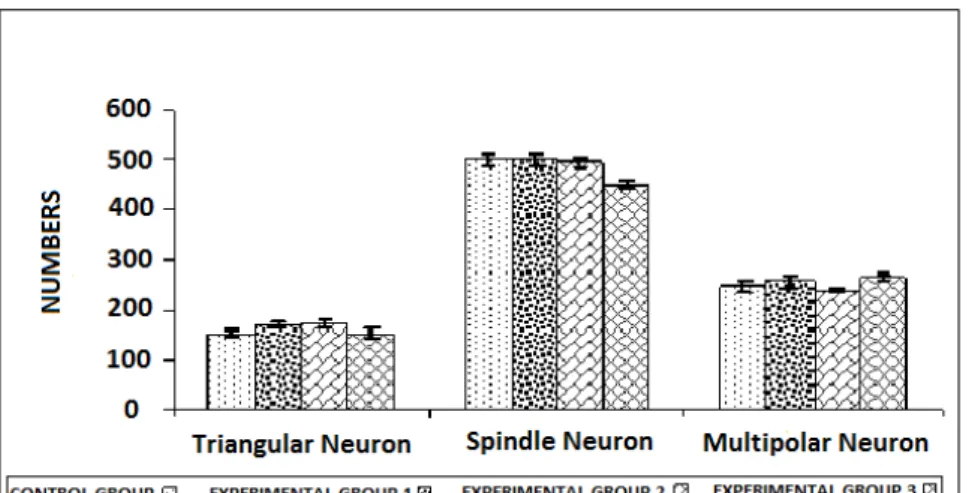
The results of this study showed that three types of morphologies are found in the magnus raphe nucleus with the following frequencies (figures 1 and 2);53 % of spindle neurons, multipolar neurons with 28 % and triangular neurons 18%. The comparison of the average numbers of the multipolar, spindle, and triangular neurons between the experimental and control groups showed no significant difference. In the different
1
.Analysis LS Starter
2.
Statistical Package for Social Sciences
3.
Least significant differences
groups, the numbers of multipolar neurons were counted between 240 and 265, spindle neurons was obtained between 451 and 501, and triangle neurons was counted between 151 and 172 (see Diagram1). Comparing the size of large diameter in the triangular neurons of the experimental group rather than the control group showed no
significant difference, however, a relative
increasing is obvious in the large diameter of the neurons in the experimental group 2 compared to the other groups.Comparison of the large diameter size in the spindle neurons in experimental groups 2 and 3 significantly increased compared to the control group (p < 0.001). The comparison of large multipolar size of the neurons demonstrated a stair increasing from the control groups to the experimental group 2 while this increase was significantly in all categories compared to the control group (p < 0.001) (Diagram2).
The comparison of small triangular size of theneurons in the experimental group 3 revealed significant differences compared to the control group (p < 0.05). Comparing the size of small diameter of the spindle neurons in experimental groups 2 and 3 showed a significant difference compared to the control group (p < 0.001). Comparing the size of small diameter in themultipolar neurons in the experimental groups showed a significant difference compared to the control group (p < 0.001) (Diagram3).
DISCUSSION AND CONCLUSION
Considering thatactive ingredients of Valeriana
officinalis are used frequently in the
Figure 1: Magnus raphe nucleus in the control group (magnification 400, Olympus microscope, stained with hematoxylin and eosin)
Figure 2: Magnus raphe nucleus in the experimental group 3 (magnification 400, Olympus microscope, stained with hematoxylin and eosin)
Diagram 1: Number of multipolar neurons, spindle and triangular nucleus raphe magnus in the studied groups.
Diagram3: size small multipolar neurons, spindle and triangular nucleus raphe magnus inthe studied groups
The results of this study showed that there are three types of morphologies in the magnus raphe nucleus including spindle neurons with53 %, multipolar neurons with 28 %, and triangular neurons with 18 %.The achieved numbers of the neurons were same with the number of neurons that Jaghataie et al., (2004) reported (19).
Kordero et al (2001) reported the sizes of multipolar neurons (Greater than 40 microns), spindle neurons (larger than 2 microns), and neurons oval (larger than 15 microns) in the magnus raphe nucleus (20).
The results of the present research showed that hydro- alcoholic extract of Valeriana officinalis L. root has significant effect on the number of neurons in the magnus raphe nucleus of rats. Since neurons had not reproducibility, then if the extract of Valeriana officinalis has toxicity then it can induce the cell death while it didn’t occur. It is probably the reason for non-toxic effect of Valeriana officinalis L. that is compliance with the results of Zahedi and colleagues (2003) study. The reason for increasing the size of neurons in the experimental groups of this research can be hypertrophy in organelles involved in the proteins synthesis including nucleus, rough and smooth endoplasmic reticulum, ribosomes and secretory granules.
Based on the conducted studies using the electron microscopy, Nissl bodies which are named conical bodies due to their special shapes in the cytoplasm
and dendritic poles, are indeed the rough endoplasmic network that are involved in the protein synthesis while changes in the size and shape of them demonstrates the changes of the metabolic activity of cells (22, 19).
Several studies have shown that Tonic Formalin pain after one week could increase the long diameter of neurons by affecting the neurons of magnus raphe nucleus significantly (23).The opposite point of this event occurs in the aging process while their size is reduced along with the reduction of cell activity (19).
According to the results, increasing the small and large diameter neurons demonstrates increase of their activity for the secretion of serotonin that also demonstrates the impact of this plant in the treatment of depression, anxiety, and insomnia. The results of the present research is consistent with the results of Houghton (1999) and Balderer & Borbely
laboratory studies showed that valerenic acid involved in the complete extracts of valerian acts as a partial agonist for the receptor of 5-HT (5a) (28).
Different researchesdemonstrated that
Valepotriates derived from the Valeriana
officinalis L. increased the spent time in the high open arm and also raised the number of entry to the open arm.Therefore, it can be suggested that Valepotriates have the anti-anxiety effect (29). Generally, it is concluded that hydro- alcoholic extract of Valeriana officinalis root has no effect on the number of neurons in the magnusraphe nucleus, however it can affect the size of these neurons that demonstrates the increased activity of these neurons. Therefore the results can be the increase of serotonin secretion and the effect of this plant in the treatment of depression and anxiety.
Finally, it is suggested that a detailed research should be conducted Using immunohistochemical techniques aimed to evaluate the morphology of neurons following the gavage of valerian.
ACKNOWLEDGEMENT
The authors gratefully acknowledge the medicinal plant research center of Yasouj University of Medical Sciences (YUMS) where the project was approved and funded and the director of the Center for Cellular and Molecular Research in order to facilitate implementation of the project for their cooperation in this study.
REFERENCES
1. K
andel ER, Schwarts JH, JJessell TM. Principles
of neural scicence. 3rd ed. Elsevier:
Netherlands; 1991; 556-60.
2. H
olzel B, Pfister C. Neuron typing of the nucleus centralis superior (nucleus raphe medianus) of the rat. J Hirnforsch 1983; 24(6): 593-7.
3. L
oizou I, Davidoff M. Age-related changes in
serotonin-immunoreactive neurons in the rat nucleus raphe dorsalis and nucleus centralis superior: a light microscope study. Mech Ageing Dev 1992; 62(3):279-89.
4. P
alkovits M, Brownstcin M, Saavedra JM. Serotonin content of the brainstem nuclei in the rat. Brain Res 1980; 25: 237-49.
5. B
enowitz LI, Routtenberg A. GAP-43: an intrinsic determinant of neuronal development and plasticity. Trands Neurosci 1997; 20(2): 84-91.
6. K
irifides ML, Lin RC, Waterhouse BD. Topographic organization and neurochemical identity of dorsal raphe neurons that project to the trigeminal somatosensory pathway in the rat. J Comp Neurol 2001; 435(3): 325-40.
7. H
ornung JP. The human raphe nuclei and the serotonergic system. J Chem Neuroanat 2003; 26(4): 331-43.
8. H
ornung JP, Lausanne CH. Raphe nuclei-The serotonin system.Proceeding of the Human Brain International Conference. Rome: Italy; 2002.
9. P
axinos G, Watson C. The rat brain in
stereotaxic coordinates. 1st ed. Sydney:
Academic Press; Orland; 1982.
10. F
asmer OB, Berge OG, Hole K. Changes in nociception after lesions of descending sercending serotonergic pathways induced with 5-6 Dihydroxytryptamine. Neuropharmacology 1985; 24(8): 729-34.
11. J
ensen SL, Light AR. Serotonergic medullary raphespinal projection to the lumbar spinal
cord in the rat: a retrograde
12. D erMarderosian A, Beutler J, the Review of Natural Products. 3rd ed. St. Louis, Mo: Facts and Comparisons; 2001; 33: 609- 11
13. G
ruenwald J, Brendler T, Jaenicke C et al, eds. PDR for Herbal Medicines. 2nd ed. Montvale, NJ: Medical Economics Company Inc; 2000; 11(11):783-789.
14. K
hoshkhoi M, Dehghanzadeh M. The effect of mediums and growth regulators on the valerian products. The First Herbal and Industry Seminar. Gorgan; 2009; 270.
15. S
antos MSF, Faro C, Pires E, Carvalho AP, Macedo T. The amount of GABA present in aqueous extract of valerian is sufficient to account for GABA release in synaptosomes. Planta Med 1994; 60: 475- 6.
16. K
arimi Gh, Hossinzadeh H, Bakhtiari H. The anticonvultive effects of Valeriana officinalis L extract in mice in compare to nitric oxide. Journal of Medicinal Plants 2003; 7: 43-48
17. L
eathwood PD, Chauffrd F, Heck E, Munoz-Box R. Aqueous extract of Valerian root improves sleep quality in man. Pharmacol Biochem Behav 1982; 17(1): 65-71.
18. N
ikbakht F, The specific destructive effect s of descending serotonergic on raphe magnous neurons morphology in rat: MSc Thesis; 1999.
19. J
oghataei MT, Behzadi Zh, Nikbakht F, The specific destructive effect s of descending serotonergic on raphe magnous neurons morphology in rat. Iranian Journal of Anatomical Sciences 2004: 1: 29-35.
20.
Kordero ME, Rodrigez A, Torres R,
Valenzuela CY. Human raphe magnus nucleus: a morphometric Gagi-Cox study with emphasis
on sex differences. Brian Res 2001; 26: 2185-92.
21. Z
ahedi M, Heidari M, Mohjeri M. The effects of Valeriana officinalis L and borago officinalis on the liver and kidney function of the rat. Journal of Kerman Medical University 2003: 1: 22-27.
22. L
undberg C,Wictorin KBA. Retrograde
degenerative changes in the substantia nigra pars compacta following an excitotoxic. Lesion of the striatum. Brain Res 1994; 644: 205-212.
23. L
iu Rll, Yamug J, Engelhardt JK, Xi MC, Morales FR, chase MH. Cell size and geometry of spinal cord motoneurons in the adult cat following the intramuscular injection of adrimycin, comparison with data from aged cats. Brian Res 1996; 731(1): 121-30.
24. H
oughton PJ. The scientific basis for the reputed activity of Valerian. J Pharm and Pharmacol 1999; 51(5): 505-12.
25. B
alderer G, Borbely AA. Effect of valerian on human sleep. Psychopharmacology (Berl) 1985; 87(4): 406-9.
26. P
opva S, Handjieva N, Marekou NA. New valepotriate: 7 Epi deacetylisovalterate from 'A-. V.Officinalis. phytochemistry 1874; 2815-18.
27. H
iller KO, Zetler G. Neuropharmacological studies on ethanol extract of valeriana officinalis L: Behavioural and anticonvulsant properties. Phytotherapy Research 1996; 10(2): 145-51.
28. D
29.Solati J, Sanaguye Motlagh H, Anxiolytic
effects of Valepotriates extracted from