A redescription of the leggiest animal,
the millipede
Illacme plenipes
, with notes
on its natural history and biogeography
(Diplopoda, Siphonophorida, Siphonorhinidae)
Paul E. Marek1, William A. Shear2, Jason E. Bond3
1 University of Arizona, Department of Entomology, Forbes Building, Tucson, Arizona, USA 2 Hampden-Sydney College, Department of Biology, Gilmer Hall, Hampden-Hampden-Sydney, Virginia, USA 3 Auburn University, Department of Biological Sciences, Funchess Hall, Auburn, Alabama, USA
Corresponding author:Paul E. Marek (paulemarek@gmail.com)
Academic editor: Pavel Stoev | Received 14 August 2012 | Accepted 29 October 2012 | Published 14 November 2012
Citation: Marek PE, Shear WA, Bond JE (2012) A redescription of the leggiest animal, the millipede Illacme plenipes, with notes on its natural history and biogeography (Diplopoda, Siphonophorida, Siphonorhinidae). ZooKeys 241: 77–112. doi: 10.3897/zookeys.241.3831
Abstract
With up to 750 legs, the millipede Illacme plenipes Cook and Loomis, 1928 is the leggiest animal known on Earth. It is endemic to the northwestern foothills of the Gabilan Range in San Benito County, Cali-fornia, where it is the only known species of the family Siphonorhinidae in the Western Hemisphere.
Illacme plenipes is only known from 3 localities in a 4.5 km2 area; the 1926 holotype locality is uncertain. Individuals of the species are strictly associated with large arkose sandstone boulders, and are extremely rare, with only 17 specimens known to exist in natural history collections. In contrast with its small size and unassuming outward appearance, the microanatomy of the species is strikingly complex. Here we provide a detailed redescription of the species, natural history notes, DNA barcodes for I. plenipes
and similar-looking species, and a predictive occurrence map of the species inferred using niche based distribution modeling. Based on functional morphology of related species, the extreme number of legs is hypothesized to be associated with a life spent burrowing deep underground, and clinging to the surface of sandstone boulders.
Keywords
California Floristic Province, paleoendemic, endemic, silk, San Benito County, Silicon Valley, Salinas Val-ley, sandstone, burrowing, conservation, Gabilan Range
Paul E. Marek et al. / ZooKeys 241: 77–112 (2012)
78
introduction
he millipede Illacme plenipes has more legs than any other known organism, with one
female individual possessing 750 legs on 192 body segments. he Siphonophorida, the order in which I. plenipes is placed, comprises a diversity of taxa that have fascinating ana-tomical features, biogeographical patterns, and very intriguing biology. Siphonophoridan species are mainly Pantropical in distribution with a few outlying taxa in the Himalayas, New Zealand, South Africa and California (Shelley and Golovatch 2011). Despite their interesting biological and life history characteristics and a relictual distribution pattern, the group has been deemed a “taxonomist’s nightmare” and is among the least popular taxa in Diplopoda (Hofman 1980; Jeekel 2001; Read and Enghof 2009). At present, two families are recognized in the order: Siphonophoridae and Siphonorhinidae. Among these families, there are three genera of Siphonophorida in the United States: Sipho-nophora, Siphonacme and Illacme. he irst two are classiied as Siphonophoridae, while
Illacme is the only known Western Hemisphere representative of Siphonorhinidae. Like many other colobognath millipedes, the Siphonophorida often occur in cryptic subterranean habitats, shun light, are infrequently encountered, and there-fore are rare in natural history collections. All known taxa are eyeless and have rela-tively large antennae. Species of the family Siphonophoridae have the front of the head drawn out into a long, narrow extension that is paralleled by a similar extension of the gnathochilarium, forming a tube that encloses reduced, stylet-like mandibles. Fungivory, the consumption of soft fungal tissues and spores, may be linked to this suite of adaptations. Siphonorhinids, in contrast, do not have this “beak” and the head is not strongly modiied. he siphonorhinid gnathochilarium has all of its ele-ments indistinguishably fused and is tightly appressed to the ventral surface of the head, leaving only a small opening anteriorly, which may be homologous to the labral indentation in eugnathan millipedes.
he cuticle of I. plenipes isadorned with a surprising diversity of peculiarly shaped
spines, teeth, setae, sensilla, and other phaneres. Numerous setae clothing the dorsum of the millipede appear to secrete a viscous silk-like substance. he posterior one-third of its gut (the metenteron) is spiraled and visible through its translucent exoskeleton.
Illacme plenipes was described by O.F. Cook and H.F. Loomis in 1928 from seven
individuals collected from a site located “a short distance after crossing the divide be-tween Salinas and San Juan Bautista…in a small valley of a northern slope wooded with oaks, under a rather large stone” (Cook and Loomis 1928: 12). Cook and Loomis described the species (and genus) without an illustration or image and provided a short diferential diagnosis distinguishing it from the other U.S. Siphonophorida species,
Siphonophora and Siphonacme. Based on specimens examined from the type series,
Shel-ley (1996b) provided the irst illustrations of the genus and species, and reviewed the current knowledge of the order Siphonophorida in North America some seventy years later. To our knowledge, the species was not seen again in the wild for almost 80 years.
Marek and Bond (2006) and included irst-ever live video of the species, natural his-tory observations and scanning electron micrographs of the external anatomy. hese recent specimens, and previously collected material conserved in various museums, form the basis of the detailed redescription provided here.
Fieldwork
Following the locality description of Cook and Loomis (1928), oak valleys in San Be-nito and Monterey counties were searched for populations of I. plenipes by P.E.M. and J.E.B in 2005. We focused collecting beside roads connecting the cities of Salinas and San Juan Bautista in the northwestern half of the Gabilan Range, from Fremont Peak northwest to Pinecate Peak and U.S. Highway 101. We thoroughly covered areas on the north slopes of the Gabilan Range closer to San Juan Bautista because the type locality speciically mentions the city, and moister conditions exist on the north-facing slopes. We also (in 2006) searched nine localities in a 67.5 km radius around the site where we rediscovered populations of I. plenipes in 2005. We visited the following localities: Frank
Raines Park, Henry Coe State Park, Fremont Peak State Park, Pinnacles National Mon-ument, Mount Madonna County Park, Alum Rock, Joseph D. Grant County Park, El Rancho Cienega del Gabilan and a private ranch near San Juan Bautista. Google Maps (Mountainview, CA), USGS geological maps, and topomaps in ACME Mapper 2.0 (Acme Labs, Berkeley, CA) were examined for suitable localities to search for popula-tions of I. plenipes. hese localities were chosen prior to estimating I. plenipes’ ecological
niche (see methods below). We focused on valleys and oak woodlands because they too are moister. he underside of decaying oak logs and stones were examined for mil-lipedes. When an individual was encountered, featherweight forceps were used to gen-tly lift the millipede and place it into a collecting vial. Geographical coordinates were recorded, and signiicant biotic and abiotic features were documented. Specimens were each given unique numbers and maintained alive in collecting vials for between 2 – 10 days to photograph, record video footage and observe behavior and locomotion.
Ecological niche modeling
Paul E. Marek et al. / ZooKeys 241: 77–112 (2012)
80
justiication of layer choice). Seven climatic layers were obtained from the WORLDCLIM data set (Hijmans et al. 2005): annual precipitation, precipitation seasonality, annual max-imum temperature, annual minmax-imum temperature, temperature seasonality, and mean precipitation during the driest and wettest quarters. A seventh layer, elevation, was con-structed from a mosaic of Digital Elevation Models (DEMs) derived from the National Elevation Dataset (USGS). DEMs were converted to raster format in ArcMap and resam-pled from 30-m resolution to 1-km resolution using bilinear interpolation. All seven layers were clipped to the same extent, cell size, and projection. Niche-based DMs were created using the computer program Maxent (Phillips et al. 2006). Maxent employs a maximum likelihood method that estimates a species’ distribution with maximum entropy subject to the constraint that the environmental variables for the predicted distribution must match the empirical average (Elith et al. 2006; Phillips et al. 2006). Parameters for all Maxent analyses used the default values: convergence threshold = 10−5, maximum iterations = 500, regularization multiplier = 1, and auto features selected. Additional larger values of the regularization multiplier were used to ensure that models were not overitting the data.
Specimen preservation
Specimens from which DNA was not extracted (typically longer females possessing more than 170 segments) were directly preserved in 80% ethanol. he posterior seven segments of two specimens (# SPC000924 and SPC001187) were dissected from live individuals with lame-sterilized forceps and stored in RNAlater (Qiagen Inc., Valencia, CA) at 10°C for 24h, and subsequently at -80°C for long-term preservation and archi-val storage of DNA and RNA. he enteron was removed from the segments to prevent contamination due to the DNA or RNA of the millipede’s gut contents. Specimens from which DNA was extracted were subsequently preserved in 70% isopropanol.
DNA barcoding
sequencer. For diagnostic identiication purposes, COI barcoding DNA from common-ly encountered colobognathan millipedes that co-occur with I. plenipes in the western
U.S. (Gosodesmus claremontus, Brachycybe producta, Brachycybe rosea, and Siphonacme lyttoni) and may be confused with the species, was extracted, ampliied and sequenced in an identical manner to provide a database of sequences against which unknown query sequences can be compared. Sense and antisense COI sequence chromatograms were processed using Phred and Phrap in the Mesquite ver. 2.75 module Chromaseq (ver. 1.0), which includes matching contiguous regions and base call quality scoring (Ewing and Green 1998; Maddison and Maddison 2011a; Maddison and Maddison 2011b). Sequences were aligned, inspected for length variation, and percent sequence diference among taxa calculated in PAUP ver. 4.0b10 (Swoford 2002). Finally, sequences were annotated and uploaded to GenBank at the NCBI website (www.ncbi.nih.gov).
Descriptive taxonomy
Illacme plenipes is represented in natural history museum collections by 17 known
speci-mens, which includes type and non-type material. hese specimens were borrowed from the following repositories: Florida State Collection of Arthropods (FSCA), Smithsonian Institution (USNM), and Virginia Museum of Natural History (VMNH). Newly col-lected material, compared with historical type specimens to conirm species identity, was subsequently georeferenced and databased. he precise locations of recently collected specimens are not plotted on the distribution map; instead, a circle around the coordi-nates is shown to preserve the conidentiality of sensitive habitat (Fig. 1). Type specimens collected by Cook in 1926 are from an imprecise location on “San Juan grade above Sali-nas, San Juan Bautista, Calif. Nov. 27, 1926”. However, based on the description, it pro-bably lies on the north side of the Gabilan Range on San Juan Grade Road or Old Stage Road in a radius of 4 km around the coordinates 36.831371°N, -121.562808°W. Due to sensitivity of the habitat and extreme rarity of individuals, locality coordinates from georeferenced material is available upon request from the corresponding author. All of the material (including types and non-type material) was measured, examined in detail and is listed in the “Material examined” section. Specimens were measured at 18 locations on the exoskeleton to summarize continuous morphological variation: (1) body length from anterior margin of labrum to posterior margin of paraprocts, BL; (2) head width, HW; (3) head length, HL; (4) interantennal socket width, ISW; (5) antennomere 6 width, AW; (6) collum width, CW; (7) metazonite width at 1/4 length of body, W1; (8) metazonite width at mid-length of body, W2; (9) metazonite width at 3/4 length of body, W3; (10)
meta-zonite length at 1/4 length of body, L1; (11) metazonite length at mid-length of body, L2;
(12) metazonite length at 3/4 length of body, L3; (13) metazonite height at 1/4 length of body, H1; (14) metazonite height at mid-length of body, H2; (15) metazonite height at 3/4 length of body, H3; (16) irst apodous metazonite width, AS1; (17) anterior gonopod article 5 width, A5W; and (18) posterior gonopod article 5 width, P5W. Body length
Paul E. Marek et al. / ZooKeys 241: 77–112 (2012)
82
a Leica M125 stereomicroscope (Wetzlar, Germany) with an iPhone 4 (Apple, Cuper-tino, CA) using the segmented line measurement tool in ImageJ64 (Rasband 2011). All measurements are recorded in millimeters and these units are omitted throughout the paper. Anatomical measurements in the variation section are given with the following four summary statistics in the following order and format: maximum-minimum (mean/ standard deviation). he mean of measurements 7–9 (average body width across three metazonites) is given as “WM”; mean of 10–12 is “LM” (average metazonite length); and
mean of 13–15 is “HM” (average body height). he number of segments were counted
and number of legs (l) then calculated according to the following formula: l = ((p + a) x
4) – (a x 4) – (10), where p is the number of podous tergites (each with four legs), a is the
number of apodous tergites (each without legs), and 10 is the number to be subtracted be-cause the irst tergite (or the collum) is legless and the second through fourth tergites (the millipede “thorax”) have only two legs apiece. he gonopods, modiied leg pairs 9 and 10
are included in the leg count, albeit non-ambulatory. he telson, which is not a segment and does not bear legs (posterior to the proliferation zone), is not included in the formula (Enghof et al. 1993). Segment architecture for the specimens is denoted by the shorthand p + a + T, where T is the telson and always 1, however always included in the notation (following Enghof et al. 1993) to indicate that it is never incorporated in the segment tally. Live material was observed through the eyepieces of a Leica 12.5 stereomicroscope to document I. plenipes motion, silk production and live habit. Videos were recorded
with a Nikon Coolpix 995 digital camera through a C-mounted phototube according to methods described by Marek and Bond (2006). he antennal sensilla nomenclature follows that of Nguyen Duy-Jacquemin (1974) and Chung and Moon (2006). All of the measured material is composed of adult males and females. Because of their rarity, and presumed sensitivity of the species to over-collection, juvenile specimens were not targeted for collection, and are therefore not included in the measurements (one juvenile specimen, listed in the materials examined, was inadvertently collected). Juveniles were identiied in the ield by a lack of gonopods, small length (≤ 10 mm) and weakly calciied cuticle. Adult males were easily identiied by the presence of gonopods, and adult females tentatively by the combination of a lack of gonopods and lengths ≥ 30 mm.
Data resources
he data underpinning the analysis reported in this paper are deposited in the Dry-ad Data Repository at http://dx.doi.org/10.5061/dryDry-ad.3b3h8 and in the National Center for Biotechnology Information’s genetic sequence database GenBank under the accession numbers: JX962721 – JX962725 (http://www.ncbi.nlm.nih.gov).
Results
Fieldwork
Individuals of I. plenipes were found at three localities, geographically separated by a
maximum of 4.5 aerial km. he irst collecting event was on 29 November 2005, the second on 8 December 2005, and the third 16 December 2007. One survey, at which time specimens were found but not collected, occurred 27 January 2006. Each locality is in the northwestern Gabilan foothills no more than 4.5 aerial km from the mission at San Juan Bautista and 3.2 aerial km southwest of the San Andreas Fault. Illacme plenipes were not found in any of the other sites investigated. Individuals were found in
Paul E. Marek et al. / ZooKeys 241: 77–112 (2012)
84
dry bottom of the stones, or on fallen logs or any other decaying organic matter or detritus. Illacme plenipes were consistently discovered by closely examining the stone
surface (approximately 10–15 cm below soil) and the edge of the crater after removing the stone. Nine additional specimens, comprising 4 males, 4 females, and a juvenile were found throughout 2005–2007 in three localities (increasing the total number of specimens for I. plenipes, which includes the type series, to 17 total: 6 males, 10 fe-males, and a juvenile). Illacme plenipes were uncommon at every locality and
individu-als were only found after one hour of two persons surveying a suitable-appearing site. Individuals were typically encountered beneath the stones singularly; no more than two individuals were ever found simultaneously.
Ecological niche modeling
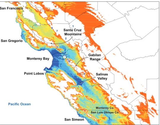
he niche-based distribution model for I. plenipes indicates the highest probability of occurrence, representing ecological suitability for the species, in the terrestrial areas on the periphery of Monterey Bay extending just past the gap between the Santa Cruz Mountains and Gabilan Range and throughout the Salinas Valley (Fig. 1). Areas of medium to high probability extend from Monterey Bay along a thin region on the coast northward to San Gregorio and southward to Point Lobos. here are other areas of medium to high probability, also restricted to the coast, between San Simeon in the north and the western boundary between Monterey and San Luis Obispo counties.
DNA barcoding
Polymerase chain reaction of the COI barcoding region, when electrophoresed and visualized on a 12% agarose gel, recovered single bands of uniform lengths in all species. Sanger sequencing resulted in sense/antisense chromatograms reads of ~600 bp in length when contiguous fragments were assembled in Mesquite. Mean Phred quality scores of individual contigs are between 73–80. When aligned and ragged ends trimmed, sequence length is invariant between species. Mean nucleotide per-cent sequence diference between species is 25% and between amino acid sequences (total diference), 17%. he NCBI GenBank accession numbers are as follows: I. plenipes (JX962724), G. claremontus (JX962723), B. producta (JX962721), B. rosea
(JX962722), and S. lyttoni (JX962725). he COI barcodes of the Siphonophorida species (I. plenipes and S. lyttoni) and the Platydesmida species (G. claremontus, B. producta, and B. rosea) are hitherto the only that exist for these two orders; there is
only one other DNA barcode for the entire subterclass Colobognatha. he following species are listed in order of increasing percent nucleotide diference from I. plenipes, indicated in parentheses (mean percent diference of amino acids proceeds after the “/”): G. claremontus (28.7% / 23.8%), B. producta (29.7% / 24.4%), S. lyttoni (29.9%
taxonomy
Class Diplopoda de Blainville in Gervais, 1844 Subclass Chilognatha Latreille, 1802/1803 Infraclass Helminthomorpha Pocock, 1887 Subterclass Colobognatha Brandt, 1834 Order Siphonophorida Hofman, 1980 Family Siphonorhinidae Cook, 1895
Genus Illacme Cook & Loomis, 1928
http://species-id.net/wiki/Illacme
Cook and Loomis 1928: 12; Chamberlin and Hofman 1958: 189; Buckett 1964: 29; Jeekel 1971: 39; Hofman 1980: 116; Shelley 1996b: 23; Shelley 1996a: 1808; Hof-man 1999: 195; Jeekel 2001: 46; Marek and Bond 2006: 707; Shelley 2010: 45.
Type species.I. plenipes Cook and Loomis 1928: 12; by original designation.
Family placement. Illacme is placed with other taxa in the family
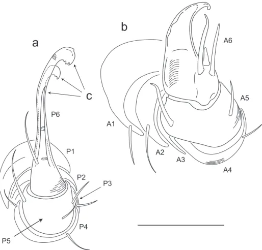
Siphonorhini-dae based on the following characters: Head pear-shaped (♂) or triangular (♀), not elongate or bird beak-shaped, as in the Siphonophoridae (Fig. 2, Morphbank 805574, Appendix I). Antennae elbowed between antennomeres 3, 4 (Fig. 3, Mb-805578). Antennomeres 5, 6 with apical dorsal cluster of 7 or 8 basiconic sensilla (Bs2) in slight depression, not deep-set into circular pits, as in the Siphonophoridae (Fig. 4, Mb-805575). Posterior gonopods with distal podomere divided into 2 or 3 branches (Fig. 5, Mb-805576, Fig. 6c). See also diagnoses of Illacme in Shelley (1996b, p. 23) and of Siphonorhinidae in Shelley and Hofman (2004, p. 218).
Diagnosis. Adults of Illacme are distinct from other siphonorhinid genera (and
commonly-encountered millipedes co-occurring with I. plenipes) based on the
Paul E. Marek et al. / ZooKeys 241: 77–112 (2012)
86
(Fig. 6a, b). Anterior gonopod thick, more robust than posterior gonopod (Fig. 10, Mb-805583, Fig. 6b). Anterior gonopodal apex (podomere 6, Fig. 6a, A6) shovel-shaped; in repose, cupped sheath-like around lagelliform posterior gonopodal apex (podomere 6, Fig. 11, Mb-805584, Fig. 6b, P6). Posterior gonopodal podomere 6
divided, comprising a bundle of 3 stylus-shaped articles (Fig. 5, Mb-805627, Fig. 6a,
acuminate distally, spike-like. Habit in life. Movement very slow, nearly imperceptible (Appendix II, III). Antennae movement rapid, independent. Terminal antennomeres held lat and rapidly tap substrate and surroundings (Appendix IV).
Illacme plenipes Cook & Loomis, 1928
http://species-id.net/wiki/Illacme_plenipes
Cook and Loomis 1928: 12. Chamberlin and Hofman 1958: 189; Buckett 1964: 29; Shelley 1996b: 23; Shelley 1996a: 1808; Hofman 1999: 195; Jeekel 2001: 46; Shelley and Hofman 2004: 221; Marek and Bond 2006: 707; Read and Enghof 2009: 554; Shelley 2010: 45; Shelley and Golovatch 2011: 26.
Material examined.Type specimens: ♂ holotype (USNM), 1♂, 3♀ paratypes (FSCA)
and 3♀ paratypes (VMNH)—from United States, California, San Benito County,
Paul E. Marek et al. / ZooKeys 241: 77–112 (2012)
88
from “near divide between Salinas and San Juan Bautista” [an imprecise location prob-ably on the north side of the Gabilan Range on San Juan Grade Road or Old Stage Road in a radius of 4 km around the coordinates 36.831371°N, -121.562808°W], 27.xi.1926 (Coll. O.F. Cook). Non-type specimens: California, San Benito County: 1♂ (SPC000924), 2♀ (SPC000930, -931), Gabilan Range, San Juan Bautista, 29.xi.2005 (Colls: P. and R. Marek); 3♂ (SPC000932, -933, -934), 1 juvenile (SPC000935), loc. ibid., 8.xii.2005, (Coll: J. Bond). 2♀ (SPC001187, MIL0020), Gabilan Range, San
Juan Bautista, 16.xii.2007, 13:00 (Colls: P. and R. Marek).
Diagnosis. (See generic diagnosis.)
Description of holotype (♂) USNM TYPE NO. 976 – Counts and measurements:
p = 143. a = 2. l = 562. (143 + 2 + T). HW = 0.30. HL = 0.34. ISW = 0.20. AW = [antennae missing]. CW = 0.42. W1 = 0.53. W2 = 0.55. W3 = 0.55. L1 = 0.20. L2 =
0.20. L3 = 0.18. H1 = 0.31. H2 = 0.30. H3 = 0.33. AS1 = 0.45. A5W = 0.05. P5W
Paul E. Marek et al. / ZooKeys 241: 77–112 (2012)
90
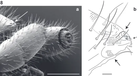
Figure 8. Lateral (right) view of antennal and cephalic apices (♂). a Scanning electron micrograph: ar-row, denticulate shelf-like carina, projecting dorsally from labrum-epistome margin. Scale bar 0.1 mm
b Line drawing: top arrow, shelf-like carina; middle arrow, triangular tooth-lined oriice; bottom arrow, gnathochilarium. Scale bar 0.01 mm.
Paul E. Marek et al. / ZooKeys 241: 77–112 (2012)
92
just dorsal to anchor-shaped spikes (Fig. 17a, Mb-805601). Anchor-shaped spikes alter-nating in size (large, small) along row. Ozopores oriented dorsally, located near limbus, absent from tergites 1 – 3 and telson. Ozopores elevated slightly (porosteles absent), with 2 stout posteriorly projecting spines and encircled by 13 – 15 robust setae (Fig. 19, Mb-805602). 3 or 4 stout lat tubercles opposite ozopore near anterior margin, lunate arrangement encircling ozopore (Fig. 17b, Mb-805603). Posterior tergites more convex, covered with a greater density of long, slender “silk”-exuding setae (Fig. 20, Mb-805604). Lunate-arranged tubercles opposite ozopores on posterior metazonites: conical and spiked, not lat. Apodous segments lacking sterna, pleurites contiguous in midline. Apodous tergites densely setose, covered with unevenly distributed spikes (Fig. 21, Mb-805605). Telson densely covered with irregularly oriented and unevenly distrib-uted stout spines; posterior margin lined with variably-shaped posterodorsally oriented anchor-shaped spikes. Tergal tubercles and spikes: consistently projecting posteriorly, occasionally posterodorsally. Prozonite highly sculptured, with 5 rows of discoidal lat tubercles; anterior 3 rows staggered and posterior 2 rows aligned (Fig. 22, Mb-805606).
Pleurites quadrate, lat, with jagged scaly lateral, posterior and medial margins (Fig. 16, Mb-805609). Pleurite medial margin broad, with scaly carina (Fig. 16b, Mb-805610). Left and right pleurites plate-like, comprising 4/5’s of ventral segment space. Left and right pleurites broadly overlapping sternite, covering spiracles (Fig. 23, Mb-805612).
Sternites free, separate from pleurites; heart-shaped, wider anteriorly. Sternal surface with broad, jagged scales. Medial sternal ridge projecting ventrally, with spiracles and legs oriented ventrally (Fig. 24, Mb-805614). Spiracles circular, oriice open; oriented dorsally above legs (Fig. 25, Mb-805615). Anterior and posterior sternites separate. Ter-gites, pleurites and sternites separated by arthrodial membrane (Fig. 20, Mb-805616). Arthrodial membrane between tergites and pleurites wider posteriorly. Telson pilose, covered with long, slender posteriorly recurved setae (Fig. 20, Mb-805628). Parap-rocts semihemispherical, anterior margins slightly scaly. Epiproct absent. Hypoproct small, one-eighth area of paraproct, with row of posterior projecting setae. Legs: six
subequally shaped podomeres, with coxa slightly shorter and tarsus slightly longer. Legs with sparse setae, appearing similar to trichoid sensilla, with 2 or 3 barbules. Coxae nearly contiguous medially, separated by thin sternal ridge. Large posteroventral D-shaped opening for eversible sac (Fig. 26, Mb-805618). Eversible sacs membranous, bulging slightly from opening (Fig. 24b, Mb-805620). Pregonopodal tarsus with stout bifurcate claw; dorsal subdivision thicker, more arcuate (Fig. 27, Mb-805621). Postgo-nopodal tarsus with two separate claws, co-terminal on tarsal apex; dorsal claw thick and arcuate, ventral claw thin and setiform (Fig. 16c, Mb-805623). 2nd leg pair with posteriorly oriented coxal gonapophyses; rounded, protuberant, one-third length of prefemur. Gonopods: 9th, 10th leg pairs modiied into gonopods, each comprising 6
Figure 16–21. 16Ventral view of segments (♂). a Lateral tergal and pleural carinae jagged, pronounced on midbody segments b Pleurite medial margin broad, with scaly carina c Postgonopodal tarsus with thin-ner claw and without bifurcation, but with stout seta. Scale bar 0.4 mm. 17Dorsal view of segments (♂).
Paul E. Marek et al. / ZooKeys 241: 77–112 (2012)
94
of P6 laminate distally, recurved laterally, denticulate posterior margins, appearance similar to a chicken foot in rigor mortis (Fig. 12, Mb-805585, Fig 6a). Ventral-most,
shortest article of P6 acuminate distally, spike-like. hin ridge-shaped sterna present between left and right gonopods, thicker between anterior gonopods.
Description of largest paratype (♀) VMNH – Counts and measurements: p =
190. a = 2. l = 750. (190 + 2 + T). HW = 0.37. HL = 0.44. ISW = 0.30. AW = anten-nae missing. CW = 0.44. W1 = 0.58. W2 = 0.58. W3 = 0.57. L1 = 0.23. L2 = 0.21. L3 = 0.23. H1 = 0.46. H2 = 0.44. H3 = 0.48. AS1 = 0.44. BL = 40.40. Anatomical
description similar to male holotype. In combination with its measurements, the fol-lowing structures difer from male holotype. Head triangular, chevron-shaped, tapered anteriorly to round point at a 135° angle anterior from antennal sockets; occipital area posterior from antennal sockets straight, not curved medially towards neck. Cyphopods
large, area 1/6 the segmental area in widest cross-section; almond-shaped, bivalvular, narrow apex oriented ventrolaterally. Valves transparent, glassy. Ventral valve thickened and clam-like, with 4 or 5 thick setae; dorsolateral valve thin and lat, with 2 or 3 spines. Oviduct connected posteriorly to cyphopod, opening oriented ventromedially and located between valves. Oviduct tube wrinkled, appearing highly expandable in width, cross-section 1/8 area of cyphopod. Receptacle, suture and operculum absent.
Etymology. Cook and Loomis (1928) named this species “in highest fulillment of
feet”. Il = “in” (Latin); acme, άκμή (Greek) = “the highest point, or culmination”; pleni
= “full” (Latin); pes = “foot” (Latin).
Variation. here is negligible variation in coloration among live specimens. (FSCA
paratype specimens that have been stored in alcohol for 86 years are dark mahogany brown, which is likely an unnatural color and a result of alcohol preservative, vial stop-per and age.) he predominant source of variation between specimens is segment and leg counts (Tables 1 – 3). Females have between 486-750 legs with a standard deviation of 78, and males between 318–562 legs with a standard deviation of 107. he seg-ments of I. plenipes (males and females) are uniform in length, width and height along
table 1.Segment and leg count, head measurements.
p l HW HL ISW AW CW
♂ (107 / 27)84–145 (410 / 107)318–562 (0.301 / 0.006)0.295–0.308 (0.382 / 0.024)0.344–0.406 (0.189 / 0.011)0.172–0.202 (0.101 / 0.002)0.098–0.103 (0.393 / 0.019)0.374–0.422
♀ 126–192(159 / 20) 486–750(619 / 78) (0.335 / 0.020)0.308–0.369 (0.446 / 0.045)0.408–0.556 (0.217 / 0.033)0.185–0.295 (0.103 / 0.006)0.098–0.113 (0.431 / 0.021)0.407–0.472
table 2.Width and length measurements.
W1 W2 W3 WM L1 L2 L3 LM
♂ (0.485 / 0.033)0.437–0.526 (0.500 / 0.036)0.467–0.554 (0.488 / 0.034)0.455–0.545 0.491 / 0.032 (0.173 / 0.021)0.148–0.203 (0.162 / 0.020)0.150–0.197 (0.159 / 0.017)0.140–0.183 0.165 / 0.019
Paul E. Marek et al. / ZooKeys 241: 77–112 (2012)
96
the trunk, and are slightly taller, and more convex, in posterior segments—potentially to accommodate the spiraled metenteron.
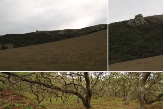
Natural history. Illacme plenipes specimens were collected during the day in a
small valley adjacent to cattle pasture. he woodland habitat was primarily composed of California live-oak, Quercus agrifolia (Fig. 28). Understory lora included ferns (bracken, Pteridium aquilinum; California polypody, Polypodium californicum; and
California maiden-hair, Adiantum jordanii), California blackberry (Rubus ursinus),
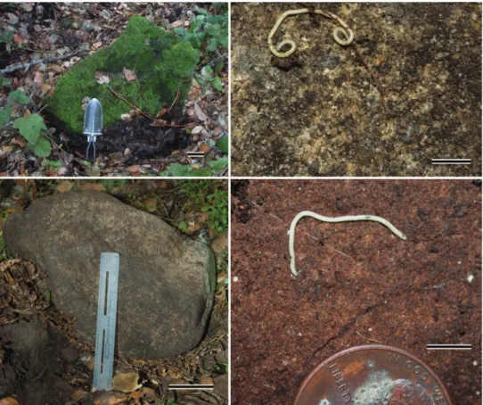
and poison oak (Toxicodendron diversilobum) (Fig. 29). Specimens were found be-neath large moss-covered boulders, typically with a mass > 30 kg (Fig. 30). he
mil-table 3.Height, apodous segment / gonopodal width, body length measurements.
H1 H2 H3 HM AS1 A5W P5W BL
♂ 0.273– 0.400 (0.350 / 0.057) 0.277– 0.418 (0.337 / 0.055) 0.295– 0.381 (0.336 / 0.036) 0.341 / 0.047 0.394– 0.445 (0.423 / 0.022) 0.047– 0.055 (0.051 / 0.003) 0.036– 0.043 (0.040 / 0.003) 13.368– 28.156 (19.251 / 6.305) ♀ 0.220– 0.486 (0.365 / 0.077) 0.289– 0.488 (0.384 / 0.064) 0.295– 0.504 (0.370 / 0.079) 0.373 / 0.071 0.412– 0.482 (0.451 / 0.024)
- - 24.541– 40.399
(31.055 / 5.474)
Figure 29. Oak forest understory habitat of I. plenipes. Top, base of sandstone pinnacle (from Fig. 28), where specimens were found. Bottom, mossy oak forest—close-up of habitat where I. plenipes individuals were encountered.
Promecogna-Paul E. Marek et al. / ZooKeys 241: 77–112 (2012)
98
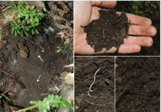
thus ground beetles (Carabidae). Edaphic setting: Specimens collected in 2007 were
found beneath a large stone (Fig 30, about 30 kg). When the stone was removed, individuals were seen corkscrewing outward into the cavity from the soil (Fig. 31). he soil, consisting of moist small-grained substrate, was dark chocolate brown in coloration and somewhat sandy (Fig. 31). he soil did not contain clay particles and seemed to drain water quickly. During the 16 December 2007 collections, soil mois-ture extended 15 cm below the surface.
Distribution. Illacme plenipes is only known from a small area, ca. 4.5 km in
diameter, in the northwestern foothills of the Gabilan Range in San Benito County, California.
Figure 30. Sandstone microhabitat of I. plenipes.Top left, 50 kg sandstone from 29.xi.2005 rediscovery locality of I. plenipes; one ♀ with 666 legs was discovered from beneath the stone (scale bar = 5 cm, hand shovel shown for scale). Bottom left, 30 kg sandstone from the 16.xii.2007 locality, two ♀ (specimen #s: SPC001187, MIL0020) were discovered below the stone (scale bar = 5 cm, 15 cm ruler shown for scale). Top right, surface close up of sandstone from 16.xii.2007 locality with ♂I. plenipes, not collected (scale bar = 5 mm). Bottom right, surface close up of sandstone from 29.xi.2005 locality with ♂I. plenipes
Discussion
“he acme of plentiful feet”
he pattern by which I. plenipes add segments and subsequently legs post-embryonical-ly between developmental stadia is referred to as anamorphosis (Enghof et al. 1993). Based on the large number of legs and considerable variation in leg and segment count among adults, anamorphosis likely continues for an indeterminate period, extending well beyond the attainment of sexual maturity (Enghof et al. 1993; Marek and Bond 2006). Millipedes generally use their numerous legs to burrow between and through obstacles that they encounter (Hopkin and Read 1992; Manton 1954). A leg pair acts to push and propel the myriapod forward, and with two leg-pairs per segment (diplosegments in millipedes represent a fusion of two primordial segments), milli-pedes create a stronger thrust for a relatively compact body. Millimilli-pedes with heavily calciied cuticles and rather incompressible bodies composed of rigid rings (e.g., the Spirobolida and Spirostreptida), burrow through the soil by brute leg force, ramming and bulldozing with a smooth rounded head and collum. In contrast, many millipedes
Paul E. Marek et al. / ZooKeys 241: 77–112 (2012)
100
with lexible cuticles and compressible bodies, which are composed of free sternites and pleurites (e.g., other Siphonophorida and I. plenipes), move through the soil by
squeez-ing lexible anterior segments forward by leg force and subsequently telescopsqueez-ing poste-rior segments forward and repeating, i.e. the borer millipedes (Hopkin and Read 1992; Manton 1961). he anterior segments in these millipedes are tapered, most noticeably in the Polyzoniida, and bore and wedge to facilitate movement through the soil. With
I. plenipes, the numerous legs presumably impart greater motive force to push within
a subterranean microhabitat, and to cling tightly to the surface of sandstone boulders (as described below).
Natural history
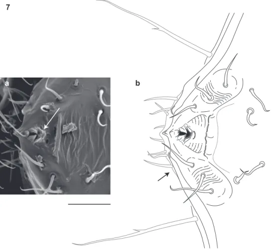
he diet of I. plenipes is unknown. Given the shape of its mouthparts, the typical mil-lipede diet in which decaying organic matter is mechanically fragmented is unlikely for the species. Illacme plenipes possesses a comb-like structure on the posterior margin of the labrum and an open triangular tooth-lined “mouth” formed by an oriice though the labrum (Fig. 7a, b; Fig. 8a, b; Mb-805580; Mb-805588). he mouthparts are composed of the stylet-like mandibles and the gnathochilarium (structures observed between 500-2000× with a scanning electron microscope and the mandibles through the translucent head capsule at 400× with a compound microscope). hese mouth-parts are tightly appressed and tapered anteriorly to a rounded point. Given that the mandibles appear stylet-like, and assuming the mouthparts are moveable, a functional hypothesis for feeding is that the gnathochilarium hinges open, the mandibles are protruded to pierce plant and/or fungal tissue, and then the tooth-lined mouth is used to suck out the luid contents. he teeth and labral comb could serve to ilter particu-lates exceeding a certain size. Other Colobognath millipedes with somewhat reduced mouthparts, for example species of the family Andrognathidae, feed on fungus or other live plant or soft organic matter (Gardner 1974). Manton (1961) described the feeding of captive siphonophorids Siphonophora portoricensis Brandt, 1837 and Siphonophora (=Siphonocybe) hartii (Pocock, 1894) and observed individuals probing decayed veg-etation with their beaked proboscises, after tapping the material with their antennae. Fungi were not observed associated with I. plenipes, as they are often with species
via the Malphigian tubules, or a combination of the metenteron and mesenteron.) Al-ternatively, a long trunk may function to store additional eggs, and potentially evolved under fecundity selection. Consistent with this hypothesis, I. plenipes are sexually size
dimorphic: female maximum length (BL) and maximum width (BM) is 1.43-fold and 1.16-fold greater than male length and width.
Based on natural history observations of I. plenipes in the ield, individuals are al-ways found approximately 10 – 15 cm beneath the soil, or clinging to the surface of large sandstones. he great number of legs may beneit a deep subterranean lifestyle clinging to sandstone. Illacme plenipes has bifurcate claws on anterior legs and two sepa-rate claws, coterminal on the tarsal apex (in lieu of the abifurcation), on posterior legs. In several millipede species, e.g. Cylindroiulus imbriatus Enghof, 1982 and Dolistenus savii Fanzago, 1874, the additional claws serve a stone-clinging function for surface
adherence and an epilithic lifestyle (Enghof 1983; Manton 1961). Illacme plenipes has
large eversible sacs, structures that have also been implicated in surface clinging in pe-trophilic colobognath millipedes (Manton 1954; 1961). On the dorsal surface of the millipede, setae secrete a silk-like substance, which appears sticky, and may be used for clinging to the stone surface. he secretions seem to increase with handling, perhaps al-ternatively indicating an anti-predatory function (Shear 2008; Youngsteadt 2008). he silk may also function as a soil shedding mechanism to allow eicient burrowing, or as a means to ensnare parasites or debris particles (Youngsteadt 2008). he chemical com-position of the silk is unknown. While millipedes in seven other orders of Diplopoda produce a silk-like substance from various body structures, its threads are not true silk composed of protein (one order produces silk from openings on the legs, one order from metatergal setae like I. plenipes, 4 orders from epiproctal spinnerets, and one order from both metatergal and epiproctal setae). In contrast with the silk’s origin from the setal tip in I. plenipes (Fig. 18, Mb-805600), the other seven orders appear to produce silk from pores located at the setal base (Shear 2008). he diverse locations where silk originates in millipedes (legs, epiprocts, metatergal setae), suggests independent origins and pre-cludes homology (Shear, 2008). he extrusive sticky appearance of I. plenipes’silk-like secretion may indicate a mucopolysaccharide identity, as is the composition of epiproc-tal silk spun by millipedes in the order Polydesmida (Adis et al. 2000; Shear 2008).
In contrast with the smooth exoskeleton of the bulldozer millipedes, I. plenipes’ has
pro-Paul E. Marek et al. / ZooKeys 241: 77–112 (2012)
102
zonital microsculpture is divided into two shape classes: a smooth scaly texture anterior to the prozonital transverse ridge and a rugged knobby surface, with discoidal tubercles or spherical knobs posterior to the ridge. he presence of spherical knobs and other cuticular ornaments in certain families of Polydesmida appear to relect major evolu-tionary groups in the order (Akkari and Enghof 2011). he function of the cuticular ornaments in I. plenipes is uncertain. Authors have suggested several hypotheses for the function of various projections including a locking mechanism for volvation, in the case of the anchor-shaped spike in Julida, and maintaining a cloak of soil for camou-lage, in the case of branching tree-shaped setae in Polydesmida (Shear, 1977).
Evolutionary relationships
he widely scattered distribution of modern Siphonorhinidae, predominately in the Southern Hemisphere except with I. plenipes in North America, indicates that their most recent common ancestor likely predates the breakup of Pangaea more than 200 million years ago. A phylogeny for Siphonorhinidae, or any taxa in the four orders of Colobognatha, does not exist, except for a recent species phylogeny of the genus
Brachycybe in the order Platydesmida (Brewer et al. 2012). Even though the number of COI barcodes for the Colobognatha is low and the region may not be ideal for recover-ing the ancient divergences between the colobognath taxa represented here (likely > 200 mya), we inferred a preliminary phylogeny with the COI nucleotides using a maximum likelihood tree search in RAxML ver. 7.0.3 (Stamatakis 2006). We recovered monophy-letic Platydesmida and Siphonophorida with S. lyttoni sister to I. plenipes. When Polyzo-nium germanicum (Polyzoniida) was included in the RAxML analysis and visualized in an unrooted tree, it occurred on an intervening branch between Siphonophorida and Platydesmida clades. (Polyzonium COI barcoding sequences from Spelda et al. 2011).
he paleoendemic species I. plenipes is the sole representative of the family in the
Western Hemisphere. Remaining genera in the family occur primarily in the Old World tropics in Wallacea, Sundaland, Himalayas (Siphonorhinus species), Indo-Burma ( Kleru-chus olivaceus and Siphonorhinus species), and Maputaland-Pondoland-Albany ( Nemato-zonium ilum). he closest relative of I. plenipes is uncertain. he present day range of
Siphonorhinidae may be the remnant of an ancient and widespread tropical distribution across Pangaea. he most likely sister taxon to I. plenipes is Nematozonium ilum from South Africa, as they share a number of anatomical similarities. Among the known spe-cies of Siphonorhinidae, a South African spespe-cies is a probable candidate for closest relative based on other close relationships between co-distributed taxa, for example the lightless Californian beetle genus Promecognathus and its close relatives in the tribe Axinidiini in
beyond 182 segments, e.g., Siphonophora millepeda Loomis, 1934 with 190 segments). Individuals of K.olivaceus and Siphonorhinus species have bifurcate posterior gonopods
(i.e. without a spine as in N. ilum), fewer segments, and a shorter and more compact
body form. Siphonorhinid millipedes, studied sporadically over the last 80 years by dif-ferent taxonomists concentrating on various geographic faunas, are ideal candidates for a modern synthesis and molecular phylogenetics. For example Siphonorhinus, as is certainly the case for Siphonophora, seems to be a taxonomic dumping ground for long and
spin-dly Siphonophorida without a bird-like beak or paranota (Jeekel 2001). he diversity of anatomical forms in the Siphonophorida, in particular the Siphonorhinidae, is quite conserved compared to other diplopod taxa. Compared to other Colobognatha, somatic anatomical diversity across lineages is low and indicates that early Siphonophorida may have appeared similar to present day species. his suggests that contemporary habitats, and current environmental factors afecting body shape, may have been similar to those in which early Siphonophorida taxa occurred. Illacme plenipes and related lineages may have persisted unchanged in a mild, constant habitat for hundreds of millions of years. his idea raises fascinating questions about climate and habitat constancy where Siphonorhinidae occur (its six regions also happen to be global biodiversity hotspots), and also important concerns about the conservation of the species and co-inhabitants that may have persisted in these mild climates that are now currently threatened by global climate change.
Local biogeography
Paul E. Marek et al. / ZooKeys 241: 77–112 (2012)
104
where individuals were encountered overlay marine arkosic sandstone deposits between the Vergeles and San Andreas faults (Dibblee et al. 1979). High probability of I. plenipes
occurrence is also present in the areas around the southern Monterey Bay and Salinas Valley that overlay more recent suricial alluvial deposits. While the probability of occur-rence is high in these unsampled areas, the edaphic setting indicates lower suitability. he soils of the Monterey Basin and Salinas Valley are composed of alluvial sediments and ine-grained deposits, lacking the large arkose sandstones and boulders that I. plenipes
may be specially adapted to. Nonetheless, there is a present-day low overall probability of occurrence of I. plenipes in the area, or of any other native soil dweller for that matter, since the Salinas Valley is heavily inluenced by agriculture and development.
Conservation
Illacme plenipes is threatened by extinction as a result of its restricted geographical distribution, narrow microhabitat requirements, seasonal rarity, and low observed population numbers. Natural populations are threatened by habitat loss due to ram-pant development and intense land use in the area (agricultural, industrial, transit and housing), climate change, invasive species, and potential for over-collecting. he restricted location of I. plenipes, limited to the gap between the Santa Cruz Mountains and Gabilan Range at the eastern fog limit, may be due to edaphic requirements (soils composed of sandstone or other native formations in the area: San Lorenzo Forma-tion or Dacitic volcanic rocks), or extirpaForma-tion due to the heavy agricultural inluence around Monterey Basin and the Salinas Valley since the 1800s. In contrast with habitat degradation from development and farming, the presence of cattle does not appear to negatively afect I. plenipes. At each locality where I. plenipes was discovered, there was noticeable inluence of cattle on the habitat. Boulders under which I. plenipes occurred
were sometimes a meter away from deep cattle hoof prints. he most serious impacts that I. plenipes faces are human-induced habitat loss and climate change. As suggested by the distribution model and I. plenipes’ apparent dependence on marine layer fog (likely inluencing moisture and stability of its habitat), the documented 33% reduc-tion in coastal California fog due to higher atmospheric and ocean temperature since the early 1900s (Johnstone and Dawson 2010) may severely impact the species and hasten its extinction. he few locations where I. plenipes exist are unique storehouses of this evolutionary relict, and potentially other ancient lineages that await discovery.
Morphbank annotations
(Published at www.morphbank.net):
Paul E. Marek et al. / ZooKeys 241: 77–112 (2012)
106
Acknowledgements
hanks to Rob Marek for assistance in the ield to collect I. plenipes. Peter Raven
recom-mended I. plenipes localities in San Benito County and shared helpful knowledge of the area and co-occurring native plants. Avery Lane, David Beamer, Amy Stockman and Mat-thew Walker assisted in the ield and laboratory. Many thanks to Patrick, Peter and Tom Breen, the Reeves Family, and Bart O’Brien who shared information and details about California mountain ranges and potential localities. he Nature Conservancy kindly al-lowed access to sites in the Gabilan Range. Dotti Marek and Katy Murphy provided lo-cal support during ieldtrips to California. hanks to anonymous reviewers and Charity Hall for reading earlier versions of the manuscript. California State Parks and the Na-tional Park Service supported research in the parks and permits for collections. Jonathan Coddington, Dana DeRoche, G.B. Edwards, Charles Whitehill, Judith Winston, and Joe Keiper provided essential type specimens and access to natural history collections in support of the project. his research was supported by a U.S. National Science Founda-tion Partnerships for Enhancing Expertise in Taxonomy Grant to P. Sierwald, J.E.B., and W.A.S. 0529715); and by a NSF Phylogenetic Systematics grant to P.E.M. (DEB-1119179). Wendy Moore and the Entomology Department at the University of Arizona is acknowledged for their support of P.E.M. and systematic entomology. his article is in memoriam of Richard Hofman (1927 – 2012), whose support for the irst author’s study of millipedes will always be appreciated. Dr. Hofman loaned the Virginia Museum of Natural History’s I. plenipes specimens shortly before he passed away. His inquisitive
naturalist spirit and scientiic legacy lives on in the 50+ organisms named in his honor, numerous scientiic contributions, and love of the natural history of Virginia.
References
Adis J, Hansen B, Wilck L, Adis I, Hirschel K (2000) A spinning apparatus documented in Polydesmida for the irst time. In: Wytwer J, Golovatch SI (Eds) Progress in Studies on Myriapoda and Onychophora. Fragmenta Faunistica, Supplement 43: 139–149.
Akkari N, Enghof H (2011) On some surface structures of potential taxonomic importance in families of the suborders Polydesmidea and Dalodesmidea (Polydesmida, Diplopoda). ZooKeys 156: 1–24. doi: 10.3897/zookeys.156.2134
Akkari N, Stoev P, Enghof H (2011) Two new cavernicolous genera of Julidae (Diplopoda, Julida), with notes on the tribe Brachyiulini and on julid subanal hooks and anchors. ZooKeys 114: 1–14. doi: 10.3897/zookeys.114.1490
Attems CG (1951) Révision systématique des Colobognata (Myriapodes Diplopodes) et de-scription d’espèces nouvelles, Mémoires du Muséum National d’Histoire Naturelle, Série A, Zoologie 3: 193–231.
ex-treme genetic divergence and geographic structuring. Systematic Biology 57: 628-646. doi: 10.1080/10635150802302443
Brandt JF (1834) Note on Colobognatha. Oken’s Isis 27: 704.
Brandt JF (1837) Note sur un ordre nouveau de la classe des Myriapodes et sur l’établissement des section de cette classe d’animaux en général. Bulletin Scientiique de l’Académie Impé-riale des Sciences de Saint-Pétersbourg 1: 178–179.
Brewer MS, Spruill CL, Rao NS, Bond JE (2012) Phylogenetics of the millipede genus Brachy-cybe Wood, 1864 (Diplopoda: Platydesmida: Andrognathidae): Patterns of deep evolution-ary history and recent speciation. Molecular Phylogenetics and Evolution 64: 232–242. doi: 10.1016/j.ympev.2012.04.003
Buckett JS (1964) Annotated list of Diplopoda of California. Simmons Publishing Co., Davis, California, 34 pp.
Chamberlin RV, Hofman RL (1958) Checklist of the millipeds of North America. Bulletin of the United States National Museum 212: 1–236. doi: 10.5479/si.03629236.212
Chung KH, Moon MJ (2006) Antennal sensory organs in the female millipede Orthomorphella pekuensis (Polydesmida: Paradoxosomatidae). Integrative Biosciences 10: 183-189. doi: 10.1080/17386357.2006.9647300
Cook OF, Loomis HF (1928) Millipeds of the order Colobognatha, with descriptions of six new genera and type species, from Arizona and California. Proceedings of the United States National Museum 72: 1–26.
Cook OF (1895) Introductory note on the families of Diplopoda, in Cook & Collins, he Craspedosomatidae of North America. Annals of New York Academy of Science 9: 1–9. doi: 10.1111/j.1749-6632.1896.tb55430.x
Dibblee TW, Nisen TH, Brabb EE (1979) Preliminary geologic map of the San Juan Bautista quadrangle, San Benito and Monterey counties, California. Department of the Interior, United States Geological Survey.
Elith J, Graham CH, Anderson RP, Dudik M, Ferrier S, Guisan A, Hijmans RJ, Huettmann F, Leathwick JR, Lehmann A, Li J, Lohmann LG, Loiselle BA, Manion G, Moritz C, Na-kamura M, Nakazawa Y, Overton JM, Peterson AT, Phillips SJ, Richardson K, Scachetti-Pereira R, Schapire RE, Soberon J, Williams S, Wisz MS, Zimmermann NE (2006) Novel methods improve prediction of species’ distributions from occurrence data. Ecography 29: 129–151. doi: 10.1111/j.2006.0906-7590.04596.x
Enghof H (1983) Adaptive radiation of the millipede genus Cylindroiulus on Madeira: habitat, body size, and morphology (Diplopoda: Iulida: Iulidae). Revue d’écologie et de biologie du sol 20: 403–415.
Enghof H, Dohle W, Blower JG (1993) Anamorphosis in millipedes (Diplopoda) - the present state of knowledge with some developmental and phylogenetic considerations. Zoological Journal of the Linnean Society 109: 103–234. doi: 10.1111/j.1096-3642.1993.tb00305.x Erwin TL (1985) he taxon pulse: a general pattern of lineage radiation and extinction among
carabid beetles. In: Ball GE (Ed) Taxonomy, Phylogeny and Zoogeography of Beetles and Ants. Junk, Dordrecht, 437–472.
Paul E. Marek et al. / ZooKeys 241: 77–112 (2012)
108
Folmer O, Black M, Hoeh W, Lutz R, Vrijenhoek R (1994) DNA primers for ampliication of mitochondrial cytochrome c oxidase subunit I from diverse metazoan invertebrates. Mo-lecular Marine Biology and Biotechnology 3: 294–299.
Gardner MR (1974) Revision of the millipede family Andrognathidae in the Nearctic region. Memoirs of the Paciic Coast Entomological Society 5: 1–61.
Gervais P (1844) Études sur les Myriapodes. Annales des Sciences Naturelles Zoologie et Bi-ologie Animale 3: 51–80.
Hijmans RJ, Cameron SE, Parra JL, Jones PG, Jarvis A (2005) Very high resolution inter-polated climate surfaces for global land areas. International Journal of Climatology 25: 1965–1978. doi: 10.1002/joc.1276
Hofman RL (1980) “1979” Classiication of the Diplopoda. Muséum d’histoire naturelle, Genève, 237 pp.
Hofman RL (1999) Checklist of millipeds of North and Middle America. Virginia Museum of Natural History, Martinsville, VA, 584 pp.
Hopkin SP, Read HJ (1992) he Biology of Millipedes. Oxford University Press, Oxford, 233 pp.
Jeekel C (1971) Nomeclator generum et familiarum Diplopodorum: A list of the genus and family-group names in the class Diplopoda from the 10th edition of Linneaus, 1758, to the end of 1957. Monograieën van de Nederlandse Entomologische Vereniging 5: 1–412. Jeekel C (2001) A bibliographic catalogue of the Siphonophorida (Diplopoda). In: Jeekel C
(Ed) Myriapod Memoranda. C.A.W. Jeekel, Amsterdam, Netherlands, 44–71.
Johnstone JA, Dawson TE (2010) Climatic context and ecological implications of summer fog decline in the coast redwood region. Proceedings of the National Academy of Sciences of the United States of America 107: 4533–4538. doi: 10.1073/pnas.0915062107
Latreille PA (1802–1803) Histoire naturelle, générale et particulière des Crustacés et des In-sectes; ouvrage faisant suite aux oeuvres de Leclerc de Bufon, et partie du cours complet d’histoire naturelle rédigé par C. S. Sonnini. Volume 2. F. Dufart, Paris, 467 pp.
Maddison DR, Maddison WP (2011a) Chromaseq: a Mesquite module for analyzing sequence chromatograms. Version 1.0. http://mesquiteproject.org/packages/chromaseq
Maddison WP, Maddison DR (2011b) Mesquite: a modular system for evolutionary analysis. Version 2.75 http://mesquiteproject.org
Manton SM (1954) he evolution of arthropodan locomotory mechanisms. Journal of the Lin-nean Society of London Zoology 42: 299–368. doi: 10.1111/j.1096-3642.1954.tb02211.x Manton SM (1961) he evolution of arthropodan locomotory mechanisms. Part 7. Functional
requirements and body design in Colobognatha (Diplopoda), together with a comparative account of diplopod burrowing techniques, trunk musculature, and segmentation. Journal of the Linnean Society of London Zoology 44: 383–462. doi: 10.1111/j.1096-3642.1961. tb01622.x
Marek PE, Bond JE (2006) Rediscovery of the world’s leggiest animal. Nature 441: 707–707. doi: 10.1038/441707a
Mesibov R (2012) New species of Prosopodesmus Silvestri, 1910 (Diplopoda, Polydesmida, Haplodesmidae) from Queensland, Australia. ZooKeys 190: 33–54. doi: 10.3897/zook-eys.190.3276
Nguyen Duy-Jacquemin M (1974) Les organes intracérébraux de Polyxenus lagurus et compari-son avec les organes neuraux d’autres diplopodes. Symposia of the Zoological Society of London 32: 211–216.
Phillips SJ, Anderson RP, Schapire RE (2006) Maximum entropy modeling of species geographic distributions. Ecological Modelling 190: 231–259 doi: 10.1016/j.ecolmodel.2005.03.026 Pocock RI (1887) On the classiication of the Diplopoda. Annals and Magazine of Natural
History 20: 283–295. doi: 10.1080/00222938709460057
Pocock RI (1894) Contributions to our Knowledge of the Arthropod Fauna of the West Indies. – Part III. Diplopoda and Malacopoda, with a Supplement on the Arachnida of the Class Pedipalpi. Journal of the Linnean Society of London, Zoology 24: 473-544.
Rasband WS (2011) ImageJ. U.S. National Institutes of Health, Bethesda, Maryland, USA. Version 1.46 http://rsbweb.nih.gov/ij/
Read H, Enghof H (2009) he order Siphonophorida - A taxonomist’s nightmare? Lessons from a Brazilian collection. Soil Organisms 81: 543–556.
Shear WA (1977) Millipedes (Diplopoda) from Caves in Mexico, Belize and Guatemala III. Subterranean Fauna of Mexico, 3. Problemi Attuali di Scienza e di Cultura, Quaderno Ac-cademia Nazionale dei Lincei 171 (3): 235–265.
Shear WA (2008) Spinnerets in the milliped order Polydesmida, and the phylogenetic signii-cance of spinnerets in millipeds (Diplopoda). International Journal of Myriapodology 2: 123–146. doi: 10.1163/187525408X395904
Shelley RM (1996a) A description of Siphonophora portoricensis Brandt (Diplopoda: Siphono-phorida: Siphonophoridae), with a catalogue of ordinal representatives in the New World. Journal of Natural History 30: 1799–1814. doi: 10.1080/00222939600771051
Shelley RM (1996b) he milliped order Siphonophorida in the United States and northern Mexico. Myriapodologica 4: 21–33.
Shelley RM (2010) Rediscovery, redescription, and illustrations of the milliped, Mitocybe
auri-portae Cook and Loomis, 1928 (Colobognatha: Platydesmida: Andrognathidae). Zootaxa
2475: 39–47.
Shelley RM, Golovatch SI (2011) Atlas of myriapod biogeography. I. Indigenous ordinal and supra-ordinal distributions in the Diplopoda: Perspectives on taxon origins and ages, and a hypothesis on the origin and early evolution of the class. Insecta Mundi 0158: 1–134. Shelley RM, Hofman RL (2004) A contribution on the South African millipede genus,
Nematozonium Verhoef, 1939 (Siphonophorida: Siphonorhinidae). African Entomology
12: 217–222.
Spelda J, Reip HS, Oliveira-Biener U, Melzer RR (2011) Barcoding Fauna Bavarica: Myriapo-da – a contribution to DNA sequence-based identiications of centipedes and millipedes (Chilopoda, Diplopoda) ZooKeys 156: 123–139. doi: 10.3897/zookeys.156.2176 Stamatakis A (2006) RAxML-VI-HPC: maximum likelihood-based phylogenetic analyses with
Paul E. Marek et al. / ZooKeys 241: 77–112 (2012)
110
Stockman AK, Bond JE (2007) Delimiting cohesion species: extreme population structur-ing and the role of ecological interchangeability. Molecular Ecology 16: 3374–3392. doi: 10.1111/j.1365-294X.2007.03389.x
Swoford DL (2002) PAUP*: Phylogenetic Analysis Using Parsimony (*and Other Methods). Version 4.0b10 Sinauer Associates, Sunderland, MA.
Verhoef K (1939) Polydesmoideen, Colobognathen und Geophilomorphen aus Südafrica, be-sonders en Drakensbergen, Natal. Annals of the Natal Museum 9: 203–224.
Walker MJ, Stockman AK, Marek PE, Bond JE (2009) Pleistocene glacial refugia across the Appalachian Mountains and coastal plain in the millipede genus Narceus: Evidence from population genetic, phylogeographic, and paleoclimatic data. BMC Evolutionary Biology 9: 1–11. doi: 10.1186/1471-2148-9-25
Youngsteadt NW (2008) Laboratory observations on the behavior of two troglobitic mil-lipede species in the genus Causeyella (Chordeumatida: Trichopetalidae) from the southern Ozarks. Transactions of the Kansas Academy of Science 111: 136–140. doi: 10.1660/0022-8443(2008)111[136:LOOTBO]2.0.CO;2
Appendix i
Movie of ♀ I. plenipes (specimen # SPC000931) with 662 legs showing live move-ment and head shape. Individual ilmed in a glass petri dish with a Nikon Coolpix 995 digital camera mounted to a Leica 12.5 stereomicroscope. (doi: 10.3897/zook-eys.241.3831.app1). File format: Apple QuickTime Movie (MOV).
Copyright notice: his video is made available under the Creative Commons
Attribu-tion License 3.0 (CC-BY) (http://creativecommons.org/licenses/by/3.0/).
Appendix ii
Movie of ♀ I. plenipes (specimen # SPC000930) with 666 legs showing very slow, nearly imperceptible locomotion. Individual ilmed on an oak leaf with a Nikon Coolpix 995 digital camera. (doi: 10.3897/zookeys.241.3831.app2). File format: Ap-ple QuickTime Movie (MOV).
Copyright notice: his video is made available under the Creative Commons
Attribu-tion License 3.0 (CC-BY) (http://creativecommons.org/licenses/by/3.0/).
Citation:Marek PE, Shear WA, Bond JE (2012) A redescription of the leggiest animal, the millipede Illacme plenipes, with notes on its natural history and biogeography (Diplopoda, Siphonophorida, Sipho-norhinidae). ZooKeys 241: 77–112. doi: 10.3897/zookeys.241.3831.app2
Appendix iii
Movie of ♀ I. plenipes (specimen # SPC000930) with 666 legs showing very slow, nearly imperceptible locomotion. Individual ilmed on a cardboard sheet with the same method described in Appendix II. (doi: 10.3897/zookeys.241.3831.app3). File format: Apple QuickTime Movie (MOV).
Copyright notice: his video is made available under the Creative Commons
Attribu-tion License 3.0 (CC-BY) (http://creativecommons.org/licenses/by/3.0/).
Citation:Marek PE, Shear WA, Bond JE (2012) A redescription of the leggiest animal, the millipede Illacme plenipes, with notes on its natural history and biogeography (Diplopoda, Siphonophorida, Sipho-norhinidae). ZooKeys 241: 77–112. doi: 10.3897/zookeys.241.3831.app3
Appendix iV
Movie of ♀I. plenipes (specimen # SPC000931) with 662 legs showing live motion
and rapid, independent antennal movement. he species is blind and presumably relies on the antennae to sense its environment. Individual ilmed in a glass petri dish with the same method described in Appendix I. (doi: 10.3897/zookeys.241.3831.app4). File format: Apple QuickTime Movie (MOV).
Copyright notice: his video is made available under the Creative Commons
Attribu-tion License 3.0 (CC-BY) (http://creativecommons.org/licenses/by/3.0/).
Citation:Marek PE, Shear WA, Bond JE (2012) A redescription of the leggiest animal, the millipede
Paul E. Marek et al. / ZooKeys 241: 77–112 (2012)
112
Appendix V
Times lapse series of visible satellite images of Monterey Bay, California, showing the occurrence of fog extending into the Monterey Basin and Salinas Valley. (doi: 10.3897/zookeys.241.3831.app5). File format: Apple QuickTime Movie (MOV).
Explanation note: Times lapse series of 330 visible satellite images of Monterey Bay,
California, recorded every 15 mins by the GOES-15, Geostationary Operational En-vironmental Satellite (U.S. National EnEn-vironmental Satellite, Data, and Information Service) from 10-18 September 2012. Contour lines = 61 m (200 ft). Images provided by the U.S. Naval Research Laboratory, Monterey, California http://www.nrlmry. navy.mil/NEXSAT.html
Copyright notice: his video is made available under the Creative Commons
Attribu-tion License 3.0 (CC-BY) (http://creativecommons.org/licenses/by/3.0/).
Citation:Marek PE, Shear WA, Bond JE (2012) A redescription of the leggiest animal, the millipede Illacme plenipes, with notes on its natural history and biogeography (Diplopoda, Siphonophorida, Sipho-norhinidae). ZooKeys 241: 77–112. doi: 10.3897/zookeys.241.3831.app5
Appendix Vi
Images of ♀I. plenipes (specimen # MIL0020) with 618 legs. Individual photographed with a Nikon D40 dSLR and a 60 mm 1:2.8 AF-S macro lens. (doi: 10.3897/zook-eys.241.3831.app6). File format: JPEG Interchange Format (JPG).
Copyright notice: his video is made available under the Creative Commons
Attribu-tion License 3.0 (CC-BY) (http://creativecommons.org/licenses/by/3.0/).