ARTICLE DOI: Xxxxxxxxxxxxxxxxxxxxx
Cyclosporin safety in a simplified rat brain
tumor implantation model
Segurança do tratamento com ciclosporina em um modelo pré-clínico de tumor cerebral
experimental em ratos
Francisco H. C. Felix1, Juvenia B. Fontenele2, Milena G. Teles3, João E. Bezerra Neto3, Márcia H. A. M.
Santiago3, Roberto L. Picanço Filho3, Dalgimar B. de Menezes4, Glauce S. B. Viana5, Manoel O. de Moraes5
here is a continuing need for experimental neuro-oncolo-gy for animal models that can be used to assess the eicacy of innovative approaches for the treatment of brain tumors. he rat has been one of the most widely used of all experimental an-imals, and rat brain tumor models have been used extensively
Laboratory of Experimental Oncology, Physiology and Pharmacology Department, Universidade Federal do Ceará, Fortaleza CE, Brazil.
1 Pediatric hemato-oncologist, Hospital Infantil Albert Sabin, Fortaleza CE, Brazil;
2 Professor of Pharmacy, Department of Pharmacy, Faculdade de Farmácia, Odontologia e Enfermagem, Universidade Federal do Ceará (UFC), Fortaleza CE,
Brazil;
3 UFC, Fortaleza CE, Brazil;
4 Professor of Pathology, Pathology and Forensics Department, Faculdade de Medicina, UFC, Fortaleza CE, Brazil; 5 Professor of Pharmacology, Physiology and Pharmacology Department, Faculdade de Medicina, UFC, Fortaleza CE, Brazil.
Correspondence: Francisco H. C. Felix; Hospital Infantil Albert Sabin; Rua Tertuliano Sales 544; 60410-790 Fortaleza CE - Brasil; E-mail: helderfelix@hias. ce.gov.br
Conflict of interest: There is no conflict of interest to declare.
Received 01 November 2010; Received in final form 05 August 2011; Accepted 12 August 2011
ABSTRACT
Brain cancer is the second neurological cause of death. A simplified animal brain tumor model using W256 (carcinoma 256, Walker) cell line was developed to permit the testing of novel treatment modalities. Wistar rats had a cell tumor solution inoculated stereotactically in the basal ganglia (right subfrontal caudate). This model yielded tumor growth in 95% of the animals, and showed absence of extra-cranial metastasis and systemic infection. Survival median was 10 days. Estimated tumor volume was 17.08±6.7 mm3 on the 7th day and
67.25±19.8 mm3 on 9th day post-inoculation. Doubling time was 24.25 h. Tumor growth induced cachexia, but no hematological or
bioche-mical alterations. This model behaved as an undifferentiated tumor and can be promising for studying tumor cell migration in the central nervous system. Dexamethasone 3.0 mg/kg/day diminished significantly survival in this model. Cyclosporine 10 mg/kg/day administration was safely tolerated.
Key words: brain neoplasms, neoplasms, experimental, carcinoma 256, Walker, cachexia, cell movement, immunosuppressive agents.
RESUMO
Neoplasias encefálicas constituem a segunda causa neurológica de morte. Foi desenvolvido um modelo animal simplificado de tumor cerebral em ratos utilizando a linhagem celular W256 (carcinoma 256 de Walker) para permitir teste de novos tratamentos. Ratos Wistar foram inoculados nos gânglios da base (caudato subfrontal direito) com uma solução celular tumoral, por via estereotáxica. Este modelo demonstrou crescimento tumoral em 95% dos animais inoculados com sucesso, além de mostrar ausência de metástases extracranianas e infecção sistêmica. A mediana de sobrevida dos animais foi de 10 dias. O volume tumoral estimado foi de 17,08±6,7 mm3 no sétimo dia e de
67,25±19,8 mm3 no nono dia após a inoculação. O tempo de duplicação foi estimado em 24,25 h. O crescimento tumoral induziu a caquexia,
mas não houve alterações bioquímicas ou hematológicas. Esse modelo permite fácil reprodução e comporta-se como um tumor indife-renciado, mostrando potencial para estudar migração celular tumoral no sistema nervoso central. Dexametasona 3,0 mg/kg/dia reduziu significantemente a sobrevida dos animais inoculados com tumor nesse modelo. Ciclosporina 10 mg/kg/dia não teve efeito na sobrevida, sendo sua administração bem tolerada.
Palavras-Chave: neoplasias encefálicas, neoplasias experimentais, carcinoma 256 de Walker, caquexia, movimento celular, imunossupressores.
since the mid 1970s. Rat tumor models have a series of advan-tages when compared to mouse models despite the recent and frequent use of knockout genetic mice models.
selectively induced in adult rats after treatment with chemi-cal carcinogens. hese studies led to the development of a number of implanted rat brain tumor models that were high-ly reproducible1,2.
W256 tumor has been used in a number of brain tumor
animal models or CNS heterotopic tumor growth models3,4.
Morreale et al.5 developed an improved technique to
trans-plant tumors to the CNS of rats, resulting in more eicient tumor growth in brain parenchyma, less invasion of adja-cent structures (meninges, skull bone, subcutaneous tissue and skin) and less distant dissemination. We had modiied the model of Morreale, manually inoculating cellular
sus-pensions obtained from in vivo solid tumors (in contrast to
the in vitro cell source usually used), aiming at the simplic-ity and easiness of reproduction of the brain tumor model. We, then, treated animals bearing brain tumors with dex-amethasone, an immunosuppressant widely used to treat tumor-related brain edema, and with cyclosporine (CS), a candidate drug for chemotherapy multidrug resistance reversal.
METHODS
wWalker carcinosarcoma tumor was maintained in vivo
by intramuscular inoculation of 1.0x10⁶ tumor cells, obtained from previous generation of tumor bearing animals in the hind limb of 150–200 g Wistar rats (Rattus norvegicus), ev-ery 9 days.
Drugs
Drugs used were of analytical grade or commercially avail-able. Dexamethasone phosphate 4 mg/mL (Prodome, Brazil) and cyclosporine 100 mg/mL (Biosintetica, Brazil) were used for treatment. Cyclosporine was extemporaneously manipu-lated for i.p. use as published elsewhere6.
Timeline of experiments
Animals were operated to implant an apparatus com-prised of a cannula for tumor inoculation, stereotactically positioned. After this, animals received saline (SHAM) or a tumor cell suspension and the indwelling cannula was oc-cluded. Treated animals received drugs and their survival was annotated as outlined in next sections. Animals were eutha-nized after a predeined time for tumor volume estimation.
Animals and inoculation
Male Wistar rats (200–250 g) had a cannula stereotac-tically implanted in the target point of the CNS (anterior-posterior=1.5 mm; lateral=3.0 mm; dorsoventral=4.0 mm; in relation to bregma). Cannulas were crafted as previously described and were occluded with a needle so that its tip
went 2.0 mm below the cannula tip6. his procedure created
a pocket where, after 5-15 days (mean 8), a cell suspension with 6.0–7.0x10⁴ viable W256 tumor cells in 1x10⁻⁶ L volume was manually inoculated using a 25 μL syringe (model 702 80400, Hamilton Co., Reno-Nevada-USA). After inoculation, we waited 5 minutes to permit difusion of cell suspension into the interstitial luid. Cell suspension was prepared from solid W256 tumors, using a method previously described7. Cell viability was tested in the cell suspension by Trypan blue staining7. Animal handling and all the procedures followed the requirements of the Institutional Ethics Board on the Use of Animals in Experimentation. Ether handling and using is not prohibited in our Institution and followed strict safety rules.
Experimental groups
Experiments were carried out with groups of six ani-mals each, in triplicate (n=18 per treatment). Only dexam-ethasone treated animals were diferent: 12 animals received 0.3 mg/kg/day and 6 animals received 3.0 mg/kg/day. A SHAM-operated group received a cell-free isotonic saline inoculated after cannula implantation. Tumor inoculat-ed animals were treatinoculat-ed with vehicle (saline or CS diluent, control group), dexamethasone 0.3 or 3.0 mg/kg/day or CS 10 mg/kg/day in daily i.p. injections beginning 7 days before tumor inoculation (dexamethasone treatment has begun 4 days after inoculation). Tumor volume estimation (TVE) was performed in 2 animal groups inoculated with tumor cell sus-pension that developed brain tumors, but were killed seven or nine days after inoculation (n=16).
Animal examination and sample handling
Animals from SHAM, control and CS treated groups had routine laboratorial exams performed (10 animals each group). Blood samples were collected from retroocular plex-us of rats with capillary tubes seven days after tumor cell in-oculation and used to blood cell counting and creatine dos-age. Automated total white blood cells count was done (C-6, Beckman Coulter, CA – USA). Neutrophil and lymphocyte count was done manually in May-Grümwald-Giemsa stained slides. Values obtained were classiied according to refer-ence values8. he main goal of these exams was testing cy-closporine safety, not dexamethasone, so the animals treated with the latter drug were not tested.
When an animal showed signs of imminent death, it suf-fered euthanasia by ether inhalation9 and its survival was registered. Animals used for TVE sufered euthanasia by in-tracardiac ixative perfusion9, after deep anesthesia by ether inhalation. SHAM animals sufered euthanasia by ether in-halation. Organs were ixed by immersion in 20 times their volumes of neutral bufered 10% formalin ixative. Two in-dependent pathologists revised the sections and described brain (tumor cell appearance, organization, normal tissue in-iltration, meningeal tumor growth, inlammatory iniltrates, hemorrhage and necrosis), lungs and liver (search for meta-static foci), kidneys (looking for interstitial or parenchyma-tous changes).
TVE group brain slides were used for calculation of tu-mor volume (TV) and doubling time (DT). Sections previous-ly compared with stereotactic atlas pictures for proper local-ization were observed with an ocular micrometer (20 times magniication). he tumor diameters were measured in mil-limeters in each section. TV was calculated assuming that inoculation point was roughly in the center of the tumor, us-ing a modiied ellipsoid volume calculation formula, as
previ-ously described10. Measures taken from animals that sufered
euthanasia 7 or 9 days after inoculation were used to calcu-late the DT, expressed as the time length in hours necessary to duplicate tumor volume. DT was calculated as previously described10.
Statistical analysis of data
Data were electronically registered and summarized us-ing Microsoft Excel (Microsoft Corporation, USA). All statis-tical tests were performed with R 2.X (R Development Core Team, 2009). Hematological, biochemical, weight and tumor volume results were showed as mean and standard deviation
of mean (mean±SD). Univariate comparison between groups
was performed with student’s t-test or one-way ANOVA
(Tukey as post-hoc test). Multivariate comparison was per-formed with mixed efects model ANOVA (Bonferroni test as post-hoc), linear regression, binomial logistic regression and multinomial logistic regression. Survival of animals was pre-sented as median and compared with log-rank test and Cox proportional hazards regression. Alpha-type error level for signiicance was 0.05.
RESULTS
Only animals that had an uneventful experimental pro-cedure were included in the study. Ninety-ive per cent of the animals successfully inoculated developed brain tumors (data not shown). Only animals that developed brain tumors were used in this study. hree animals were excluded because of wound infection. he median of survival time for control group was 10 days. No dissemination was identiied in lungs
and liver. Fourteen of the control animals were examined for tumor growth outside the inoculation point. Tumor growth related to the inoculation site was seen in bony skull in four animals. he animals that sufered euthanasia for TVE mea-surements showed no tumor growth outside the inoculation point.
Brain tumor and TVE
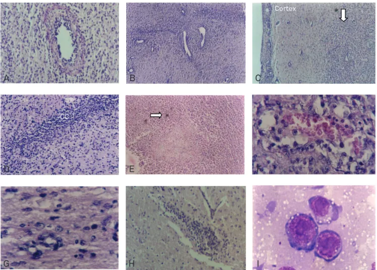
Brain tumors were formed by small and medium-sized round cells with regular cell shape, nucleus with reticular chromatin and prominent nucleoli. Cells were undiferenti-ated and highly mitotic (30 mitosis by high power magniica-tion ield). Tumors formed nodules centered at the inocula-tion point with necrotic foci and many satellite nodules. hey were not encapsulated, and tumor cells iniltrated deeply into normal brain, perivascular spaces and white matter tracts, like corpus callosum (Fig 1). here were tumor necrosis and hemorrhagic areas (41.7% of tumors), as well as deposits of a ibrinoid proteinaceous material and vascular neoformation. here were tumor cell iniltrations in the meninges of 97.5% of the analyzed animals. Some tumors showed intense inil-tration by neutrophils (33.3%).
Estimated TV at day 7 (n=12) was 17.08±6.7 mm3, and at day 9 was 67.25±19.8 mm3 (n=4). We assumed that tu-mor growth was log linear between days 7 and 9 in order to estimate DT. Calculated value was 24.25 h. he measure of largest tumor diameter, but not estimated volume, was correlated signiicantly with neutrophil iniltration, but not with other histological features observed (linear regression, p<0.05, R2=0.6, F value=3.2).
Hematological and biochemical results
Comparison of hematological values between groups by multinomial logistic regression disclosed no statistically sig-niicant diference (Table 1). Mean serum creatine levels were not signiicantly diferent between groups (ANOVA).
Weight of animals and organs
Fig 1. (A) Perivascular cell infiltration and fibrinoid deposits. (X100) (B) Brain tumor angiogenesis. Blood vessels of variable diameters can be observed in the caudate area inoculated with tumor. Usually there’s no blood vessel of this size in this area. (X40) (C) Meningeal tumor infiltration. Tumor focal mass grows without a clear border below cortex (arrow) and shows pial meninx neoplastic infiltration. (X40) (D) Tumor dissemination along white matter tract (callosum). Tumor cells invade a section of corpus callosum (CC) ipsilateral to the tumor inoculation point. (X100) (E) Necrosis. Central tumor necrosis, polymorphonuclear cell infiltration and scattered hemosiderin deposits (arrow). (X40) (F) Intratumoral focal hemorrhage. (X400) (G) Perineuronal infiltration. Detail of normal surrounding brain tissue densely infiltrated by migrating tumor cells. (400X) (H) Satellite nodule. Focal tumor nodule grows at the contralateral brain side. (X100) (I) Tumor cell. W256 tumor cell in imprinting preparation. (X1000, oil immersion). Paraffin embedded, hematoxylin-eosin (HE) stained.
A
D
G
H
I
E
F
B
C
WBC Neutrophils Lymphocytes
nx103 nx103 nx103
SHAM (n=10)
11.1±3.0 1.5±0.4 (15%)
9.2±2.7 (83%)
Control (n=10)
13.2±2.6 2.2±1.3 (17%)
10.6±2.6 (80%)
CS (n=10)
10.7±2.1 2.2±0.8 (21%)
8.1±2.1 (76%) Table 1. Neutrophil and lymphocyte counts in animals with and without brain tumor.
Automated total WBC count was done (Coulter C-6). Neutrophil and lymphocyte count was done manually in May-Grümwald-Giemsa stained slides. We report mean±SD and relative frequencies to WBC (between brackets). CS: cyclosporine treated. Differences were not statistically significant (multinomial logistic regression). WBC: white blood cell. SHAM: isotonic saline.
Weight (g)
Surgery 5d
(SHAM: 15d)
Death or euthanasia (SHAM: 21–27d) SHAM
(n=7) 218±23 224±28 249±31
Control
(n=7) 214±25
210±20 (-2%)
193±32 (-10%)* CS
(n=7) 216±14
185±14 (-25%)†*
181±24 (-26%)†*
Dexa
(n=7) 204±9
231±14 (13%)†
204±19 (0%)* Table 2. Weigh evolution in animals inoculated with tumor (mean ± SD).
Animals were weighted on surgery day, 5 days after inoculation (5d) and on death or euthanasia day. Variation rates (%) within groups comparing to weigh on surgery day are shown between brackets. Weigh averages were compared between and within groups in each occasion. CS: cyclosporine treated. Dexa: dexamethasone treated. SHAM: isotonic saline.
*p<0.05 between groups, comparing to SHAM, Mixed-model ANOVA, Bonferroni.
†p<0.05 within groups, comparing to initial weigh, Mixed-model ANOVA,
Pharmacological treatment
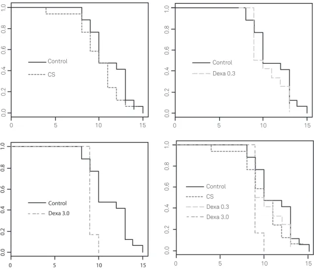
Dexamethasone and CS treated animals median survival was 9.0 (dexa 3.0), 9.5 (dexa 0.3) and 10 (CS) days. Comparison of survival functions by log rank test disclosed a signiicant diference to control only for dexamethasone 3.0 mg/kg/day. A Cox proportional hazards regression conirmed that, of all the treatments, only the highest dose of dexamethasone di-minished survival of animals in this model (HR=4.2, p<0.01, DF=3, likelihood ratio=7.1). he frequency of histological features did not vary signiicantly with CS treatment (logis-tic model). Cyclosporine did not modify signiicantly mean brain weight comparing to control group (Fig 2).
DISCUSSION
We obtained a highly malignant and invasive brain tumor model. Treatment with CS, a speciic T lymphocyte inhibitor, was safely tolerated and did not afect survival in this model despite its induction of an accelerated cachexia. Cyclosporine is an immunosuppressant that has been shown to possess in-trinsic antitumor activity11 and to inhibit p-glycoprotein, a multidrug resistance protein that is a candidate target for brain tumor chemosensitivity modulation12. We may con-clude that further pre-clinical and clinical testing with CS in brain tumors is warranted.
he model presented here showed good reproducibility. Meningeal dissemination seems to be a late occurrence in this model, and did not modify survival median time (data not shown). he time between surgery and inoculation pro-cedure permitted blood-brain barrier (BBB) healing and re-active local inlammation resolution. All animals had lesion points localized to the right cerebral hemisphere. Left cortical brain lesions, but not right cortex ones, can induce immune deicits13. he short DT during growth log phase indicated that the tumor was fast growing and highly malignant. he presence of satellite nodules near the main tumor mass in-dicated tumor cell dissemination through any of these three ways: brain parenchyma, perivascular spaces and white mat-ter tracts. Contralamat-teral hemisphere satellite nodule showed that there had been tumor dissemination by cerebrospinal
luid (CSF) circulation routes. Presence of proteinaceous i-brinoid material deposits could indicate ii-brinoid necro-sis areas. Increase in brain weight was due to local tumor growth, as well as peritumoral brain edema. Lung weight can be used as a quantitative measure of tumor burden in
meta-static lung tumorigenesis models14. here were no evidence
of signiicant lung or liver metastasis, no macroscopic chang-es, no organ weight diferences and no microscopic altera-tions in control or CS treated animals. his showed that CS did not enhance metastatic potential in this model.
Primary malignant brain tumors and some extent meta-static tumors show a layer of brain adjacent to tumor (BAT) consisting of tumor cells iniltrating deeply into normal brain tissue, inducing neovascularization. Capillary permeability and blood perfusion in this region are lower than in tumor
layer, and drug penetration is impaired15. he mechanisms of
invasion are similar to that of metastatic process of non-neu-rologic solid tumors, depend on adhesion molecules expres-sion and are related to induction of angiogenesis16,17. In vitro
and in vivo laboratory models had been developed aiming
the study of CNS tumor invasion. Many transplanted tumors show perivascular invasion, probably related to the presence of the vascular basal layer, rich in extracellular matrix (ECM) structural proteins. Some tumor models show cellular mi-gration along white matter tracts or through CSF circulation routes18.Leptomeningeal carcinomatosis is a complication afecting up to 20% of the patients with some histological types of cancer. Animal models of leptomeningeal metastasis are important to study the physiopathology of meningeal
in-vasion by primary and secondary brain tumors4.
It was observed in our model a region probably equivalent to BAT layer. It showed to have intense tumor cell activity, with parenchymatous iniltration, markedly cell tumor mi-gration through white matter tracts (as callosum) and along perivascular spaces. Also, our model showed consistent lep-tomeningeal metastasis. Intracerebral W256 tumor model
developed by Morreale et al.5 did not show important
inva-sion of adjacent brain parenchyma. Tumors formed compact, well delimited, masses in contrast to our tumor model. he fact that tumor cells used in this study were obtained in vivo
is probably important. Supposedly, these tumor cells from
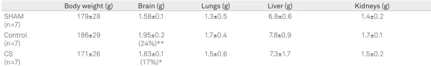
Body weight (g) Brain (g) Lungs (g) Liver (g) Kidneys (g)
SHAM (n=7)
179±28 1.58±0.1 1.3±0.5 6.8±0.6 1.4±0.2
Control (n=7)
186±29 1.95±0.2
(24%)**
1.7±0.4 7.8±0.9 1.7±0.1
CS (n=7)
171±26 1.83±0.1
(17%)*
1.5±0.6 7.3±1.7 1.5±0.2
Table 3. Brain and lung weigh in animals inoculated with tumor (control and CS-treated) and SHAM.
Animals had their bodies weighted and their organs dissected and weighted after death or ether euthanasia. We report mean±SD. Ratio from control or CS groups to SHAM group (%) between brackets. CS: cyclosporine treated. SHAM: isotonic saline.
Fig 2. Survival function plots. Survival function was estimated with Kaplan-Meier method and compared with log-rank test. Only treatment with dexamethasone 3.0 mg/kg/day (Dexa 3.0) was statistically different from control. Cox proportional hazards regression confirmed this.
0
0.
0
0.2
0.4
0.6
0.8
1.
0
5 Control
CS
10 15
0.
0
0.2
0.4
0.6
0.8
1.
0
0 5 10 15
Control Dexa 0.3
0.0
0.2
0.4
0.6
0.8
1.0
0 5 10 15
Control
Dexa 3.0
0.
0
0.2
0.4
0.6
0.8
1.
0
0 5 10 15
CS Control
Dexa 0.3 Dexa 3.0
animal tumors are geno and phenotypically diferent from in
vitro cultivated cells, with diverse potential to invasiveness.
he present model constitutes, thus, a potential tool for the research of tumor cell migration and invasion in CNS.
Neoplastic disease related cachexia is caused by the pro-duction of a number of cytokines by tumor and inlamma-tory cells, the most important of them being IL-6, TNF-α and PGE2
19.W256 tumor implantation in extraneural sites
is associated with cachexia and modiied dietetic glucose and lipid metabolism in host animals, as well as induced hy-percalcemia, bone mineral density loss and osteoporosis20,21. Moreover, W256-induced cachexia is associated with
im-munosuppression caused by lymphocyte apoptosis.22 hese
metabolic efects of W256 tumor could have contributed to the cachexia presented by the animals of the control group. As far as we are concerned, this is the irst experimental ac-count of intracerebral W256 induced cachexia and one of the irst experimental brain tumor animal models to show signiicant cachexia so far. Because of the intracerebral focal growth of the tumor in basal ganglia in this model, there is
also the possibility that the animals had developed dienceph-alic syndrome23.
possible direct efect of dexamethasone in glioblastoma cells, it has been suggested that it could protect tumor cells from chemotherapy, hence afecting patient survival26. We could show a direct relationship between polymorphonuclear cell iniltrate and tumor diameter in this model. One can postu-late tentatively that possibly the tumor iniltrating inlamma-tory cells play a role in this tumor model, an event already observed in other tumor types27. Neutrophil iniltration could possibly have had inluence in survival modulation by dex-amethasone. Unfortunately, we did not prospectively com-pared histology in all the treatments, so we are unable to answer whether dexamethasone reduced neutrophil iniltra-tion as well. he lack of proteciniltra-tion from dexamethasone in this model and, additionally, its detrimental efect in surviv-al deserve further experimentsurviv-al clariication in order to ind the mechanisms of action of dexamethasone in this particu-lar model.
In conclusion, rat brain simpliied implantation of W256 tumor model showed to be highly reproducible, yielding un-diferentiated highly malignant tumor with local invasive char-acteristics similar to that of primary brain tumors, and should be a potential model to the study of tumor cell migration in CNS. he model could be used as a tool to investigate biologi-cal behavior and novel treatment options for both brain par-enchymatous tumor growth and leptomeningeal carcinoma-tosis. Brain tumor growth induced signiicant cachexia, and this model could potentially be used to study diencephalic syndrome. Its attractive points are in vivo tumor origin, easy
manual inoculation technique and high intracerebral tumor growth yielding. Dexamethasone, a drug widely used in brain tumor patients, reduced survival in this model. he same did not happen with CS treatment. Cycloporine treatment did not afect adversely tumor-bearing animals. Cyclosporine could thus be used for further testing against brain tumor models.
1. Barth RF, Kaur B. Rat brain tumor models in experimental neuro-oncology: the C6, 9L, T9, RG2, F98, BT4C, RT-2 and CNS-1 gliomas. J Neurooncol 2009;94:299-312.
2. Peterson DL, Brown Jr WE. Animal models for brain tumors. In: Black PM, Loeffler JS (Eds). Cancer of the nervous system. USA: Blackwell Science, 1997.
3. Huang TY, Arita N, Hayakawa T, Ushio Y. ACNU, MTX and 5-FU penetration of rat brain tissue and tumors. J Neurooncol 1999;45:9-17.
4. Nakagawa H, Yoshioka K, Miyahara E, Fukushima Y, Tamura M, Itoh K. Intrathecal administration of Y-27632, a specific rho-associated kinase inhibitor, for rat neoplastic meningitis. Mol Cancer Res 2005;3:425-433.
5. Morreale VM, Herman BH, Der-Minassian V, et al. A brain-tumor model utilizing stereotatic implantation of a permanent cannula. J Neurosurg 1993;78:959-965.
6. Félix FHC. Modelo de implante de tumor de Walker no cérebro de ratos [dissertação]. Fortaleza: Faculdade de Medicina. Universidade Federal do Ceará; 2001.
7. Freshney RI. Culture of animal cells. New York: Alan R. Liss, 1987. 8. Smith CA, Andrews CM, Collard JK, Hall DE, Walker AK. A color atlas of
comparative diagnostic and experimental hematology. Barcelona: Wolfe Publishing, 1994.
9. American Veterinary Medical Association Panel on Euthanasia. 2000 Report of the AVMA Panel on Euthanasia. J Am Vet Med Assoc 2001;218:669-696.
10. Steel GG. Growth Kinetics of Tumours. Oxford: Claredon Press, 1977. 11. Sliwa M, Markovic D, Gabrusiewicz K, et al. The invasion promoting effect
of microglia on glioblastoma cells is inhibited by cyclosporin A. Brain 2007;130:476-489.
12. Aller SG, Yu J, Ward A, et al. Structure of P-glycoprotein reveals a molecular basis for poly-specific drug binding. Science 2009;323:1718-1722. 13. Dunn AJ. Interactions between the nervous system and the immune
system. Implications for psychopharmacology. In: Bloom FE, Kupfer DJ (Eds). Psychopharmacology: The Fourth Generation of Progress. New York: Raven Press, 1995.
14. Sava G, Clerici K, Capozzi I, et al. Reduction of lung metastasis by ImH[trans-RuCl4(DMSO)Im]: mechanism of the selective action investigated on mouse tumors. Anticancer Drugs 1999;10:129-138. 15. Giese A, Bjerkvig R, Berens ME, Westphal M. Cost of migration: invasion
of malignant gliomas and implications for treatment. J Clin Oncol 2003;21:1624-1636.
16. Silva RG, Tavora B, Robinson SD, et al. Endothelial alpha3beta1-integrin represses pathological angiogenesis and sustains endothelial-VEGF. Am J Pathol 2010;177:1534-1548.
17. Gao CF, Xie Q, Su YL, et al. Proliferation and invasion: plasticity in tumor cells. Proc Natl Acad Sci U S A 2005;102:10528-10533.
18. Shelton LM, Mukherjee P, Huysentruyt LC, Urits I, Rosenberg JA, Seyfried TN. A novel pre-clinical in vivo mouse model for malignant brain tumor growth and invasion. J Neurooncol 2010;99:165-176.
19. Rebeca R, Bracht L, Noleto GR, et al. Production of cachexia mediators by Walker 256 cells from ascitic tumors. Cell Biochem Funct 2008;26: 731-738.
20. Fernandes LC, Machado UF, Nogueira CR, Carpinelli AR, Curi R. Insulin secretion in Walker 256 tumor cachexia. Am J Physiol 1990;258: 1033-1036.
21. Waki Y, Miyamoto K, Kasugai S, Ohya K. Osteoporosis-like changes in Walker carcinoma 256-bearing rats, not accompanied with hypercalcemia or parathyroid hormone-related protein production. Jpn J Cancer Res 1995;86:470-476.
22. Lima TM, Lima MM, Almeida DC, Mendonça JR, Curi R. Cachexia induced by Walker 256 tumor growth causes rat lymphocyte death. Cancer Immunol Immunother 2005;54:179-186.
23. Fleischman A, Brue C, Poussaint TY, et al. Diencephalic syndrome: a cause of failure to thrive and a model of partial growth hormone resistance. Pediatrics 2005;115:742-748.
24. Kotsarini C, Griffiths PD, Wilkinson ID, Hoggard N. A systematic review of the literature on the effects of dexamethasone on the brain from in vivo human-based studies: implications for physiological brain imaging of patients with intracranial tumors. Neurosurgery 2010;67:1799-1815. 25. Gu Y, Qin L, Qin X, Xu F. The molecular mechanism of
dexamethasone-mediated effect on the blood–brain tumor barrier permeability in a rat brain tumor model. Neurosci Letters 2009;452:114-118.
26. Qian YH, Xiao Q, Chen H, Xu J. Dexamethasone inhibits camptothecin-induced apoptosis in C6-glioma via activation of Stat5/Bcl-xL pathway. Biochim Biophys Acta 2009;1793:764-771.
27. Hillen F, Baeten CIM, van de Winkel DC, Van der Schaft DWJ, Winnepenninckx V, Griffioen AW. Leukocyte infiltration and tumor cell plasticity are parameters of aggressiveness in primary cutaneous melanoma. Cancer Immunol Immunother 2008;57:97-106.