Pathological changes in turkeys liver associated with Histomoniasis in
Duhok City,Kurdistan Region, Iraq
M.A. Abdullah
1*, E.K. Zankana
2and V.J. Ameen
11 Department of Pathology and Microbiology, 2 Department of Medical Science, Faculty of Veterinary Medicine,
University of Duhok, Iraq, * E-mail: [email protected]
(Received November 19, 2013; Accepted December 18, 2013)
Abstract
Histomoniasis were detected and described among naturally affected of twenty three young Turkeys poult compared with the adults. Sample for study were collected from different areas of Duhok city in Kurdistan region/Iraq. Giemsa stain where used for identification of parasite from specimens of liver and cecum samples, then specimen, where fixed in 10% neutral buffered formalin for routine histopathological study. The affected birds showed clinical signs of lethargic, drooping of head and wings with progressive emaciation and a clear distinctive signs in live ones, and appearance of continuous yellowish diarrhea. Pathologically there is an enlargement and discoloration of the liver associated with appearance of white to yellow multifocal nodules in the surface of the liver. While the result of histolpathological changes showed severs inflammatory reaction around necrotic tissues with degenerative and necrotic changes of the liver cells.
Keywords: Pathology; Histomonasis; Turkeys Available online at http://www.vetmedmosul.org/ijvs
ا يغتلا
زساينوموتسھلا
ءادل
بحاص لا
يمو لا
ابكا
يض لا
كوھ
نيدم
يف
،
ا علا
اتس ك
ميلقا
ﷲدبع
لع
دھم
،
ر اق
اسحا
نكن
و
نيما
عج
ليھف
يرھج لا
ءايحااو
ارماا
عرف
،
يريرسلا
و علا
عرف
،
ر ي لا
ب لا
ي لوكاف
،
وھ
عماج
،
ارعلا
صا لا
ساينومو سھلا
ءا
فصوو
صي شت
مت
،ايعي
با لاو
غلا لا
يمورلا
عم
نرا لاب
يمورلا
ارفا
راغص
نم
وشعو
اث
يف
م
ق انم
نم
ساردلا
انيع
تع ج
ثيح
ع
فرع ل
ا ي لا
غ ص
تمد سا
ثيح
، ارعلا
ا س رك
مي قا
يف
وھ
نيدم
نم
عااو
د لا
انيع
نم
ي ي لا
و ب
انيعلا
تي ثت
مت
اھدعبو
,
نيرو
نيلامروف
%
اع م
را
روي لا
رھ ا
.
يجيسنلا
سارد ل
يضر لا
ساردلا
رھ أو
ا ك
.
و لا
ر صا
ر سم
اھس ب
ا فار م
ر س لا
ا ھلا
عم
نجااو
ارلا
يلدتو
نھولا
اماع
با لا
ع ب
روھ
عم
د لا
ول
يف
اريغتو
ا
ت
وجو
اريغ ل
ساردلا
جئا ن
ت ضوا
ا نيب
.
د لا
ح س
ع
و لا
ءار ص
لا
ءا يب
دع م
يباھ لا
لعا ت
وجو
يجيسنلا
ديدش
روھ
عم
ر ن لا
جسناا
وح
اريغ لا
ر نلا
و
ي
يس ن لا
.
Introduction
Histomoniasis is also called blackhead, enterohepatitis, is an infectious disease of gallinaceous birds caused by the protozoan Histomonas meleagridis belonging to the trichomonais group which occurs at all over the world (1). In 1893, Cushman first brief described histomoniasis (2).
The parasite causes high morbidity and mortality in turkeys and the disease is manifested by a diphtheroid inflammation of the caeca and by necrosis in the liver (3,4).
exposed young turkeys to other in absence of the caecal worm vector (6,7).
Clinically the Symptoms include depression, reduced appetite, poor growth, increased thirst, sulphur-yellowish diarrhea, listlessness and dry, ruffled feathers. The head may become cyanotic (bluish in color) (8).
The diagnosis of disease depend mainly on pathological appearance of liver, presence of small nodules in the surface of liver which representing a necrotic changes and regarded a pathognomic lesions of histomoniasis (9).
Due to the scant information about the disease in Duhok area as well as occurrence of disease, therefore, the aim of our research is to study the pathological observation of Histomoniasis, further study is needed to know about incidence, epidemiology and the role of vector and caecal worm eggs in the transmission of disease.
Material and methods
Sample collection
A total of 42 cases of turkey affected clinically were obtained from different flocks in Duhok governorate, 23 of death birds was examined by post-mortem examination after manifestation a clinically and the changes were recorded, organs which showed lesions were fixed especially liver and cecum. The mortality rate was recorded among young poult rather than adults. Diagnosis of disease was done according to history, clinical signs and detection of histomonas by Giemsa staining smears from liver and cecum, in addition to gross and histopathological changes. Histopathology was performed in Pathology and Molecular Laboratory at Faculty of Veterinary Medicine, Duhok Research Center. Specimen were collected and fixed in neutral buffered formalin saline solution 10%. Tissues were then dehydrated in different concentration of ethanol, cleared in xylen, embedded in white paraffin wax at melting point 56-58°C for preparation of paraffin block. Sections were made at 3-4 micrometers with leica microtome (leica, Germany). Haematoxylin and Eosin were used for staining tissue as per standard staining technique according to (10). The stained slides were examined under light microscopes with photography (leica, Germany).
Results
Clinical signs
During this study clinical signs were recorded in an infected birds just be a sudden rise in mortality especially among poult, the infected bird was appeared lethargic, with drooping head and wings most of birds showing marked decrease of body weight with progressive emaciation and a clear distinctive signs in the live bird is the appearance of Sulphur-yellow droppings diarrhea.
Post-Mortem Observations
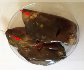
The typical important lesions associated with Histomoniasis are thickened cecal wall pouches containing yellowish, firm cores, and enlargement of livers (hepatomegally) with spotted nodules target lesions representing the area of necrosis which appears as yellowish to yellowish-green with depressed below the surface especially in advanced cases as showing in (figure 1).
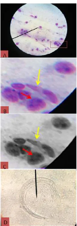
To confirm the diagnosis smears from liver and cecum stain with Giemsa showing a typical feature known as double-eyed with infected cells as well as flagellated form of parasite as showing in (figure 2A, B and C). Result also showing a cecal worm parasite Heterakis gallinarum from direct smears of cecum as showing in (figure 2D).
Figure 1: Showing enlargement of liver (hepatomegaly) spotted target lesions representing an areas of necrosis red arrow which appears as nodules in the surface of liver infected with Histomonas meleagridis.
Histopathological appearance
Figure 2: Giemsa staining from smear of liver showing typical characteristic features of Histomonas meleagridis double-eyed appearance B and C red arrow, flagellated form B and C yellow arrow and larvae of parasite heteraks gallinarum D.
Figure 3: Histopathological examination of liver section showing a focal area of necrosis red arrow which surrounded by a zone of inflammatory cells black arrow, H & E 20x.
Figure 5: Histopathological examination of liver section showing cellular infiltration of lymphocytes red arrow, macrophages black arrow and giant cells green arrow, H & E 40x.
Figure 6: Histopathological examination of liver section showing coagulative necrosis red arrow and intracellular parasite black arrow, H & E 40x.
Discussion
Histmoniasis is regarded as a systemic disease in turkey (3), therefore, the invading of histomonads go deeper through the caecal mucosa, then take the hepatic portal route and carried out to the liver, where they cause multi focal necrosis which is clear in this study as a many reaction in the area of portal artery and this was agreement with other study (11). Histopathological studies have shown that many individual and clustered histomonads are visible in the periphery of lesions confirming the results of (12). Liver
necrosis and degeneration with histomoniasis might be due to infilammatory reaction and toxic damage of protozoan. Since the discovery of H. meleagridis in 1895, researchers have used a variety of approaches for the prevention and diagnosis of histomoniasis. Diagnosis is initially made at onset of clinical symptoms and pathologic changes. However, the use of traditional methods of pathogen detection or epidemiologic investigations. Furthermore, microscopic examination which helpful for identification of lesions as well as characterization of histmoniaisis, Further study is needed to confirm the diagnosis of histomoniasis such as molecular taxonomy of parasites and molecular genetics.
Figure 7: Histopathological examination of liver section showing amoeboid shape of Histomonas meleagridis red arrow and fatty changes black arrow, H & E 40x.
References
1. McDougald LR. Blackhead disease Systemic histomoniasis associated with high (Histomoniasis) in poultry, a critical review. Avian mortality and unusual lesions in the bursa Avain Dis. 2005;49:462-76.
2. Smith T. An infectious disease among turkeys caused by protozoa (infectious enterohepatitis). USDA, Bureau Animal Industry Bull. 1995;8:1-38
3. Sentier-Cue G, Chin RP, Shivaprasada HL. Systemic histomoniasis associated with high mortality and unusual lesions in the bursa of Fabricius and lungs in commercial turkeys. Avian Dis. 2009;53:231-238.
4. Popp C, Hauck R, Blazey B, Hanel A, Hafez HM. An unusual outbreak of histomoniasis in a commercial turkey flock. Berl Munich Tierraztl Wschr, 2011;125:153-158.
5. Saif YM. Bacterial Infection. In: Diseases of Poultry, Saif, YM. (Ed.). Blackwell Publi Inc., USA. 2001.
6. McDougald LR, Fuller L. Blackhead disease in Turkeys: Direct transmission of Histomonas meleagridis from bird to bird in a laboratory model. Avian Dis. 2005;49:328-331
7. Hue J, McDougald LR. Direct lateral transmission of Histomonas meleagridis in Turkeys. Avian Dis. 2003;47:489-492.
8. Merck M. The Merck Veterinary Manual: http//www.Merckvet maual.com/mvm/index.jsp http//www.Merck. Cited on 2010.
9. Al-Sadi HI, Basher HA, Qubih TS. A retrospective study of clinically diagnosed poultry diseases in Nenevha Province, Iraq. Iraqi J Vet Sci.2000;13:107–113.
10.Luna, Lg. Manual of histologic staining methods of the armed forces Institute of pathology 3 rd Ed. Mc Gran.Hill book Comp. 1968; pp:34. 11.Bon Durant RH, Wakenell PS. Histomonas meleagridis and relatives.
In: Parasitic protozoa, vet.Q. 2nd Edn, Krier, JP, JR Baker (Eds.), Acad.Press, San diego, CA.1994;pp: 189-206.