Discovery of New Candidate Genes Related
to Brain Development Using Protein
Interaction Information
Lei Chen2‡, Chen Chu3‡, Xiangyin Kong4, Tao Huang4
*, Yu-Dong Cai1*
1Institute of Systems Biology, Shanghai University, Shanghai 200444, People’s Republic of China, 2College of Information Engineering, Shanghai Maritime University, Shanghai 201306, People’s Republic of China,3Institute of Biochemistry and Cell Biology, Shanghai Institutes for Biological Sciences, Chinese Academy of Sciences, Shanghai 200031, People’s Republic of China,4Institute of Health Sciences, Shanghai Institutes for Biological Sciences, Chinese Academy of Sciences, Shanghai 200025, People’s Republic of China
‡These authors contributed equally to this work. *[email protected](TH);[email protected](YDC)
Abstract
Human brain development is a dramatic process composed of a series of complex and fine-tuned spatiotemporal gene expressions. A good comprehension of this process can assist us in developing the potential of our brain. However, we have only limited knowledge about the genes and gene functions that are involved in this biological process. Therefore, a sub-stantial demand remains to discover new brain development-related genes and identify their biological functions. In this study, we aimed to discover new brain-development related genes by building a computational method. We referred to a series of computational meth-ods used to discover new disease-related genes and developed a similar method. In this method, the shortest path algorithm was executed on a weighted graph that was con-structed using protein-protein interactions. New candidate genes fell on at least one of the shortest paths connecting two known genes that are related to brain development. A ran-domization test was then adopted to filter positive discoveries. Of the final identified genes, several have been reported to be associated with brain development, indicating the effec-tiveness of the method, whereas several of the others may have potential roles in
brain development.
Introduction
Beginning with the segregation of neural and glial cells from other types of tissues, brain devel-opment is a dramatic process composed of a series of complex and fine-tuned spatiotemporal gene expressions. These gene expressions contribute to brain transformation that involves both microscopic and macroscopic changes [1,2]. Although all mammalian embryos develop in a fundamentally similar way, human brains show more sophisticated neural structures and cir-cuits than other species, which gives rise to human intelligence. Therefore, understanding the OPEN ACCESS
Citation:Chen L, Chu C, Kong X, Huang T, Cai Y-D (2015) Discovery of New Candidate Genes Related to Brain Development Using Protein Interaction Information. PLoS ONE 10(1): e0118003. doi:10.1371/journal.pone.0118003
Academic Editor:Yu Xue, Huazhong University of Science and Technology, CHINA
Received:October 10, 2014
Accepted:January 3, 2015
Published:January 30, 2015
Copyright:© 2015 Chen et al. This is an open access article distributed under the terms of the
Creative Commons Attribution License, which permits unrestricted use, distribution, and reproduction in any medium, provided the original author and source are credited.
Data Availability Statement:All relevant data are within the paper and its Supporting Information files.
process and mechanism of brain development is important. To explore this mystery, molecular and cellular biology approaches have widely been applied over the past decades and greatly ad-vanced our understanding in this field. Many genes and pathways have been identified that play essential roles in the development of human brains. For example, the Wnt-Frizzled signal-ing pathway was shown to be the key element in physiological neural-stem cell proliferation and differentiation. Additionally, this pathway could also lead to the pathological generation of brain cancers [1,3]. Sox family transcription factors also provided important clues about the differentiation of neural stem cells into neurons and glia [2].
Despite these advances, the intricate brain development process remains largely unveiled, and a great demand is present to discover new genes and identify their functions. However, the discovery of new genes related to brain development only by biochemical experiments is chal-lenging because we have to investigate each gene, resulting in a high cost and substantial time. Fortunately, with the existing knowledge of genes and pathways that relate to brain develop-ment, we can unearth common features among the genes and develop computational methods to screen for candidate genes that have stronger associations with brain development. This screening reduces the search space and provides new clues for future research. Various biologi-cal problems have been completely or partly settled by designing effective computational meth-ods [4,5,6,7,8,9,10,11,12]. Encouraged by the success of these studies, the design of effective computational methods is a useful process to discover new candidate genes related to brain development.
Recently, a group of computational methods was proposed to discover new disease genes ac-cording to the current known disease genes. These methods have been applied to find new genes related to various diseases including lung cancer [13] and hepatocellular carcinoma [14]. We proposed a similar computational method to discover new candidate genes related to brain development. Additionally, several newly found genes have high probabilities of being novel genes related to brain development because they have been shown to be expressed in the brain and involved in several brain diseases. The findings in this study will introduce new directions for the investigation of the biological process of brain development.
Materials and Methods
Materials
The 94 human genes related to brain development, with experimental evidence from Gene On-tology (GO:0007420), were retrieved fromhttp://amigo.geneontology.org/amigo/term/ GO:0007420(Accessed 2014 May 10) [15]. These genes were used to discover new candidate genes for brain development and are available inS1 File.
In addition, we also downloaded the 610 human genes that are related to brain development with evidence from Gene Ontology (GO:0007420) at the aforementioned website. These genes included the genes mentioned in the above paragraph. The remaining 516 genes were shown to have associations with brain development by other methods instead of direct experiments. The 516 genes were termed as inferred genes for brain development and are listed inS2 File. These genes will be used to compare with genes discovered in our method.
Construction of the weighted graph based on protein-protein interactions
Protein-protein interactions provide useful information to investigate protein-related problems [16,17,18,19,20] because the interaction can reflect the association between proteins from near-ly all cellular functions such as regulation of signaling and metabolic pathways, protein synthe-sis, DNA replication, gene translation, and immunological recognition [21]. In this study, we employed protein-protein interactions retrieved from STRING (Search Tool for the Retrieval Competing Interests:The authors have declaredof Interacting Genes/Proteins,http://string.embl.de/) [22], which have been successfully applied to previously construct several computational methods [16,20,23]. The obtained pro-tein-protein interactions were derived from genomic contexts, high-throughput experiments, (conserved) coexpression and previous knowledge. Thus, they contain the direct and indirect associations between proteins. For each obtained protein-protein interaction, the entry con-tains two proteins and one score, in which the score can reflect the strength of the interaction from many aspects of proteins. For later formulation, the score of the interaction containing proteinsp1andp2was denoted byS(p1,p2).
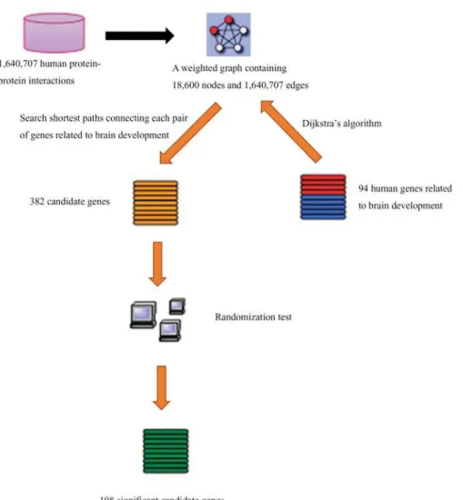
According to the protein-protein interactions retrieved from STRING, we constructed a weighted graphG= (V,E) as follows:Vrepresented all human proteins occurring in protein-protein interactions retrieved from STRING, andEcontained all pairs of nodes such that the corresponding proteins comprised a protein-protein interaction in STRING. Because the range of interaction scores is between 150 and 999, a weight was assigned to each edge,e, inGby 1000-S(p1,p2), wherep1andp2were two corresponding proteins of the endpoints of edgee. The constructed graphGconsisted of 18,600 nodes and 1,640,707 edges.
Methods for discovery of new candidate genes
The discovery method was executed on the weighted graphGconstructed in Section“ Con-struction of the weighted graph based on protein-protein interactions”, which consisted of two stages: (I) execute the shortest path algorithm onGfor finding new candidate genes and (II) construct a randomization test to filter these candidate genes. Readers can refer to previous studies [13,14] for the detailed procedures of the method and its principle. Here, we only pro-vide a brief description.
Stage I. The well-known shortest path algorithm, Dijkstra’s algorithm [24], was executed onGto find all of the shortest paths which connect any two genes related to brain develop-ment,i.e., genes listed inS1 File. Certain gene occurring in at least one of these paths as an inner node was selected as a candidate gene for brain development. For the later randomization test, we also counted the number of paths containing each candidate gene as an inner node. This value was termed as betweenness in this study.
Stage II. The randomization test was executed by constructing 500 gene sets whose sizes were equal to that of the set consisting of human genes related to brain development. For each gene set, we executed the procedure inStage Iand counted the betweenness for each candidate gene. Another value, named the permutation FDR (False Discovery Rate), was then computed for each candidate gene, which was defined as“the number of gene sets on which the between-ness exceeded that on the gene set related to brain development”/500. Finally, candidate genes with permutation FDRs smaller than 0.05 were selected as significant candidate genes for brain development.
Results and Discussion
According to the procedures (including the construction of the weighted graph) described in Section“Construction of the weighted graph based on protein-protein interactions”and
“Methods for discovery of new candidate genes”, some candidate genes for brain development can be identified. The workflow of these procedures is illustrated inFig. 1.
Candidate genes related to brain development
Significant candidate genes related to brain development
According to the method, the 382 candidate genes were filtered by a randomization test, and the permutation FDR of each candidate gene was calculated, the results of which are available inS1 Table. We selected 108 candidate genes with permutation FDRs smaller than 0.05 as the significant candidate genes (see the first 108 genes inS1 Table).
Gene ontology enrichment analysis of the significant candidate genes
Gene Ontology (GO) is a widely used bioinformatics tool, which represents gene product prop-erties across all species using GO terms [15,25]. Here, we analyzed the 108 significant candidate genes by investigating their GO enrichment terms. This was performed by a functional annota-tion tool, DAVID (Database for Annotaannota-tion, Visualizaannota-tion and Integrated Discovery) [26]. The results are shown inTable 1.The 108 significant candidate genes were significantly enriched in Biological Process (BP) terms related to neuronal cell proliferation and development: GO:0042127 (Regulation of cell proliferation), GO:0008284 (Positive regulation of cell proliferation), GO:0060284 (Regulation
Fig 1. The workflow of the procedures for discovery of candidate genes related to brain development.
of cell development), GO:0010720 (Positive regulation of cell development), GO:0051960 (Reg-ulation of nervous system development) and GO:0035270 (Endocrine system development).
Analysis of the relationship between several significant candidate genes
and the biological process of brain development
In this study, we obtained 108 genes that possibly relate to brain development, among which 16 genes (14.81%, 16/108) were GO inferred genes. These 16 genes are listed inTable 2. In the
Table 1. The GO BP enrichment analysis of the 108 significant candidate genes.
Term Count FDR
GO:0042127~regulation of cell proliferation 25 3.15E-06
GO:0008284~positive regulation of cell proliferation 17 9.54E-05
GO:0060284~regulation of cell development 12 5.43E-04
GO:0010720~positive regulation of cell development 8 0.0012
GO:0048010~vascular endothelial growth factor receptor signaling pathway 5 0.0015 GO:0044093~positive regulation of molecular function 18 0.0021 GO:0006357~regulation of transcription from RNA polymerase II promoter 20 0.0022 GO:0008285~negative regulation of cell proliferation 14 0.0041
GO:0048729~tissue morphogenesis 10 0.0121
GO:0035270~endocrine system development 7 0.0194
GO:0051960~regulation of nervous system development 10 0.0204 GO:0010604~positive regulation of macromolecule metabolic process 20 0.0236
GO:0051098~regulation of binding 9 0.0289
GO:0045944~positive regulation of transcription from RNA polymerase II promoter 13 0.0305 GO:0051173~positive regulation of nitrogen compound metabolic process 17 0.0313
GO:0007183~SMAD protein complex assembly 4 0.0359
GO:0045927~positive regulation of growth 7 0.0456
GO:0043085~positive regulation of catalytic activity 15 0.0459
doi:10.1371/journal.pone.0118003.t001
Table 2. Detailed information for the 16 significant candidate genes that were also inferred genes.
Ensembl ID Gene name Betweenness Permutation FDR
ENSP00000287934 FZD1 88 0
ENSP00000354607 FZD5 87 0.02
ENSP00000363826 FZD8 88 0
ENSP00000305769 SMAD1 252 0.016
ENSP00000176195 SCT 147 0.048
ENSP00000330633 CNTN2 88 0.004
ENSP00000354478 DLX1 88 0.002
ENSP00000320147 EZH2 88 0.008
ENSP00000354859 DRD2 6 0.016
ENSP00000329623 BCL2 570 0.002
ENSP00000396219 MEF2C 88 0.014
ENSP00000366413 POU4F1 88 0
ENSP00000261349 LRP6 174 0.026
ENSP00000353059 APAF1 250 0.018
ENSP00000237527 GHRH 4 0.02
ENSP00000320180 GHRHR 2 0.03
following paragraphs, previously reported experimental evidence are provided for the expres-sion and functions of these inferred genes in brain development, indicating that our method is effective for the discovery of new candidate genes.
The Frizzled proteins were identified to be the receptors for the Wnt signaling molecules [27,28]. Wnt proteins are highly expressed in the developing central nervous system and were shown to play essential roles in brain development, adult neurogenesis and brain disorders [3,29,30]. In our study, Frizzled receptorsFZD1,FZD5andFZD8were predicted to be involved in brain development, which is consistent with previous observations.
The growth hormone releasing hormoneGHRHand its receptorGHRHRwere also pre-dicted to be related to brain development. The expression of both GHRH and GHRHR were detected in the brain [31,32]. GHRHR was highly expressed in the hypothalamus and pituitary, but not in the olfactory bulb, caudate putamen, cerebral cortex, hippocampus, cerebellum or brainstem, suggesting possible sites of GHRH action. GHRH stimulates the release of growth hormone, which has been shown to alter neurogenesis, myelin synthesis and dendritic branch-ing [33].
SMADs are important signaling molecules of cytokine-mediated signaling pathways and are involved in a series of physiological and pathological processes. One of the family members,
SMAD1, was reported to form a complex with STAT3 (signal transducers and activators of transcription), and the formation process was bridged by the transcriptional coactivator EP300. This complex was involved in the cooperative signaling of LIF (leukemia inhibitory fac-tor) and BMP2 (bone morphogenetic protein-2) and the subsequent induction of astrocytes from neural progenitors [34]. Our study predicted roles forSMAD1,EP300and other SMAD family members such asSMAD2andSMAD4.
TheSCTgene encodes a gastrointestinal peptide secretin, which is widely expressed during mouse embryonic development [35]. Several lines of evidence indicate that the expression of secretin or secretin-like peptides were also present in several developing brain regions such as the cephalic mesenchyme, cerebellar primordium and choroid plexus [35,36,37,38]. Secretin deficient mice demonstrated impairment in synaptic plasticity in the CA1 area of the hippo-campus, which implied the potential neuroactive role of secretin in the brain [39].
Among the contactin family,CNTN1(also known as contactin) has been extensively investi-gated for its role in the brain. The GPI-linked neural cell recognition molecule F3/contactin is clustered at the paranodal region during development, a vital site for axoglial interaction. F3/ contactin was reported to act as a functional ligand of Notch, and this trans-extracellular inter-action triggered gamma-secretase-dependent nuclear translocation of the Notch intracellular domain. The F3/contactin was shown to specifically initiate a Notch/Deltex1 signaling pathway that promoted oligodendrocyte maturation and myelination [40]. Our study predicted a poten-tial role ofCNTN2, which was expressed on the surface neuronal cells [41] in
brain development.
The POU class 4 homeobox 1 protein POU4F1 has been shown to regulate dorsal-root gan-glion-sensory neuron specification and axonal projection into the spinal cord [42]. Additional-ly, high expression of POU4F1 and other POU-homeodomain transcription factors were detected in developing projection neurons within the retina, inner ear and trigeminal ganglion with an established function in controlling cell differentiation and survival [43].
Besides the above-mentioned GO inferred genes, mounting evidence has suggested the po-tential roles of other significant candidate genes in brain development, such asCNOT1,
CNOT2,CNOT4,CNOT6,MED8,MED10,MED12,CDK8,LAMA2,LAMA4,CASP1andetc. The CNOT subunits form the CCR4-NOT complex, which is a highly conserved global transcriptional regulator. This complex has been implicated in a number of different aspects of mRNA and protein expression, including mRNA degradation, transcription initiation and elongation, ubiquitination and protein modification [46], which is essential in the context of development [47]. Previous studies described the spatiotemporal expression of CNOTs during differentiation of neural stem cells, implying their function in brain development [48].
The mediator complex aids in transcriptional activation through interaction with RNA po-lymerase II and gene-specific transcription factors. Here, subunits CDK8, MED8, MED10 and MED12 were predicted to be related to brain development. The cyclin-dependent kinase CDK8 served as a partner of MED12 in the mediator complex that functioned in developmen-tal gene regulation [49]. And a recent study unveiled a new role of the mediator complex in epi-genetic silencing of neuronal gene expression, which is linked to neuronal development and function [50].
Laminin subunits LAMA2 and LAMA4 were also predicted to be involved in brain develop-ment. The extracellular protein laminin is a major component of the basement membrane and mediate the attachment, migration, and organization of cells into tissues during embryonic de-velopment. Laminin has been shown to regulate neural precursor cells by enhancing migration, expansion, and differentiation into neurons and astrocytes [51]. Additionally, short laminin peptides were reported to promote in vitro neural stem cell proliferation and survival [52].
The caspases are a family of cysteine-aspartic acid protease, which play crucial roles in pro-grammed cell death [53]. Recent evidence indicated that caspase family also participated in vari-ous developmental processes, including the development of thymocyte [54], kidney [55], retina [56], tooth [57], and the central nervous system [58]. Several caspase family members such as caspase 3, 8 and 9 have been shown to regulate the postnatal brain development [59,60], and caspase 1 (CASP1) was predicted by our algorithm to be also involved in this process.
Here, we have provided some experimental evidence for eleven candidate genes of their functions in brain development. These results are consistent with our prediction and suggest the effectiveness and efficiency of our algorithm. Other candidate genes also serve as a useful resource for further investigation on brain development and function.
Conclusions
This study utilized the shortest path algorithm to discover new candidate genes that are related to brain development. Furthermore, a randomization test was adopted to filter the positive dis-coveries. Among the final obtained genes, several have direct evidence for their expression and functions in the brain, whereas several others may play potential roles in brain development. The findings in this study provide new insights for the discovery of novel genes related to brain development, thereby promoting the comprehension of the overall developmental process. The software is available upon the request.
Supporting Information
S1 File. 94 human genes related to brain development, which are with experimental evi-dence from Gene Ontology (GO:0007420).
(PDF)
S2 File. 516 human genes related to brain development, which are with evidence, instead of experimental evidence, from Gene Ontology (GO:0007420).
(PDF)
S1 Table. 382 candidate genes for brain development and their betweenness and permuta-tion FDRs.
(PDF)
Author Contributions
Conceived and designed the experiments: TH YDC. Performed the experiments: LC YDC. An-alyzed the data: LC CC XK. Contributed reagents/materials/analysis tools: LC CC XK TH YDC. Wrote the paper: LC CC.
References
1. Patapoutian A, Reichardt LF (2000) Roles of Wnt proteins in neural development and maintenance. Curr Opin Neurobiol 10: 392–399. PMID:10851180
2. Wegner M, Stolt CC (2005) From stem cells to neurons and glia: a Soxist’s view of neural development. Trends Neurosci 28: 583–588. PMID:16139372
3. Malaterre J, Ramsay RG, Mantamadiotis T (2007) Wnt-Frizzled signalling and the many paths to neural development and adult brain homeostasis. Front Biosci 12: 492–506. PMID:17127312
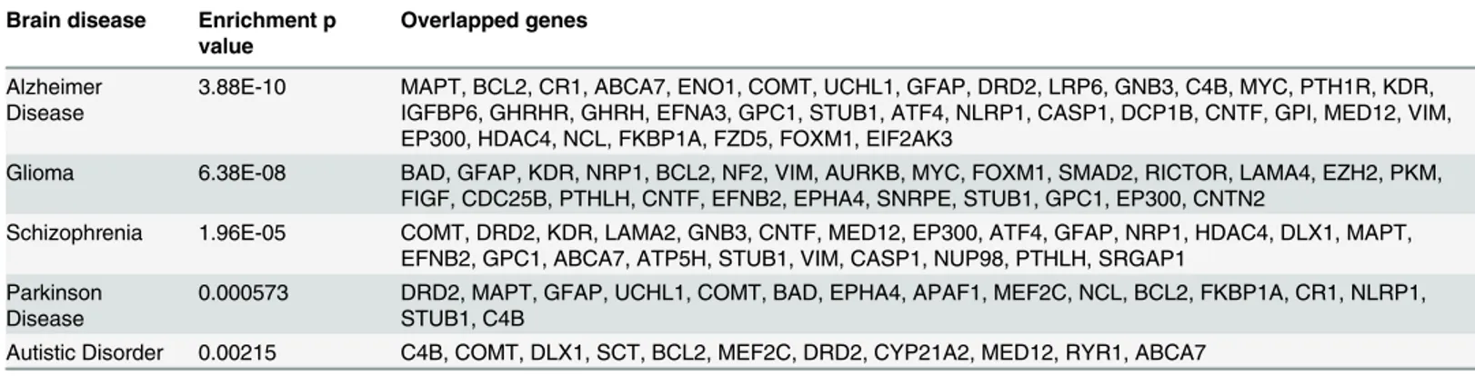
Table 3. The enrichment between the 108 candidate genes and the genes of well-known brain diseases.
Brain disease Enrichment p value
Overlapped genes
Alzheimer Disease
3.88E-10 MAPT, BCL2, CR1, ABCA7, ENO1, COMT, UCHL1, GFAP, DRD2, LRP6, GNB3, C4B, MYC, PTH1R, KDR, IGFBP6, GHRHR, GHRH, EFNA3, GPC1, STUB1, ATF4, NLRP1, CASP1, DCP1B, CNTF, GPI, MED12, VIM, EP300, HDAC4, NCL, FKBP1A, FZD5, FOXM1, EIF2AK3
Glioma 6.38E-08 BAD, GFAP, KDR, NRP1, BCL2, NF2, VIM, AURKB, MYC, FOXM1, SMAD2, RICTOR, LAMA4, EZH2, PKM, FIGF, CDC25B, PTHLH, CNTF, EFNB2, EPHA4, SNRPE, STUB1, GPC1, EP300, CNTN2
Schizophrenia 1.96E-05 COMT, DRD2, KDR, LAMA2, GNB3, CNTF, MED12, EP300, ATF4, GFAP, NRP1, HDAC4, DLX1, MAPT, EFNB2, GPC1, ABCA7, ATP5H, STUB1, VIM, CASP1, NUP98, PTHLH, SRGAP1
Parkinson Disease
0.000573 DRD2, MAPT, GFAP, UCHL1, COMT, BAD, EPHA4, APAF1, MEF2C, NCL, BCL2, FKBP1A, CR1, NLRP1, STUB1, C4B
Autistic Disorder 0.00215 C4B, COMT, DLX1, SCT, BCL2, MEF2C, DRD2, CYP21A2, MED12, RYR1, ABCA7
4. Ashburn TT, Thor KB (2004) Drug repositioning: identifying and developing new uses for existing drugs. Nat Rev Drug Discov 3: 673–683. PMID:15286734
5. Chen L, Lu J, Zhang N, Huang T, Cai Y-D (2014) A hybrid method for prediction and repositioning of drug Anatomical Therapeutic Chemical classes. Molecular BioSystems 10: 868–877. doi:10.1039/ c3mb70490dPMID:24492783
6. Brouwers L, Iskar M, Zeller G, van Noort V, Bork P (2011) Network neighbors of drug targets contribute to drug side-effect similarity. PLoS ONE 6: e22187. doi:10.1371/journal.pone.0022187PMID: 21765950
7. Chen L, Zeng WM, Cai YD, Feng KY, Chou KC (2012) Predicting Anatomical Therapeutic Chemical (ATC) Classification of Drugs by Integrating Chemical-Chemical Interactions and Similarities. PLoS ONE 7: e35254. doi:10.1371/journal.pone.0035254PMID:22514724
8. Yamanishi Y, Araki M, Gutteridge A, Honda W, Kanehisa M (2008) Prediction of drug-target interaction networks from the integration of chemical and genomic spaces. Bioinformatics 24: i232–i240. doi:10. 1093/bioinformatics/btn162PMID:18586719
9. Chen L, Zeng W-M, Cai Y-D, Huang T (2013) Prediction of Metabolic Pathway Using Graph Property, Chemical Functional Group and Chemical Structural Set. Current Bioinformatics 8: 200–207. 10. Chen M, Hofest dt R (2005) An algorithm for linear metabolic pathway alignment. In silico biology 5:
111–128. PMID:15972016
11. Kitiporn P, Jan-Phillip M, Marcus O, Fabian S, Victor S, et al. (2008) Machine learning based analyses on metabolic networks supports high-throughput knockout screens. BMC Systems Biology 2: 67. doi: 10.1186/1752-0509-2-67PMID:18652654
12. Chen L, Lu J, Huang T, Yin J, Wei L, et al. (2014) Finding Candidate Drugs for Hepatitis C Based on Chemical-Chemical and Chemical-Protein Interactions. PLoS ONE 9: e107767. doi:10.1371/journal. pone.0107767PMID:25225900
13. Li B-Q, You J, Chen L, Zhang J, Zhang N, et al. (2013) Identification of Lung-Cancer-Related Genes with the Shortest Path Approach in a Protein-Protein Interaction Network. BioMed Research Interna-tional 2013: 267375. doi:10.1155/2013/267375PMID:23762832
14. Jiang M, Chen Y, Zhang Y, Chen L, Zhang N, et al. (2013) Identification of hepatocellular carcinoma re-lated genes with k-th shortest paths in a protein—protein interaction network. Mol BioSyst 9: 2720–2728. doi:10.1039/c3mb70089ePMID:24056857
15. Ashburner M, Ball CA, Blake JA, Botstein D, Butler H, et al. (2000) Gene ontology: tool for the unifica-tion of biology. The Gene Ontology Consortium. Nat Genet 25: 25–29. PMID:10802651
16. Gao YF, Chen L, Cai YD, Feng KY, Huang T, et al. (2012) Predicting Metabolic Pathways of Small Mol-ecules and Enzymes Based on Interaction Information of Chemicals and Proteins. PLoS ONE 7: e45944. doi:10.1371/journal.pone.0045944PMID:23029334
17. Sharan R, Ulitsky I, Shamir R (2007) Network-based prediction of protein function. Molecular systems biology 3: 88. PMID:17353930
18. Ng KL, Ciou JS, Huang CH (2010) Prediction of protein functions based on function-function correlation relations. Computers in Biology and Medicine 40: 300–305. doi:10.1016/j.compbiomed.2010.01.001 PMID:20089249
19. Bogdanov P, Singh AK (2010) Molecular Function Prediction Using Neighborhood Features. IEEE-ACM Transactions on Computational Biology and Bioinformatics 7: 208–217.
20. Gao P, Wang QP, Chen L, Huang T (2012) Prediction of Human Genes Regulatory Functions Based on Proteinprotein Interaction Network. Protein and Peptide Letters 19: 910–916. PMID:22486617 21. Alberts B (1989) Molecular biology of the cell: Garland Pub.
22. Jensen LJ, Kuhn M, Stark M, Chaffron S, Creevey C, et al. (2009) STRING 8-a global view on proteins and their functional interactions in 630 organisms. Nucleic acids research 37: D412–416. doi:10.1093/ nar/gkn760PMID:18940858
23. Hu LL, Huang T, Shi X, Lu WC, Cai YD, et al. (2011) Predicting functions of proteins in mouse based on weighted protein-protein interaction network and protein hybrid properties. PLoS ONE 6: e14556. doi: 10.1371/journal.pone.0014556PMID:21283518
24. Gormen TH, Leiserson CE, Rivest RL, Stein C, editors (1990) Introduction to algorithms: MIT press Cambridge, MA.
25. Altshuler D, Daly MJ, Lander ES (2008) Genetic mapping in human disease. Science 322: 881–888. doi:10.1126/science.1156409PMID:18988837
27. Bhanot P, Brink M, Samos CH, Hsieh JC, Wang Y, et al. (1996) A new member of the frizzled family from Drosophila functions as a Wingless receptor. Nature 382: 225–230. PMID:8717036
28. Rulifson EJ, Wu CH, Nusse R (2000) Pathway specificity by the bifunctional receptor frizzled is deter-mined by affinity for wingless. Mol Cell 6: 117–126. PMID:10949033
29. McMahon AP, Bradley A (1990) The Wnt-1 (int-1) proto-oncogene is required for development of a large region of the mouse brain. Cell 62: 1073–1085. PMID:2205396
30. Lako M, Lindsay S, Bullen P, Wilson DI, Robson SC, et al. (1998) A novel mammalian wnt gene, WNT8B, shows brain-restricted expression in early development, with sharply delimited expression boundaries in the developing forebrain. Hum Mol Genet 7: 813–822. PMID:9536085
31. Miranda LA, Strobl-Mazzulla PH, Somoza GM (2002) Ontogenetic development and neuroanatomical localization of growth hormone-releasing hormone (GHRH) in the brain and pituitary gland of pejerrey fish Odontesthes bonariensis. Int J Dev Neurosci 20: 503–510. PMID:12392754
32. Takahashi T, Okimura Y, Yoshimura K, Shigeyoshi Y, Kaji H, et al. (1995) Regional distribution of growth hormone-releasing hormone (GHRH) receptor mRNA in the rat brain. Endocrinology 136: 4721–4724. PMID:7664697
33. Waters MJ, Blackmore DG (2011) Growth hormone (GH), brain development and neural stem cells. Pediatr Endocrinol Rev 9: 549–553. PMID:22397139
34. Nakashima K, Yanagisawa M, Arakawa H, Kimura N, Hisatsune T, et al. (1999) Synergistic signaling in fetal brain by STAT3-Smad1 complex bridged by p300. Science 284: 479–482. PMID:10205054 35. Siu FK, Sham MH, Chow BK (2005) Secretin, a known gastrointestinal peptide, is widely expressed
during mouse embryonic development. Gene Expr Patterns 5: 445–451. PMID:15661652
36. Lossi L, Bottarelli L, Candusso ME, Leiter AB, Rindi G, et al. (2004) Transient expression of secretin in serotoninergic neurons of mouse brain during development. Eur J Neurosci 20: 3259–3269. PMID: 15610158
37. Chang TM, Berger-Ornstein L, Chey WY (1985) Presence of biologically and immunologically active secretin-like substance in the mammalian brain. Peptides 6: 193–198. PMID:4034410
38. O’Donohue TL, Charlton CG, Miller RL, Boden G, Jacobowitz DM (1981) Identification, characteriza-tion, and distribution of secretin immunoreactivity in rat and pig brain. Proc Natl Acad Sci U S A 78: 5221–5224. PMID:6946469
39. Yamagata T, Urano H, Weeber EJ, Nelson DL, Nishijima I (2008) Impaired hippocampal synaptic func-tion in secretin deficient mice. Neuroscience 154: 1417–1422. doi:10.1016/j.neuroscience.2008.04. 037PMID:18534766
40. Hu QD, Ang BT, Karsak M, Hu WP, Cui XY, et al. (2003) F3/contactin acts as a functional ligand for Notch during oligodendrocyte maturation. Cell 115: 163–175. PMID:14567914
41. Gautam V, D’Avanzo C, Hebisch M, Kovacs DM, Kim DY (2014) BACE1 activity regulates cell surface contactin-2 levels. Mol Neurodegener 9: 4. doi:10.1186/1750-1326-9-4PMID:24405708
42. Zou M, Li S, Klein WH, Xiang M (2012) Brn3a/Pou4f1 regulates dorsal root ganglion sensory neuron specification and axonal projection into the spinal cord. Dev Biol 364: 114–127. doi:10.1016/j.ydbio. 2012.01.021PMID:22326227
43. Deng M, Yang H, Xie X, Liang G, Gan L (2014) Comparative expression analysis of POU4F1, POU4F2 and ISL1 in developing mouse cochleovestibular ganglion neurons. Gene Expr Patterns 15: 31–37. doi:10.1016/j.gep.2014.03.001PMID:24709358
44. Chu CS, Lo PW, Yeh YH, Hsu PH, Peng SH, et al. (2014) O-GlcNAcylation regulates EZH2 protein sta-bility and function. Proc Natl Acad Sci U S A 111: 1355–1360. doi:10.1073/pnas.1323226111PMID: 24474760
45. Piper M, Barry G, Harvey TJ, McLeay R, Smith AG, et al. (2014) NFIB-mediated repression of the epi-genetic factor Ezh2 regulates cortical development. J Neurosci 34: 2921–2930. doi:10.1523/ JNEUROSCI.2319-13.2014PMID:24553933
46. Xu K, Bai Y, Zhang A, Zhang Q, Bartlam MG (2014) Insights into the structure and architecture of the CCR4-NOT complex. Front Genet 5: 137. doi:10.3389/fgene.2014.00137PMID:24904637 47. Temme C, Simonelig M, Wahle E (2014) Deadenylation of mRNA by the CCR4-NOT complex in
Dro-sophila: molecular and developmental aspects. Front Genet 5: 143. doi:10.3389/fgene.2014.00143 PMID:24904643
48. Chen C, Ito K, Takahashi A, Wang G, Suzuki T, et al. (2011) Distinct expression patterns of the subunits of the CCR4-NOT deadenylase complex during neural development. Biochem Biophys Res Commun 411: 360–364. doi:10.1016/j.bbrc.2011.06.148PMID:21741365
50. Ding N, Zhou H, Esteve PO, Chin HG, Kim S, et al. (2008) Mediator links epigenetic silencing of neuro-nal gene expression with x-linked mental retardation. Mol Cell 31: 347–359. doi:10.1016/j.molcel.2008. 05.023PMID:18691967
51. Flanagan LA, Rebaza LM, Derzic S, Schwartz PH, Monuki ES (2006) Regulation of human neural pre-cursor cells by laminin and integrins. J Neurosci Res 83: 845–856. PMID:16477652
52. Li X, Liu X, Josey B, Chou CJ, Tan Y, et al. (2014) Short laminin peptide for improved neural stem cell growth. Stem Cells Transl Med 3: 662–670. doi:10.5966/sctm.2013-0015PMID:24692587
53. Kuranaga E (2011) Caspase signaling in animal development. Dev Growth Differ 53: 137–148. doi:10. 1111/j.1440-169X.2010.01237.xPMID:21338340
54. Doerfler P, Forbush KA, Perlmutter RM (2000) Caspase enzyme activity is not essential for apoptosis during thymocyte development. J Immunol 164: 4071–4079. PMID:10754300
55. Hayashi M, Araki T (2002) Caspase in renal development. Nephrol Dial Transplant 17 Suppl 9: 8–10. 56. Laguna A, Aranda S, Barallobre MJ, Barhoum R, Fernandez E, et al. (2008) The protein kinase
DYRK1A regulates caspase-9-mediated apoptosis during retina development. Dev Cell 15: 841–853. doi:10.1016/j.devcel.2008.10.014PMID:19081073
57. Matalova E, Vanden Berghe T, Svandova E, Vandenabeele P, Healy C, et al. (2012) Caspase-7 in molar tooth development. Arch Oral Biol 57: 1474–1481. doi:10.1016/j.archoralbio.2012.06.009PMID: 22858065
58. Waters EM, Simerly RB (2009) Estrogen induces caspase-dependent cell death during hypothalamic development. J Neurosci 29: 9714–9718. doi:10.1523/JNEUROSCI.0135-09.2009PMID:19657024 59. Yakovlev A, Khafizova M, Abdullaev Z, Loukinov D, Kondratyev A (2010) Epigenetic regulation of
cas-pase-3 gene expression in rat brain development. Gene 450: 103–108. doi:10.1016/j.gene.2009.10. 008PMID:19909801
60. Chang LR, Liu JP, Song YZ, Lu T, Lu G, et al. (2011) Expression of caspase-8 and caspase-9 in rat hip-pocampus during postnatal development. Microsc Res Tech 74: 153–158. doi:10.1002/jemt.20886 PMID:21275003
61. Maki RA, Tyurin VA, Lyon RC, Hamilton RL, DeKosky ST, et al. (2009) Aberrant expression of myelo-peroxidase in astrocytes promotes phospholipid oxidation and memory deficits in a mouse model of Alzheimer disease. J Biol Chem 284: 3158–3169. doi:10.1074/jbc.M807731200PMID:19059911 62. Bauer-Mehren A, Rautschka M, Sanz F, Furlong LI (2010) DisGeNET: a Cytoscape plugin to visualize,
integrate, search and analyze gene-disease networks. Bioinformatics 26: 2924–2926. doi:10.1093/ bioinformatics/btq538PMID:20861032
63. Hadjihannas MV, Bruckner M, Jerchow B, Birchmeier W, Dietmaier W, et al. (2006) Aberrant Wnt/beta-catenin signaling can induce chromosomal instability in colon cancer. Proc Natl Acad Sci U S A 103: 10747–10752. PMID:16815967
64. Inestrosa NC, Montecinos-Oliva C, Fuenzalida M (2012) Wnt signaling: role in Alzheimer disease and schizophrenia. J Neuroimmune Pharmacol 7: 788–807. doi:10.1007/s11481-012-9417-5PMID: 23160851
65. Frenzel A, Grespi F, Chmelewskij W, Villunger A (2009) Bcl2 family proteins in carcinogenesis and the treatment of cancer. Apoptosis 14: 584–596. doi:10.1007/s10495-008-0300-zPMID:19156528 66. Liao WT, Ye YP, Zhang NJ, Li TT, Wang SY, et al. (2014) MicroRNA-30b functions as a tumour
sup-pressor in human colorectal cancer by targeting KRAS, PIK3CD and BCL2. J Pathol 232: 415–427. doi: 10.1002/path.4309PMID:24293274
67. Sheikh AM, Malik M, Wen G, Chauhan A, Chauhan V, et al. (2010) BDNF-Akt-Bcl2 antiapoptotic signal-ing pathway is compromised in the brain of autistic subjects. J Neurosci Res 88: 2641–2647. doi:10. 1002/jnr.22416PMID:20648653
68. Gordan JD, Thompson CB, Simon MC (2007) HIF and c-Myc: sibling rivals for control of cancer cell me-tabolism and proliferation. Cancer Cell 12: 108–113. PMID:17692803