161
Oral & Maxillofacial Pathology Journal [ OMPJ ] Vol. 2 No. 2 Jul-Dec -2011 ISSN 0976 - 1225
FLORID CEMENTO-OSSEOUS DYSPLASIA – AN UNUSUAL ENTITY
A CASE REPORT
1 2 2 3
Vandana Shah Nishit Soni B.S Manjunatha Swati Kumar
1 2 3
Professor Reader Senior Lecturer, Department of Oral Pathology, KM Shah Dental College & Hospital, Gujarat,India
Corresponding Author : Vandana Shah, Professor & Incharge,Department of Oral Pathology, Sumandeep Vidyapeeth, KM Shah Dental College & Hospital, Piparia Post, Waghodia Road, Vadodara, Gujarat.
E mail – brijclinic@hotmail.com
Abstract
Florid cemento-osseous dysplasia (FLCOD) is a rare, but well recognized condition that characteristically affects the jaws of middle aged women. It usually manifests radiographically as a diffuse, lobulated and irregularly shaped radio-opacities distributed throughout the alveolar processes which are usually bilaterally symmetrical. This condition has been classified as sclerosing osteitis, multiple enostoses, diffuse chronic osteomyelitis and gigantiform cementoma. The lesion is usually benign and requires no treatment unless cosmetically concerning or becomes symptomatic. For the asymptomatic patient the best management consists of regular recall examination with prophylaxis and maintenance of good oral hygiene According to the recent review of literature in 2006, only five patients of florid cemento-osseous dysplasia from India have been reported (less than 2%). Here we present such a rare case occurring in a 37 year old female who came with a swelling and dull pain in the lower right back teeth region since 1 and 1/2 years. Radiographically, it showed mixed radiolucent / radio-opaque mass in both right and left premolar to molar region.
Key Words: Fibro-Osseous Lesions; Florid Cemento Osseous Dysplasia.
Introduction
Case Report
The term fibro–osseous lesion (FOL) is a
generic designation of a group of jaw disorders A 37-year-old female patient presented to KM characterized by replacement of bone by benign Shah Dental College & Hospital, Vadodara,
1
with a complaint of swelling and dull pain in connective tissue matrix . The classification of
lower right back teeth region since 1 and ½ cemento-osseous lesions of the jaws has long
years. On extra-oral examination facial been a matter of discussion for pathologists and
asymmetry was noted on lower right side of the clinicians. A review of the literature shows a
face (Figure 1). Overlying skin was normal with wide range of terminologies used to describe
2 st no change in color, temperature and
what seem to be similar lesions . In 1 Edition of
consistency. Intraoral examination revealed a Wo r l d H e a l t h O r g a n i z a t i o n ( W H O )
localized swelling measuring about 2 X 1.5 cm classification of Odontogenic tumors 1971,
extending from distal margin of right second FLCOD was categorized under the Neoplasms
premolar anteriorly to mesial margin of right & other tumors related to odontogenic
3 third molar posteriorly. The mandibular
apparatus . The current classification of
vestibule was obliterated. On palpation, the cementomatous lesions, released in 1992 by the
swelling was hard in consistency. Teeth WHO includes FLCOD under cemento –
associated with the lesion were vital and the osseous dysplasia of the Neoplasms and other
1,3 overlying gingiva and mucosa were normal.
lesions related to bone . This paper describes
Ortho pantomogram was taken and such a rare case diagnosed on the basis of
162
Florid Cemento-osseous Dysplasia – An Unusual Entity A Case Report
Oral & Maxillofacial Pathology Journal [ OMPJ ] Vol. 2 No. 2 Jul-Dec -2011 ISSN 0976 - 1225 tooth bearing areas of posterior mandible in
relation to premolar and molar regions. Irregularly shaped radio opacities were distributed diffusely throughout the alveolar process enmeshed in poorly defined zones of decreased radio density. The borders of the lesion were indistinct and blended into the affected surrounding bone (Figure 2). Occlusal view showed no buccal or lingual cortical plate expansion (Figure 3). Posterior Anterior view of the skull showed mixed radio opaque and radio lucent areas on both sides of mandible. The left side of mandible was asymptomatic and apparently normal on clinical examination.
Surgical recontouring of the lesion on the right side was done for esthetic reasons. Multiple soft and hard tissue bits of varying sizes were received.
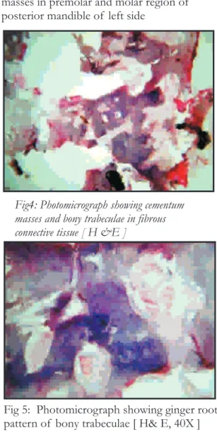
Microscopically, the H (hematoxylin) and E (eosin) stained sections showed fragments of soft and hard tissues. The soft tissue composed of cellular mesenchymal tissue with spindle shaped fibroblasts intermingled in thick bundles of collagen fibers and numerous small blood vessels. The hard tissue contained mixture of lamellar bone and globular shaped acellular, basophilic cementum like structures (Figure 4). Bony trabeculae were thick curvilinear resembling ginger root pattern (Figure 5). Few hemorrhagic areas were seen in close association with bony trabeculae. A diagnosis of florid cemento-osseous dysplasia was given.
Fig 1: Clinical photograph
Fig 2: Ortho Panto-mograph showing multiple RO-RL masses in premolar and molar region of posterior mandible of left side
Fig4: Photomicrograph showing cementum masses and bony trabeculae in fibrous connective tissue [ H &E ]
Fig 5: Photomicrograph showing ginger root pattern of bony trabeculae [ H& E, 40X ]
Discussion
163
Florid Cemento-osseous Dysplasia – An Unusual Entity A Case Report
Oral & Maxillofacial Pathology Journal [ OMPJ ] Vol. 2 No. 2 Jul-Dec -2011 ISSN 0976 - 1225
12
(sclerotic symmetrical masses) and focal (single symptomatic. Treatment of FLCOD with lesion) cemental dysplasias. Florid cemento- complete resection of the lesion would be osseous dysplasia clearly appears to be a form of impractical because it usually occupies most of bone and cemental dysplasia that is limited to the mandible and maxilla. When surgical
4
jaws. intervention is indicated, a remodelling
5
Florid cemento-osseous dysplasia was resection is recommended for esthetic reasons .
2
first described by Melrose et al. in 1976 . This
condition has been interpreted as a dysplastic C h r o n i c d i f f u s e s c l e r o s i n g lesion or developmental anomaly arising in osteomyelitis may resemble FLCOD, which
tooth-bearing areas. appears as a single, poorly delineated opaque
segment of the mandible. Clinically it involves FLCOD is more commonly seen in the body of the mandible from the alveolus to middle-aged black women, although it also may the inferior border and may extend into the
2,5
ramus. FLCOD may be familial with an occur in Caucasians and Asians. In some cases,
5 autosomal dominant inheritance pattern, but
a familial trend can be observed. These lesions
there are only a few examples in the literature in have a striking tendency toward bilateral
5
which the familial pattern has been confirmed . symmetrical location, and it is not unusual to
find extensive lesions in all four posterior
Only five Indian patients of florid (molar-premolar region) quadrants of the jaws.
cemento-osseous dysplasia have been reported The radiographic appearance can vary from
(less than 2%), according to the recent review of areas of radiolucency to mixed lesions and to
12,13
opaque masses. Over the time the lesions tend recent literature. This makes the occurrence
6,7
of FCOD a relatively rare phenomenon. In the to become increasingly radio opaque .
present case, no familial aspects of the disease FLCOD blends with adjacent bone and
were reported. therefore pathologists often receive multiple
8
curetted fragments . The lesions can be found
Florid cemento-osseous dysplasia is a in multiple quadrants in both the maxilla and
6,9 self limiting disease restricted to alveolar
mandible . FLCOD is occasionally expansile
9 processes. It is a distinct entity representing an
and patients may experience pain rarely .
exuberant variant of cemento-osseous dysplasia normally seen in multiple quadrants of both the Histopathologically, FLCOD consists
jaws. Normally, a diagnosis of florid cemento-of dense, sclerotic masses that have been
osseous dysplasia in the jaws is made by clinical interpreted either as cementum or bone which
4
findings, radiographic features and histology. are anastomosing and layers of cementum-like
These lesions have to be included in the calcifications embedded in a fibro cellular
2,5 differential diagnosis of mixed radiolucent radio
background.
opaque lesions of the jaws. Management of FLCOD may be
Many lesions that occur in the jaw have difficult and not very satisfactory. When
a similar radiographical appearance and it is symptomatic, there may be pain alone or in
often difficult to differentiate among them. association with sinus tracks with minimal
10 Differential diagnosis is crucial especially when
purulent drainage. Although in most situations
there are coincidental findings like odontogenic the lesion is not treated, treatment is required
infections or chronic diffuse osteomyelitis, when infection of the lesion occurs. For the
neoplasia, bone dysplasia that can cause changes asymptomatic patient the best management
in the mandible with similar radiographic consists of regular recall examination with
characteristics. Hence we must consider all the prophylaxis and maintenance of good oral
11 available diagnostic infor mation, the
hygiene. Extensive surgical resection and
radiographic characteristics which would saucerization are proposed treatment options
164
Florid Cemento-osseous Dysplasia – An Unusual Entity A Case Report
Oral & Maxillofacial Pathology Journal [ OMPJ ] Vol. 2 No. 2 Jul-Dec -2011 ISSN 0976 - 1225
8. Speight PM, Carlos R. Maxillofacial
Fibro-References
osseous Lesions. Cur Diagn Pathol. 2006;12(1):1- 10.
1. MacDonald–Jankowski. Focal cemento-osseous dysplasia: a systematic review. Dentomaxillofac
9. White SC, Pharoah MJ. Oral Radiology - Radiol. 2008;37(6):350-60.
th
Principles and Interpretation 4 edition. Saint Louis: Mosby Publishing; 2000.
2. Melrose RJ, Abrams AM, Mills BG. Florid osseous dysplasia. A clinical-pathologic study of
10. Waldron CA, Giansanti JS, Browand BC. thirty-four cases. Oral Surg Oral Med Oral Pathol.
Sclerotic cemental masses of the jaws (so-called 1976; 41(1): 62-82
chronic sclerosing osteomyelitis, sclerosing osteitis, multiple enostosis, and gigantiform cementoma). 3. Kramer IRH, Pindborg JJ, Shear M. Neoplasma
O r a l S u r g O r a l M e d O r a l Pa t h o l and others lesions related to bone. In: World Health
1975;39(4):590-604. Organization (Editors)-Histologic Typing of
Odontogenic Tumours. Berlim: Springer-Verlag;
11. Gündüz K, Avsever H, Karaçayli U, Senel B,
1992;28-31p.
Piºkin B. Florid cemento-osseous dysplasia: a case
4. Daðistan S, Tozoðlu Ü, Göregen M, Çakur B. report. 2009;20(4):347-50.
Florid cemento-osseous dysplasia: A case report.
Med Oral Patol Oral Cir Bucal. 2007;12:E348- 12. Mangala M, Ramesh DN, Surekha PS, Santosh
50. P. Florid cemento-osseous dysplasia: Review and report of two cases. Indian J Dent Res 5. Waldron CA. Fibro-osseous lesions of the jaws. J 2006;17:131-4.
Oral Maxillofac Surg. 1985;43(4):249-262.
13. Singer SR, Mupparapu M, Rinaggio J. Florid 6. Beylouni I, Farge P, Mazoyer JF, Courdet JL. cemento-osseous dysplasia and chronic diffuse Florid cemento osseous dysplasia: report of a case osteomyelitis Report of a simultaneous presentation with computed tomography and 3D imaging, Oral and review of the literature. J Am Dent Assoc. Surg Oral Med Oral Pathol Oral Radiol Endod. 2005;136(7):927-31.
1998;85(6):707–11.
7. Marx RE. Stern D. Oral and Maxillofacial Pathology: A rationale for diagnosis and treatment. London: Quintessence Publishing; 2004:57– 63p.
Braz Dent J.