J of Evidence Based Med & Hlthcare, pISSN- 2349-2562, eISSN- 2349-2570/ Vol. 2/Issue 35/Aug. 31, 2015 Page 5527
CONGENITAL RADIAL DYSPLASIA: A CASE REPORT
K. Venkatram Reddy1, Shruthi2, Mopur James Premsagar3, Aarthi Reddy4, Kaushik5
HOW TO CITE THIS ARTICLE:
K. Venkatram Reddy, Shruthi, Mopur James Premsagar, Aarthi Reddy, Kaushik. “Congenital Radial Dysplasia: A Case Report”. Journal of Evidence based Medicine and Healthcare; Volume 2, Issue 35, August 31, 2015; Page: 5527-5537, DOI: 10.18410/jebmh/2015/766
ABSTRACT: Congenital radial dysplasia, also referred to as radial club hand, means deficiency along the preaxial or radial side of the extremity. It ranges from hypoplasia of the thumb to various degrees of radial hypoplasia. We present one such rare case of type 4 congenital unilateral isolated radial dysplasia with carpel anomaly, reported to our department in SVS medical College, Mahabubanagar, Telangana state.
KEYWORDS: Radial Clob Hand, thumb dysplasia, radio-ulnar synostosis, hypoplasia.
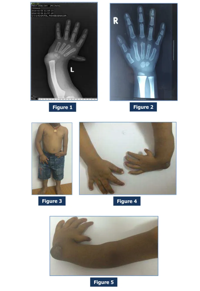
CASE REPORT: A 4year (Approx) old male child was referred for evaluation of left upper limb deformity. He was born at term by normal delivery following an uneventful pregnancy and there was no significant past medical or surgical history. He was the first child of non-consanguineous parents and there was no family history of congenital anomalies.
Physical examination of the child revealed deformity of the left forearm which appeared shorter than contralateral side with radial deviation of the wrist. Thumb of the affected arm was hypoplastic
Physical examination revealed no other abnormalities.
Hemoglobin, total and differential leucocytes count, platelet count, and renal function tests were all within normal limits in more than one occasion.
Radiographs of left upper limb revealed complete aplasia of the radius, shortening of the ulna, and hypoplastic 1st metacarpal along with carpel anomaly.(Bone age is not in
correlation with chronological age as there are only two carpels-capitate and hamate in a 4 year old child along with radial epiphysis)
Skeletal survey, ultrasonogram of abdomen and cardiac echocardiogram were done to rule any other associated congenital anomalies.
J of Evidence Based Med & Hlthcare, pISSN- 2349-2562, eISSN- 2349-2570/ Vol. 2/Issue 35/Aug. 31, 2015 Page 5528
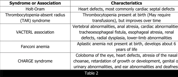
Figure 1 Figure 2
Figure 3 Figure 4
J of Evidence Based Med & Hlthcare, pISSN- 2349-2562, eISSN- 2349-2570/ Vol. 2/Issue 35/Aug. 31, 2015 Page 5529
DISCUSSION: Radial club hand isa deficiencyalong the preaxialor radialside of the extremity. Although considerable forearm andhand anomaliesare the classic findings, proximal deficiencies also can occur throughout the arm and shoulder girdle. The elbow abnormalities can include deficiencies of the olecranon, capitellum, coronoid fossa, and medial epicondyle. This article summarizes the history of radial deficiencies, lists potential etiologies, highlights relevant pathoanatomy, discusses treatment regimens, reviews expected outcome, and details potential complications
History of the Procedure: In 1733, Petit first described radial clubhand in an autopsy of a neonate with bilateral clubhands and absent radii. Subsequent autopsy observations detailed the anomalous anatomy associated with radial clubhand and the associated malformations of other body systems.
Initial surgical treatment of radial clubhand involved an ulnar osteotomy to correct the bow, along with splitting of the distal ulna for insertion of the carpus. Reconstruction of the radius with a bone graft to support the carpus was reported in the 1920s, and nonvascularized epiphyseal transfer was reported in 1945. Results of these procedures were disappointing. They had multiple causes of failure, including disruption of the ulnar growth plate and subsequent increase in limb-length discrepancy, inadvertent ankylosis or arthrodesis of the wrist and loss of motion, and failure of the transplanted bone to grow, with eventual loss of radial support.
Centralization of the carpus on the distal ulna has emerged as the preferred surgical technique for correcting radial clubhand. Pioneers in congenital hand surgery developed the basis for this procedure. Numerous modifications have been described to obtain or maintain correction of the wrist on the ulna.
Radialization is a technique that involves overcorrection of the carpus on the ulna combined with tendon transfer to further rebalance the wrist.[1]
Despite over 250 years of investigation, current treatment regimens are still unable to restorenormality:Thelimbremainsshort,andthewristlacksfullfunction
Frequency: The reported incidence of radial clubhand ranges from 1/55,000 to 1/100,000 live births. Most cases are sporadic without any definable cause. However, exposure to teratogens, such as thalidomide and radiation, can yield radial deficiencies.
Radial deficiency is bilateral in 50% of cases and is slightly more common in males than in females (3:2). The incidence of radial deficiency within the same family is low, ranging from approximately 5% to 10% of reported cases; it is most common in radial aplasia associated with cardiac abnormalities
Etiology: In the 19th century, the etiology of radial clubhand was theorized to be either a congenital absence or an acquired defect secondary to syphillis. In 1895, Kummel proposed the cause to be abnormal pressure upon the embryo along the radial bud between the third and seventh week of gestation.
J of Evidence Based Med & Hlthcare, pISSN- 2349-2562, eISSN- 2349-2570/ Vol. 2/Issue 35/Aug. 31, 2015 Page 5530
produced anomalies similar to radial clubhand.[2] Therefore, a defect of the AER is the most
probable cause of radial clubhand, with the extent of deformity related to the degree and extent of the AER absence.[3]
Pathophysiology: The forearm is foreshortened, and the wrist is positioned in radial deviation. This malalignment assumes a perpendicular relation over time. The right-angled position further shortens the limbandlimits the ability toreach into space. Theawkward angulationbetween the wrist and forearm places the extrinsic flexors and extensors at a mechanical disadvantage. The tendons must traverse this angle to elicit finger motion, and this limits the ability to move the digits
Presentation: Radial Clubhand is classified into the following four types according to the amount of radius present:
Type I deficiency: The mildest type, this is characterized by mild radial shortening of the radius without considerable bowing; minor radial deviation of the hand is apparent, though considerable thumb hypoplasia may be evident.
Type II deficiency: This is characterized by a miniature radius with distal and proximal physeal abnormalities and moderate deviation of the wrist.
Type III deficiency: This is characterized by a partial absence of the radius (most commonly the distal portion) and severe wrist radial deviation.
Type IV deficiency: The most common variant (see the image below), this is characterized by a complete absence of the radius; the hand tends to develop a perpendicular relation to the forearm.
A modified classification of radial longitudinal deficiency has been developed to combine thumb, carpal anomalies, and forearm into a single scheme (Table 1).[4] This scheme grades both
thumb and radius deficiencies on the basis of radiographic findings. The delayed ossification of the radius and carpus in preaxial deficiency must be considered during application of this scheme. Radial-side carpal bones appear even later than the ulna, which delays definitive determination of carpal anomalies.
Type Thumb
Anomaly Carpal Anomaly* Distal Radius Proximal Radius
N Absence or
hypoplasia Normal Normal Normal
O Absence or hypoplasia
Absence, hypoplasia,
or coalition Normal
Normal, radioulnar synostosis, or radial head dislocation
1 Absence or hypoplasia
Absence, hypoplasia, or coalition
>2 mm shorter than ulna
Normal, radioulnar synostosis, or radial head dislocation
2 Absence or hypoplasia
Absence, hypoplasia,
or coalition Hypoplasia Hypoplasia
3 Absence or hypoplasia
Absence, hypoplasia,
J of Evidence Based Med & Hlthcare, pISSN- 2349-2562, eISSN- 2349-2570/ Vol. 2/Issue 35/Aug. 31, 2015 Page 5531
4 Absence or hypoplasia
Absence, hypoplasia,
or coalition Absence Absence
*Carpal anomaly implies hypoplasia, coalition, absence or bipartite carpal bones. Hypoplasia and absence are more common on the radial side of the carpus, and coalitions are more frequent on
the ulnar side
Table 1: Global classification of radial longitudinal deficiency
Radiographs must be taken after the age of 8 years to allow for ossification of the carpal bones.
Clinical presentation of radial clubhand varies with the degree of radial deficiency and the presence of associated anomalies. Radial deficiency is the classic anomaly that is associated with systemic conditions.[5] All forms, regardless of the degree of expression, warrant systemic
evaluation. The prominent syndromes are as follows:
Holt-Oram syndrome.
TAR (thrombocytopenia-absent radius) syndrome.
VACTERL (vertebral, anal, cardiac, tracheal, esophageal, renal, and limb) syndrome.
CHARGE (coloboma of the eye, heart defects, atresia of the nasal choanae, retardation of growth or development, genital or urinary abnormalities, and ear abnormalities and deafness) syndrome.
Fanconi anemia.[6]
The principal organ systems involved in these are the cardiac, renal, and hematology cell lines (Table 2). Children with VACTERL syndrome can also have vertebral, tracheoesophageal, and anal problems. Isolated radial deficiency is estimated to account for only 8-30% of the cases.[7]
Syndrome or Association Characteristics
Holt-Oram Heart defects, most commonly cardiac septal defects Thrombocytopenia-absent radius
(TAR) syndrome
Thrombocytopenia present at birth (May require transfusions), but improves over time
VACTERL association
Vertebral abnormalities, anal atresia, cardiac abnormalities, tracheoesophageal fistula, esophageal atresia, renal
defects, radial dysplasia, lower-limb abnormalities
Fanconi anemia Aplastic anemia not present at birth, develops about 6 years of life
CHARGE syndrome
Coloboma of the eye, heart defects, atresia of the nasal choanae, retardation of growth or development, genital or urinary abnormalities, and ear abnormalities and deafness
Table 2
J of Evidence Based Med & Hlthcare, pISSN- 2349-2562, eISSN- 2349-2570/ Vol. 2/Issue 35/Aug. 31, 2015 Page 5532
FA in children with limb anomalies is still evolving. Testing every child with isolated thumb or hand abnormalities should be considered. I recommend a chromosomal breakage test on all children with deficiencies of the thumb and radial border of the forearm. Additional findings, such as abnormal skin pigmentation (café-au-lait spots), kidney abnormalities, growth retardation, and microcephaly, add to the suspicion of FA.
Careful clinical examination is used to assess the degree of involvement. The shoulder, elbow, wrist, and digital range of movement are evaluated for active and passive motion. This establishes a baseline for assessing treatment outcome. The ability to flex the elbow for hand-to-mouth function is examined; this influences the treatment algorithm. The position of the wrist with respect to the ulna and the ability to passively correct the radial deviation also are measured.
The thumb is examined for hypoplasia and graded accordingly.[8] Thumb deficiency
contributes to functional impairment. Stiffness of the fingers is assessed, and the ability to grasp and release is determined via functional tasks. Compensatory movements are noted to prevent inadvertent disruption of these adaptive mannerisms with surgical intervention.
Indications
The objectives of treatment in radial clubhand are to reduce the functional deficit incurred by a short or absent radius, a short ulna, an abnormal muscular anatomy, and a radial deviation of the wrist. Type I radial clubhands have minor radial deviation of the wrist, which creates less of a functional problem than types II, III, and IV. In those children with considerable absence of the radius, the wrist assumes severe radial deviation that increases to 90° over time. This further compromises the flexor and extensor tendons, creating functional difficulty.
Thumb hypoplasia also requires consideration when formulating a treatment plan for radial clubhand. An absent or deficient thumb inhibits use of the hand. Reconstruction or pollicization is necessary to optimize hand function.[9] Thumb reconstruction is usually delayed
until after forearm treatment.
The basic goals of treatment are as follows[10]:
Correct radial deviation of the wrist.
Balance the wrist on the forearm.
Maintain wrist and finger motion.
Promote growth of the forearm.
Improve function of the extremity.
Enhance limb appearance for social and emotional benefit.
Centralization is indicated in radial clubhand types II, III, and IV, in which there is severe radial wrist deviation and insufficient support of the carpus.
Relevant Anatomy:
J of Evidence Based Med & Hlthcare, pISSN- 2349-2562, eISSN- 2349-2570/ Vol. 2/Issue 35/Aug. 31, 2015 Page 5533
in flexion than in extension. The forearm is always decreased in length, and the ulna is approximately 60% of the normal length at the time of birth. This discrepancy persists throughout the growth period and into adulthood. True forearm rotation is absent in patients with partial or complete aplasia of the radius.
The wrist is radially deviated and develops a perpendicular relationship to the forearm over time. The articulation between the carpus and ulna is usually fibrous and abnormal, although some hyaline cartilage can be found. Wrist motion is primarily in the radial/ulnar plane, with some flexion/extension. Ossification of the carpal bones is delayed, with the scaphoid and trapezium often absent or hypoplastic. The capitate, hamate, and triquetrum are usually present but ossify late.
The fingers are often stiff, with limited motion at the metacarpophalangeal and interphalangeal joints. The preaxial index and long fingers are more affected than the postaxial ring and small digits.
Muscle and Tendon Abnormalities: Numerous muscular abnormalities are found throughout the upper extremity. The deltoid or pectoralis major muscle can be hypoplastic, can be partially absent, or can have an abnormal insertion. The biceps may be absent or fused to the underlying brachialis muscle.
The forearm demonstrates the most severe abnormalities, involving any muscle that originates or attaches to the radius. This includes the extensor carpi radialis longus, extensor carpi radialis brevis, pronator teres, flexor carpi radialis, palmaris longus, flexor pollicis longus, pronator quadratus, and supinator muscles. The extrinsic flexors and extensors of the fingers are usually adherent, with abnormal origins and insertions. The flexor and extensor carpi ulnaris, as well as the interossei, lumbricals, and hypothenar muscles, are often normal, whereas abnormalities of the thumb muscles are more related to the degree of thumb hypoplasia.
Nerve and Artery Abnormalities: The radial nerve usually terminates at the elbow, and the ulnar nerve is normal. An enlarged median nerve substitutes for the absence of the radial nerve and supplies a dorsal branch for dorsoradial sensibility. This subcutaneous branch is positioned in the fold between the wrist and forearm and must be protected during surgery. The vascular anatomy demonstrates a normal brachial and ulnar artery. The radial artery is often absent, and the interosseous arteries usually remain patent.
Associated Abnormalities: Radial deficiency is associated with numerous systemic conditions,[11] including Holt-Oram syndrome (cardiac septal defects); TAR syndrome; Fanconi
anemia (aplastic anemia); and VACTERL syndrome.[5,12] In addition to these conditions, a variety
of associated musculoskeletal deformities appear sporadically. These include cleft palate, clubfoot, kyphosis, scoliosis, torticollis, and rib deformities.
J of Evidence Based Med & Hlthcare, pISSN- 2349-2562, eISSN- 2349-2570/ Vol. 2/Issue 35/Aug. 31, 2015 Page 5534
Laboratory Studies: The appropriate workup for associated conditions necessitates referral to pediatric subspecialists.[5] The heart is evaluated by auscultation and echocardiography. The
kidneys are examined by means of ultrasonography, and the platelet status is assessed by means of blood count and peripheral blood smear.
The most devastating associated condition is Fanconi anemia. Children with Fanconi anemia do not have signs of bone marrow failure at birth; therefore, the diagnosis is not initially apparent. The majority of children experience signs of aplastic anemia between the ages of 3 and 12 years (Median age, 7 years). However, a chromosomal challenge test is available that allows detection of the disease before the onset of bone marrow failure. This assay subjects a sample of
the child’s lymphocytes to diepoxybutane or mitomycin C, which cause chromosomes within
Fanconi anemia cells to break and rearrange. In contrast, lymphocytes in unaffected children are stable to these agents.
Because bone marrow transplant is the only cure for Fanconi anemia, this prefatory diagnosis is crucial for the child and family. Early diagnosis provides ample time to search for a suitable bone marrow donor or consider preimplantation genetic diagnosis (PGD). PGD is a sophisticated technique that involves in vitro fertilization, sampling of the blastocytes to ensure human leukocyte antigen (HLA) similarity without Fanconi disease, and reimplantation until birth. At delivery, cord blood is harvested from the newborn and used as a source of stem cell transplant to the affected sibling.
Since PGD takes time, early detection via a chromosomal challenge test is critical and may ultimately save the affected child. The pancytopenia can be treated with bone marrow transplantation, but even with these efforts, life expectancy is only estimated at 30 years (range, 0-50 years).
Medical Therapy: Medical treatment is directed at any of the aforementioned associated syndromes. Appropriate treatment for these conditions requires referral to pediatric subspecialists.
Correction of radial clubhand requires a combination of nonoperative and operative management that begins shortly after birth. Instruction in passive stretching of the taut radial structures is provided at the initial visit. This stretching is performed at each diaper change and at bedtime. A stiff elbow with limited motion also is stretched during this time. Splint fabrication is difficult in the newborn, especially with a shortened forearm. Therefore, splint use is delayed until the forearm is long enough to accommodate a splint. Serial casting can also be used to gradually stretch the tight radial structures.
Surgical Therapy: Centralization of the wrist on the ulna is the standard treatment to correct radial deviation. This procedure is performed in patients aged approximately 1 year. Surgery at this time allows improvement in forearm length and provides a foundation for the development of motor function within the hand.
J of Evidence Based Med & Hlthcare, pISSN- 2349-2562, eISSN- 2349-2570/ Vol. 2/Issue 35/Aug. 31, 2015 Page 5535
Tendon and bony procedures are performed simultaneously to provide better alignment of the forearm and to balance the wrist. Tendon transfers are used to attempt to correct the muscular imbalance and include advancing the extensor carpi ulnaris to increase its moment arm for ulnar deviation and transfer of the flexor carpi ulnaris to the extensor carpi ulnaris.
Other, less common options are transfer of the index and long flexor digitorum superficialis around the ulna to the dorsum of the wrist, transfer of the flexor and extensor carpi radialis to the ulnar side, and proximal advancement of the hypothenar muscles to the ulna.
Bony correction of the ulna consists of a closing-wedge osteotomy when there is considerable (ie, >30°) ulnar bow. Bony reconstruction of the distal radius is more difficult; an attempt is made to provide osseous support to the radial side of the carpus. Initial efforts consisted of nonvascularized transfer of a bone graft (tibia, fibula). These efforts were unsuccessful; continued ulnar growth resulted in loss of support. However, innovative procedures involving microsurgical transfer of a vascularized bone graft along with its growth plate (fibula, second toe) have been encouraging.[13]
The basic goals of treatment in radial deficiency are as follows which were described in a nutshell are as follows[10]:
Correct the radial deviation of the wrist.
Balance the wrist on the forearm.
Maintain wrist and finger motion.
Promote growth of the forearm.
Improve the function of the extremity.
PROGNOSIS:
Numerous modifications and advances have been made in the technique of centralization. Improved methods have been developed to balance the wrist with additional tendon transfers or overcorrection of the wrist into ulnar deviation (ie, radialization). Better attempts at stretching the soft tissue with distraction techniques and bone-lengthening procedures also are used today. In addition, microsurgical transfer of a viable growth plate (fibula, second toe) to the radial side of the forearm provides a support of the radial carpus that continues to grow over time.
A successful centralization still results in a shortened forearm segment secondary to altered growth of the ulna. The short forearm is both a cosmetic and a functional problem for the teenager with radial deficiency. Lengthening of the ulna can be accomplished by using distraction osteogenesis. Uniplanar and multiplanar devices have been employed depending on the deformity, forearm size, and surgeon preference.
Successful lengthening results in functional improvement because an increased volume of space becomes available for the hand, though complications are common.[14] Restoration of
near-equal forearm length promotes use of the extremity during activities of daily living.[15]
J of Evidence Based Med & Hlthcare, pISSN- 2349-2562, eISSN- 2349-2570/ Vol. 2/Issue 35/Aug. 31, 2015 Page 5536
These procedural changes represent new concepts to correct radial clubhand. These technologic advances in limb lengthening and microsurgery add innovative methods to better correct the deformity and provide osseous support. Follow-up results of vascularized second metatarsophalangeal joint transfer to stabilize the carpus are encouraging with respect to motion and less recurrence.
REFERENCES:
1. Buck-Gramcko D. Radialization as a new treatment for radial club hand. J Hand Surg [Am]. 1985 Nov. 10(6 Pt 2): 964-8. [Medline].
2. Saunders JW Jr. The proximo-distal sequence of origin of the parts of the chick wing and the role of the ectoderm. 1948. J Exp Zool. 1998 Dec 15. 282(6): 628-68. [Medline].
3. Kozin SH. Congenital anomalies. Trumble TE, ed. Hand Surgery Update. Rosemont, Ill: American Society for Surgery of the Hand; 2003. 599-624.
4. James MA, McCarroll HR Jr, Manske PR. The spectrum of radial longitudinal deficiency: a modified classification. J Hand Surg [Am]. 1999 Nov. 24(6): 1145-55. [Medline].
5. Kozin SH. Upper-extremity congenital anomalies. J Bone Joint Surg Am. 2003 Aug. 85-A(8): 1564-76. [Medline].
6. Wilks DJ, Kay SP, Bourke G. Fanconi's anaemia and unilateral thumb polydactyly--don't miss it. J Plast Reconstr Aesthet Surg. 2012 Aug. 65(8): 1083-6. [Medline].
7. Koskimies E, Lindfors N, Gissler M, Peltonen J, Nietosvaara Y. Congenital upper limb deficiencies and associated malformations in Finland: a population-based study. J Hand Surg Am. 2011 Jun. 36(6): 1058-65. [Medline].
8. Manske PR, McCarroll HR Jr, James M. Type III-A hypoplastic thumb. J Hand Surg [Am]. 1995 Mar. 20(2): 246-53. [Medline].
9. Kozin SH, Weiss AA, Webber JB, et al. Index finger pollicization for congenital aplasia or hypoplasia of the thumb. J Hand Surg [Am]. 1992 Sep. 17(5): 880-4. [Medline].
10.Damore E, Kozin SH, Thoder JJ, et al. The recurrence of deformity after surgical centralization for radial clubhand. J Hand Surg [Am]. 2000 Jul. 25(4):745-51. [Medline]. 11.Goldfarb CA, Wall L, Manske PR. Radial longitudinal deficiency: the incidence of associated
medical and musculoskeletal conditions. J Hand Surg Am. 2006 Sep. 31(7): 1176-82. [Medline].
12.Goldfarb CA, Wall L, Manske PR. Radial longitudinal deficiency: the incidence of associated medical and musculoskeletal conditions. J Hand Surg Am. 2006 Sep. 31(7): 1176-82. [Medline].
13.Vilkki SK. Distraction and microvascular epiphysis transfer for radial club hand. J Hand Surg [Br]. 1998 Aug. 23(4): 445-52. [Medline].
14.Catagni MA, Szabo RM, Cattaneo R. Preliminary experience with Ilizarov method in late reconstruction of radial hemimelia. J Hand Surg [Am]. 1993 Mar. 18(2): 316-21. MEDLINE. 15.Raimondo RA, Skaggs DL, Rosenwasser MP, et al. Lengthening of pediatric forearm
J of Evidence Based Med & Hlthcare, pISSN- 2349-2562, eISSN- 2349-2570/ Vol. 2/Issue 35/Aug. 31, 2015 Page 5537
5. Post Graduate Resident, Department of Radio-diagnosis, SVS Medical College & Hospital, Yenugonda, Mahabubnagar, Telangana.
NAME ADDRESS EMAIL ID OF THE CORRESPONDING AUTHOR:
Dr. Mopur James Premsagar, Assistant Professor,
Department of Radio-diagnosis, SVS Medical College & Hospital, Yenugonda,
Mahabubnagar-509001, Telangana.
E-mail: [email protected]
Date of Submission: 18/08/2015. Date of Peer Review: 19/08/2015. Date of Acceptance: 22/08/2015. Date of Publishing: 31/08/2015.
AUTHORS:
1. K. Venkatram Reddy 2. Shruthi
3. Mopur James Premsagar 4. Aarthi Reddy
5. Kaushik
PARTICULARS OF CONTRIBUTORS:
1. Professor & HOD, Department of Radio-diagnosis, SVS Medical College & Hospital, Yenugonda, Mahabubnagar, Telangana.
2. Post Graduate Resident, Department of Radio-diagnosis, SVS Medical College & Hospital, Yenugonda, Mahabubnagar, Telangana.
3. Assistant Professor, Department of Radio-diagnosis, SVS Medical College & Hospital, Yenugonda, Mahabubnagar, Telangana.