Carriage of
λ
Latent Virus Is Costly for Its
Bacterial Host due to Frequent Reactivation
in Monoxenic Mouse Intestine
Marianne De Paepe1*, Laurent Tournier2, Elisabeth Moncaut1, Olivier Son1, Philippe Langella1, Marie-Agnès Petit1
1Micalis Institute, INRA, AgroParisTech, Université Paris-Saclay, Jouy-en-Josas, France,2MaIAGE, INRA, Université Paris-Saclay, Jouy-en-Josas, France
*marianne.depaepe@jouy.inra.fr
Abstract
Temperate phages, the bacterial viruses able to enter in a dormant prophage state in bacte-rial genomes, are present in the majority of bactebacte-rial strains for which the genome sequence is available. Although these prophages are generally considered to increase their hosts’ fit-ness by bringing beneficial genes, studies demonstrating such effects in ecologically rele-vant environments are relatively limited to few bacterial species. Here, we investigated the impact of prophage carriage in the gastrointestinal tract of monoxenic mice. Combined with mathematical modelling, these experimental results provided a quantitative estimation of key parameters governing phage-bacteria interactions within this model ecosystem. We used wild-type and mutant strains of the best known host/phage pair,Escherichia coliand phageλ. Unexpectedly,λprophage caused a significant fitness cost for its carrier, due to an induction rate 50-fold higher thanin vitro, with 1 to 2% of the prophage being induced. How-ever, when prophage carriers were in competition with isogenic phage susceptible bacteria, the prophage indirectly benefited its carrier by killing competitors: infection of susceptible bacteria led to phage lytic development in about 80% of cases. The remaining infected bac-teria were lysogenized, resulting overall in the rapid lysogenization of the susceptible line-age. Moreover, our setup enabled to demonstrate that rare events of phage gene capture by homologous recombination occurred in the intestine of monoxenic mice. To our knowl-edge, this study constitutes the first quantitative characterization of temperate phage-bacte-ria interactions in a simplified gut environment. The high prophage induction rate detected reveals DNA damage-mediated SOS response in monoxenic mouse intestine. We propose that the mammalian gut, the most densely populated bacterial ecosystem on earth, might foster bacterial evolution through high temperate phage activity.
Author Summary
Dormant bacterial viruses, or prophages, are found in the genomes of almost all bacteria, but their impact on bacterial host fitness is largely unknown. Through experiments in mice, OPEN ACCESS
Citation:De Paepe M, Tournier L, Moncaut E, Son O, Langella P, Petit M-A (2016) Carriage ofλLatent Virus Is Costly for Its Bacterial Host due to Frequent Reactivation in Monoxenic Mouse Intestine. PLoS Genet 12(2): e1005861. doi:10.1371/journal. pgen.1005861
Editor:Diarmaid Hughes, Uppsala University, SWEDEN
Received:August 14, 2015
Accepted:January 22, 2016
Published:February 12, 2016
Copyright:© 2016 De Paepe et al. This is an open access article distributed under the terms of the Creative Commons Attribution License, which permits unrestricted use, distribution, and reproduction in any medium, provided the original author and source are credited.
Data Availability Statement:All relevant data are within the paper and its Supporting Information files.
Funding:MDP was funded by the "Fondation pour la Recherche Médicale":http://www.frm.org/, grant number DMI20091117319. The funders had no role in study design, data collection and analysis, decision to publish, or preparation of the manuscript.
Introduction
Bacterial viruses, called bacteriophages or phages, are present in all bacterial communities and have profound impact on bacteria either by killing them or by mediating horizontal gene trans-fer through lysogeny. Lysogeny retrans-fers to the ability of temperate phages, as opposed to virulent ones, to repress their lytic multiplication after infection and stably segregate with the bacteria. In most cases, the repressed phage, or prophage, is integrated into the bacterial chromosome, but it can also replicate as an extrachromosomal element in the bacterium. Nearly all bacterial genomes contain one or multiple prophages, which can constitute up to 14% of the genome for
Escherichia colistrains [1]. Active prophages can be induced, i.e. switch back to lytic multiplica-tion in response to a signal such as DNA damage and subsequent SOS response (reviewed in [2]). Induction rates are usually too low to result in a cost to their host, and prophages were generally found to have positive impacts on lysogenic bacteria [3–6]. The benefits of lysogeny can result from three distinct mechanisms: (i) lysogenic conversion, by which phages bring useful bacterial accessory traits [4,7]; (ii) immunity, i.e. protection against other phages, as the prophage protects its carrier bacterium against the same, and sometimes other, phages [8]; and (iii) allelopathy, by releasing infectious virions that are able to kill susceptible bacterial compet-itors. While induction results in the death of the lysogen, it can provide a competitive advan-tage for the remaining lysogenic population. A large number of major bacterial toxins, such as the diphtheria, Panton-Valentine, cholera, Shiga- or scarlatin toxins are encoded on temperate phage genomes (reviewed in [7]). However, pathogenicity does not always increase bacterial fitness in a human host, suggesting that some pathogenic traits can be coincidental (reviewed in [9]). To our knowledge, except forStaphylococcus aureus, only a small proportion of pro-phages were demonstrated to carry beneficial traits for their bacterial host, such as improve-ment of the colonization of body surfaces—like intestine [10,11], nasopharynx [12], or skin [13]—or resistance to protozoa grazing [14,15]. The allelopathic character of temperate phages has been demonstrated byin vitroexperiments and mathematical modelling [16,17], but also recently during insect infection [18]. However, very few data exist concerning the impact of prophages on the fitness of their hosts in the most densely populated bacterial ecosystem, the intestine of mammals. Metagenomic studies have shown that gut bacteria harbor many tem-perate phages [19], but whether carrying a prophage is generally costly or advantageous for its host has been rarely investigated in the intestinal environment [20,21]. A well documented case of beneficial interaction is the filamentous temperate phage ofVibrio choleraeVPIF, which encodes factors essential for bacterial adherence and intestine colonization [10,11].E.
The costs or benefits of lysogeny in the gastrointestinal tract cannot be inferred fromin vitro
studies, since the parameters that rule phage-bacteria interactions vary greatly with the environ-ment, bacterial physiology and medium structure. For example, the lysogenization rate of phage λ, i.e. the proportion of infectedE.colibacteria that are lysogenized upon infection, varies from
10−3when infecting cells in optimal growth conditions, to 0.5 when infecting starved cells [25]. This rate also varies with temperature and multiplicity of infection [26]. Three other main inter-action parameters can be distinguished: (i) the induction rate, (ii) the adsorption rate onto the bacterial host, i.e. affinity of the phage for its receptors, a parameter that greatly depends on ionic conditions [27], and (iii) the multiplication rate within the host. Up to now, none of these param-eters has been determined for a temperate phage in the gut environment. Yet, characterizing tem-perate phage activity is essential to estimate their impact on lysogenic bacteria, and to evaluate the extent of the horizontal gene transfer they mediate in this environment. This point is of para-mount importance because temperate phages are major actors of bacterial genome evolution, and as such they participate to the emergence of new pathogenic strains. Moreover they are sus-pected to be important disseminators of antibiotic resistance genes [28].
The extreme complexity of the gut microbiota prevents any exhaustive characterization of all the virus-host systems it hosts. It is thus necessary to first characterize specific virus–host systems in a controlled microbiota to bridge the existing gap betweenin vitrostudies and the functional characterization of natural gut microbial communities. We used monoxenic mice, i.e. mice associated with a single bacterial species, to perform competition experiments between two isogenicE.colistrains, one carrying theλprophage and the other devoid of it. These exper-iments, supported by a mathematical model consisting of five ordinary differential equations, allowed disentangling the different components of the impact of the prophage on bacterial reproductive fitness. We obtained quantitative estimations of the main parameters driving phage-bacteria interactions in monoxenic mouse intestine. Moreover, we demonstrate that effi-cient phage spreading enabled rare events of phage-mediated gene capture by homologous recombination, and transmission to new bacteria.
Results
Monoxenic mouse gut environment allows for rapid phage multiplication
To characterize phage-bacteria interactions in the mouse digestive tract, we colonized germ-free mice with two isogenicE.coliMG1655 strains, except for antibiotic resistance markers and the presence of theλprophage (λblephage confering phleomycin resistance to the lysogen). Populations of free phage (V), bacteria from the lysogenic lineage (L), from the susceptible line-age (S) and newly lysogenized byλ(SL) were quantified in mouse feces for one week, based on their differential antibiotic resistance levels. During the first day of colonization, phage propa-gation,viafree phage production and lysogenization of the susceptible bacteria, was highly effi-cient: after 24 hours of colonization, an average 73% of the bacteria from the initially
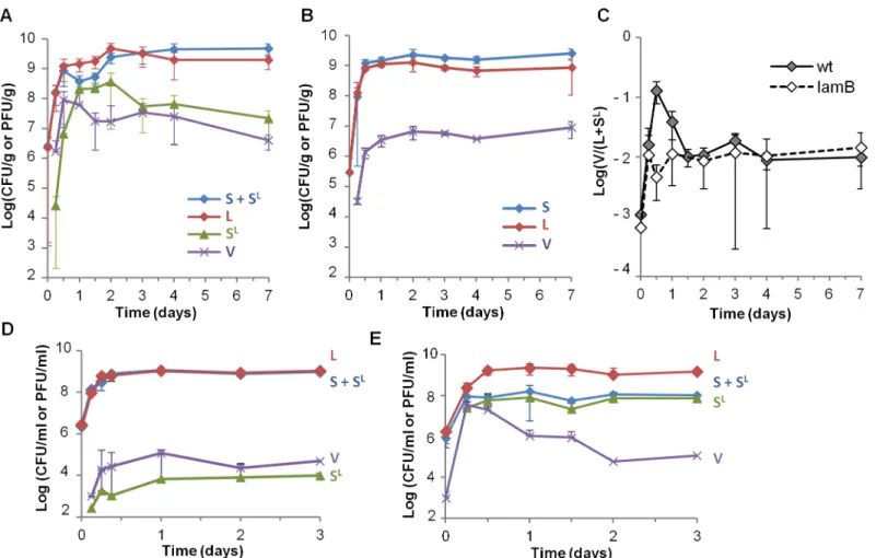
By comparison, when the same S and L strains were co-culturedin vitro, in standard rich LB medium, phage propagation was almost undetectable (Fig 1D), in line with previously pub-lished results [29]. This absence of propagation was due to low Mg2+concentration in LB, dras-tically limitingλadsorption ([27] andFig 1D and 1E). Addition of maltose did not improve phage propagation, suggesting that LamB expression in LB is sufficient for phage infection [30].
Phage propagation is impeded by
malT
mutations after 1.5 days of
colonization
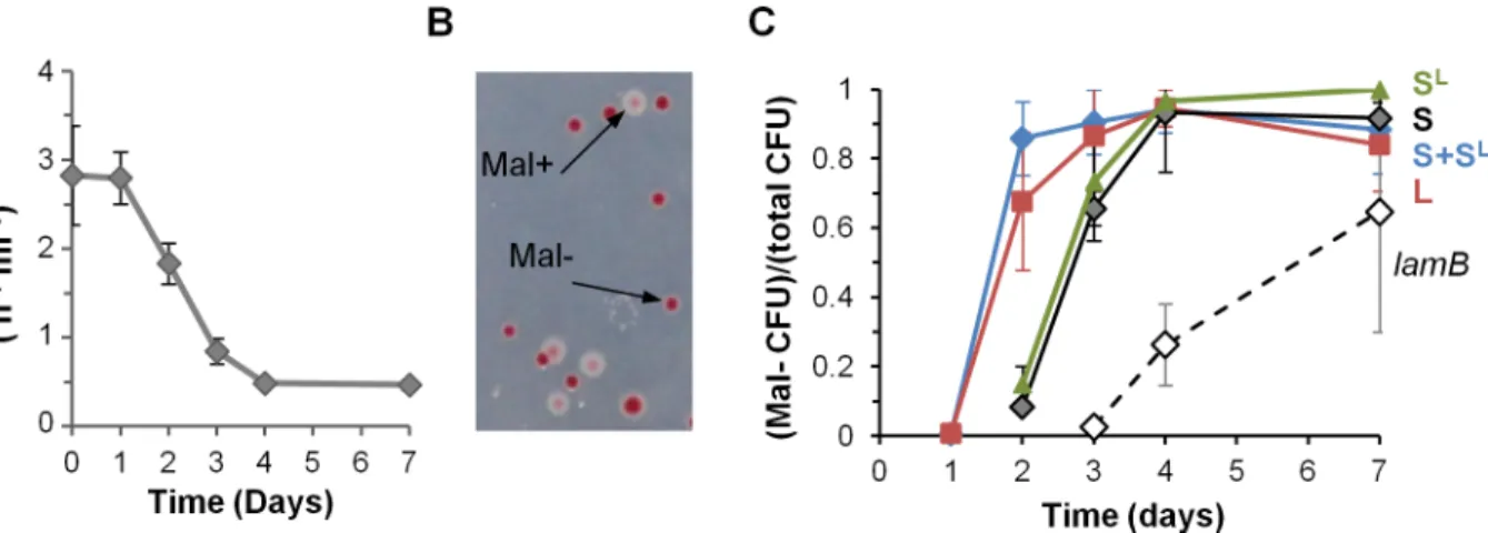
In mice, phage propagation stopped before the complete lysogenization of the S lineage. To test whether this resulted from changes in gut bacteria impairing infection, mice were monocolonized with the susceptible strain S only, and bacteria from feces were tested for affinity toλ(Fig 2A). After one day,λadsorption rate on bacteria from mouse feces was measured at 3.10−7ml h-1,
Fig 1. Rapid phage propagation occurs during the first day of colonization.A) Temporal evolution of bacterial lineages S, L and SLand free phage V densities in mouse feces: L bacteria (red), bacteria from the susceptible lineage (S + SL, blue), SLbacteria (green), free phage (purple, V). Means
+/-standard deviation from 8 mice in 2 independent experiments are indicated. B) Temporal evolution of S, L and free phage V densities in feces of mice colonized withlamBderivatives of bacterial strains. Means +/- standard deviations from 6 mice are indicated. C) Temporal dynamics of the free phage (V) over total lysogen (L + SL) ratio in mouse feces. Mice were colonized either with the two strains of panels A (wt, full line), or with theirlamBderivatives described in panel B (dashed line). Means +/- standard errors from 8 and 6 mice for the wt andlamBstrains respectively are indicated. D) Temporal dynamics of bacterial lineages S, L, SLand free phage V subjected to serial transfers in LB flasks. Phage propagation is very limited, as observed by the low density of
SLbacteria and free phage V. E) Same populations in LB supplemented with 5 mM MgSO
4. Means +/- standard deviations of ratios on 4 independent
cultures are indicated.
which is similar to the value measured at day 0in vitro. Therefore the phage receptor LamB is highly expressed in the mouse gut, and favorable ionic conditions allow for efficient binding. Later on however, the adsorption rate diminished continuously, suggesting a decrease in LamB expression (Fig 2A). To investigate this phenomenon further, we determined the susceptibility to λof S clones isolated from mouse feces two days after colonization. Nine out of the twelve clones
tested turned out to be genetically resistant toλ, and were moreover unable to use maltose, as revealed by their inability to grow on minimal medium containing maltose as the unique energy source. In subsequent colonization experiments, we quantified the increase in maltose-deficient bacteria (Mal-) by using maltose agar plates containing a tetrazolium dye that turned red in Mal -colonies (Fig 2B). Mal-bacteria were selected in the S and L lineages (Fig 2C). A similar rise in Mal-bacteria occurred in mice monocolonized with the phage-free strain S, demonstrating unambiguously that their selection is not caused byλ(Fig 2C).
Mal-andλresistance phenotypes, as well as previously published results [31], guided our identification of mutations in themalTgene. MalT is the transcriptional activator of the malt-ose regulon. It notably controls expression of theλreceptor LamB. All six resistant clones stud-ied carrstud-ied a mutation inmalT, among which three led to a truncated protein (S1 Fig), which explains that the selected mutations prevent phage infection. The reason for the selection of these mutants might be linked to the LamB-induced envelope stress associated with osmoregu-lation [30] since bacteria in the gastrointestinal lumen are continuously exposed to osmotic stress (reviewed in [32]). They are specific to monoxenic mice, asmalTmutations are not selected for in the MG1655E.colistrain when colonizing mice with a conventional microbiota [33].
Mathematical modelling of phage-bacteria interactions in monoxenic
mouse gut
The rise ofmalTmutants was nevertheless sufficiently delayed to permit observation phage infection of the majority of S bacteria during the first two days. In order to provide quantitative estimations of the parameters governing phage-bacteria interactions, we developed a
Fig 2. Resistant mutants invade independently of phage presence.A) Evolution of the phage adsorption rate on bacteria from mouse feces. On day 1 the adsorption rate was close to its maximal theoretical value. Means +/- standard errors from three mice are indicated. B) Detection and enumeration of Mal
-colonies on tetrazolium maltose plates after 2 days of colonization: white, wild-type Mal+colonies; red, Mal-colonies. Mal- colonies have mutations inmalT, the positive regulator of the maltose operon, and do not express the phage receptor LamB. C) Evolution of the proportion of Mal- bacteria with time in the S and L lineages. In addition to the results of the co-colonization experiments, the proportion of Mal-bacteria during two independent monocolonization
experiments are indicated: black line, monocolonization with the S strain; dotted line, monocolonization with alamBstrain. Means +/- standard deviations of four mice in each experiment are indicated.
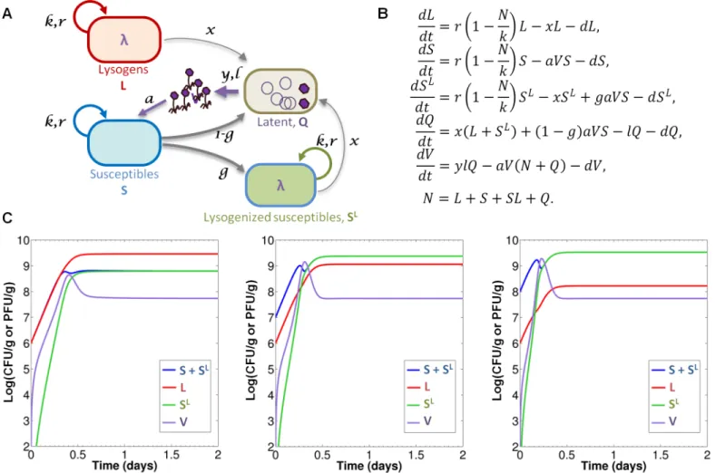
mathematical model representing the dynamics of the different microbial populations in this model ecosystem (Fig 3). The model is based on the one in [17] and adapted to take into account our experimental settings. It consists of five coupled differential equations, represent-ing time evolution of five population densities: S (susceptibles), L (lysogens), SL (newly-lysoge-nized susceptibles), V (free phage), as well as latent bacteria Q in which the phage undergoes lytic multiplication. Invasion ofmalTmutants is not included in the model.S1 Textgives a detailed description of the main modeling assumptions behind its construction, as well as a mathematical analysis of its dynamical behavior.
A careful examination of the effect of the eight model parameters onto the dynamics enabled the quantitative estimation of six of them from our experimental datasets (Table 1). With these estimated values, numerical simulations of the model (Fig 3C) are in good agree-ment with experiagree-mental observations on the first two days, before invasion ofmalTmutants, suggesting it captures most of the relevant information contained in our data. The main dis-crepancy observed is in the initial velocity of the temporal evolutions, faster in the model than
Fig 3. Quantitative mathematical modelling of temperate phage-bacteria interactions.A) Diagram representing the interactions between the different populations. S and L lineages grow with a common maximal growth raterand carrying capacityk. Lysogenic populations (L and SL) switch to lysis at a ratex
entering latent population Q. Phage binding on bacteria is determined by the adsorption constanta. Infection of susceptible leads either to lysogenization with probabilityg, or to lytic cycle with probability 1-g. Latency rateland burst sizeydetermine the production of free phage V. The dilution termdaccounts for the mouse gastrointestinal flux. B) System of differential equations governing the dynamics of the five populations (variable N denotes total population L+S+SL). C) Numerical simulations of the model with estimated values ofTable 1: the lines represent the temporal evolution of population densities S+SL(blue), L
(red), SL(green) and V (purple), with different initial conditions: L
0= 1x10⁶cfu/g and, from left to right, S0= L0, S0= 10×L0, S0= 100×L0.
in experimental data. This might result either from incorrect estimation of some parameters, or from the neglect of a phenomenon not taken into account in the model, such as the binding of free phage on some intestinal components. Such binding would result in a“loss”of phage that would slow the dynamics, as exemplified by the effect of reduced burst size (S2 Fig).
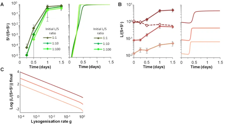
High lysogenization limits the final gain of the original prophage carrier
strain
Upon infection of a susceptible bacterium,λgoes to lysogenization with a high probability around 19%, leading to a very rapid rise of SLbacteria both in data and in numerical simula-tions (Fig 4A). The remaining infected susceptibles were lysed, resulting in an increase of the L lineage relative to the S one, independently of the initial L/S ratios (Fig 4B).LamBdeletion abolished the competitive advantage of the L lineage (Fig 4B, dotted line), confirming that the advantage of lysogens only stemmed from the lysis of susceptible competitors, and not from the presence of putative bacterial fitness genes in theλgenome that would improve growth in mice. However, the gain of the L lineage over the S one is limited by the lysogenization of sus-ceptible, independently from the rise ofλresistant mutants. Its final value, as predicted by the model, seems to be directly proportional to the inverse ofgat population equilibrium (Fig 4C). Interestingly, other parameters governing phage-bacteria interaction have very modest impact on the final gain of the L lineage (S3 FigandS1 Text).
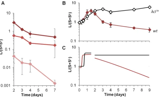
A high prophage induction rate in the gut decreases lysogen fitness
Prophage induction rate in the mouse gastrointestinal tract was estimated to be 1.6%, several orders of magnitude higher than usually assumed. This high induction leads to a slight but sys-tematic decrease of lysogens (L and SLlineages) in mice when bacteria are resistant to infection, either because ofmalTorlamBmutations (Figs1A and 1Band5A). By contrast,in vitro, induction rate is 3 x 10⁻⁴(Table 2), and competitions under conditions that did not permit phage infection resulted in a stable proportion over time of lysogenic (L and SLlineages) and non-lysogenic S bacteria (Figs1DandS3A). In mouse, model-based estimation of the induc-tion rate was derived from latent Q cell counts in mouse colonized withlamBstrains. Because of the small data set available (one experiment with three mice), the confidence interval is rela-tively large (0.6%-3.6%, seeTable 1andS1 Text). In order to strengthen the estimation, we also computed the induction rate from the relative fitness of L compared to SlamBlineages (Mate-rial & Methods). The value found (1.7% ± 0.5%) was very close to that estimated by the model.
To examine experimentally the impact of high induction rate, we used a non-inducibleλ prophage,λcIind-, which has a mutation in the repressor of the lytic cycle, CI, preventing its
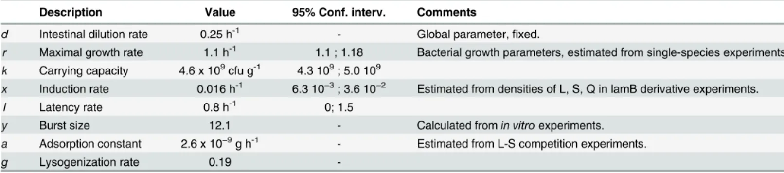
Table 1. Estimated values of the model’s parameters.
Description Value 95% Conf. interv. Comments
d Intestinal dilution rate 0.25 h-1 - Global parameter,fixed.
r Maximal growth rate 1.1 h-1 1.1 ; 1.18 Bacterial growth parameters, estimated from single-species experiments.
k Carrying capacity 4.6 x 10⁹cfu g-1 4.3 109; 5.0 109
x Induction rate 0.016 h-1 6.3 10−3; 3.6 10−2 Estimated from densities of L, S, Q in lamB derivative experiments.
l Latency rate 0.8 h-1 0; 1.5
y Burst size 12.1 - Calculated fromin vitroexperiments.
a Adsorption constant 2.6 x 10⁻⁹g h-1 - Estimated from L-S competition experiments.
g Lysogenization rate 0.19
RecA activated auto-cleavage upon DNA damage. As expected, in standardin vitroconditions, thecIind-mutation decreased the induction rate 1,000-fold (Table 2). In the mouse gastrointes-tinal tract, the mutation abolished the decrease in proportion of lysogens (Fig 5B), demonstrat-ing unambiguously that high prophage induction explains the disadvantage of lysogens. Moreover, in alamBgenetic background, S andλcIind-lysogenic strains presented no repro-ductive fitness differences over 9 days (S4B Fig), validating the model hypothesis that in the absence of lysis and induction, the presence of the prophage makes no difference in growth rate. Interestingly,λcIind-experiments also validated the absence of a rarity threshold to phage multiplication in the mouse gut: since phage amplify on susceptible bacteria, even a very low initial number of phage can lead to killing of a significant part of S lineage (Fig 5B and 5C).
Transient selection of
λ
virulent mutants in
λ
cI
ind-The switch from lysogenic to lytic cycle requires CI autocleavage, catalyzed by RecA nucleofila-ment formed by DNA damage [36,37]. In theλcIind-lysogens, RecA mediated CI autocleavage is prevented, and the few phage produced are CI low expression mutants [36]. Indeed, free phage isolated from feces of mice colonized withλcIind-lysogens formed clearer plaques thanλ wild-type, which suggest they have a lower lysogenization rate. Sequencing of thecIgene from 9 phages isolated at day 2 either from free phage in feces or from SLbacteria revealed they all had a point mutation in the -35 box of CI promoter, PRM(G->T, -33 relative to thecIstart of
Fig 4. High lysogenization limits the final gain of the original prophage carrier strain.A) Temporal dynamics of the proportion of newly lysogenized bacteria (SL/(S+SL)) for three different initial L/S ratios, in the experimental data (left panel) and with numerical simulations of the model (right panel). Means
+/- standard deviations from 8, 13 and 4 mice are indicated for L/S initial ratios 1:1, 1:10 and 1:100 respectively. In simulations, initial conditions for L and S densities are those of respective data. B) Temporal dynamics of the ratio of L over S lineages (L/(S+SL)), depending on the three initial L/S ratios (1:1, 1:10
and 1:100), in the experimental data (left panel) and in the simulation (right panel). Means +/- standard deviations from 8, 13 and 4 mice are indicated for L/S initial ratios 1:1, 1:10 and 1:100 respectively. In simulations, initial L and S densities are the mean of respective data. C) Impact of the lysogenization rate g on the final L over S lineages ratio, as predicted by the mathematical model.
transcription). Interestingly, this PRMmutation was previously shown to enableλprophage induction in the absence of SOS activation, by decreasing by 80% intracellular CI levels, leading to much higher switching rates from the lysogenic to the lytic states [36]. Indeed, these PRM mutants, namedλcI, had an induction rate 50,000-fold higher than that of the ancestralλcI ind-phage and 300-fold higher than the wild-type (Table 2). Measurement of induction rate from 12 other SLbacteria revealed they were all lysogenized byλcI. The high induction of this viru-lent mutant enabled its propagation during the first days of colonization. However, in agree-ment with evolutionary epidemiology theory, that predicts that selection for virulence
decreases with the pool of susceptible hosts [38], the virulentλcImutant was counter selected later on in the prophage form, due to killing of its host through induction (Fig 6).
Fig 5. A highin vivoinduction rate penalizes lysogens in the absence of susceptible bacteria.A) Evolution of the ratio of lysogen over susceptible lineages (L/(S+SL)) in mice feces after day 2. Means
+/-standard deviations from 8, 13 and 4 mice for initial L/S ratios (1:1, 1:10 and 1:100 respectively) are indicated. B) Evolution over 9 days of L/(S+SL) ratio for wild-type
λprophage (brown line) or forλcIind-deficient for induction (black line). Means +/- standard error of the mean from 7 and 12 mice forλcIind-and wild-type respectively. C) Two-phase temporal simulation of L/(S+SL) ratio, with a switch at 48h to a lamB-version of
the model (i.e. with a = 0). The ratio is shown for wild-type phage (red line) orλcIind-mutant (blue line, with
x= 2x10⁻⁷). The initial condition for the first phase is taken from data at time 0, and the initial condition for the
second phase (“mutant model”) is taken from experimental data at 48h.
doi:10.1371/journal.pgen.1005861.g005
Table 2. Induction rate per lysogenic bacteria growing on rich medium (LB).The induction rate was measured on ampicillin plates by scoring infective centers, as described in the Materials and Methods sec-tion. Since bile salts were shown to cause DNA damage in bacteria [34], and to induce aSalmonellaprophage [35], we measured their effect onλinduction rate, but no change was detected. Bile salts were added at a final concentration of 0.8%. Mean±standard deviation of three independent experiments are indicated.
λgenotype induction ratein vitro
λwt 3.4 x 10⁻⁴±6.8 x 10⁻⁵
λcI
ind-2.4 x 10⁻⁷±8.9 x 10⁻⁸
λcIind* 1.3 x 10⁻²±1.0 x 10⁻³
λwt + bile salts 1.9 x 10⁻⁴±2.6 x 10⁻⁵
High phage propagation permits
de novo
horizontal gene transfer
detection
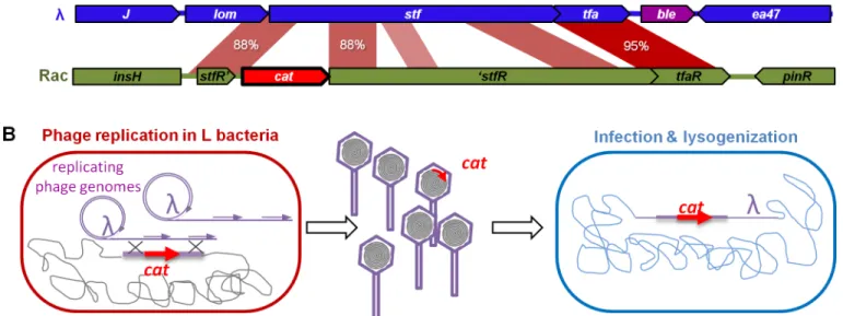
We next investigated whether this high phage activity allowed for gene exchange between the phage and bacterial genomes. We have previously reported thatλcaptures bacterial genes by homologous recombination during the lytic cycle, at frequencies ranging between 10−4and 10−6depending on the extent of homology between the DNA segments (Fig 7and [20]). In our experimental system, recombination can lead to the incorporation of the chloramphenicol resistance gene (cat) of the L strain into the phage genome, since L bacteria have thecatgene in a chromosomal region of partial homology withλ(88% identity). We investigated the occur-rence of this phenomenon in mice. Recombinant phages can be detected in their lysogenic form as they confer chloramphenicol resistance to the bacteria they are integrated in. On days 1 and 2, recombinant prophages were detected in all mice, at frequencies around 5.10−8relative
Fig 6. Transient advantage of virulentλcI*phage.Evolution with time of the proportion of bacteria carrying the virulentλcI*prophage (SL) on bacteria carrying the
λcind-prophage (L). Means +/- standard error of the mean from 7 mice.
doi:10.1371/journal.pgen.1005861.g006
Fig 7. Horizontal gene transfer mediated byλ. A)Thecatgene conferring chloramphenicol resistance CmR(red arrow, cat) of the lysogenic strain is
inserted in the defective prophage Rac. The gene is flanked by regions that share homology with the phage lambda.B)Upon induction, theλphage replicates and can recombine with the bacterial chromosome in such a way that thecatgene is captured by homologous recombination. Bacteria lysogenized by a recombinant phage are resistant to chloramphenicol.
to the number of new lysogens (SL). PCR analysis confirmed that thecatgene was placed at the expected position in theλprophage. No recombinants were detected with aλphage deleted of its main recombination gene,bet(orredβ), indicating the importance of phage recombination function for gene acquisition.
Discussion
Complete genome sequencing of thousands of gut bacteria has shown that most harbor pro-phages, yet their impact on strain fitness in the gastrointestinal tract has rarely been investi-gated. Colonization experiments, supported by a mathematical model of phage/bacteria interactions, show that the advantage ofλlysogeny in monoxenic mice gut is valid only when susceptible bacteria are present; a situation that might be only occasional in the gut microbiota. Indeed, it is supposed that only one or twoE.colistrains cohabit at the same time in the human gastrointestinal tract [39,40], and moreover, to our knowledgeλphage infects only a small proportion ofE.colistrains. In the absence of susceptible competitors, the prophage was costly for its host, due to frequent induction caused by DNA damage. Prophages were generally shown to positively impact their host fitness, and our study is, to our knowledge, the first dem-onstration that a prophage can be detrimental to bacteria in the gastrointestinal tract.
The level of phageλinduction observed in monoxenic mice was remarkable: 1 to 2% of lyso-genic bacteria were lysedpergeneration, which is almost two orders of magnitude higher than in standard laboratory conditions, in which induction was too low to constitute a measurable fitness cost (Figs1CandS4). This result is reinforced by another study showing that the induc-tion rate of 933W lambdoïd prophage is higher in the mouse gastrointestinal tract thanin vitro, and constant over time [41]. However, the reporter assay used did not permit direct esti-mation of the induction rate and the associated cost for the bacteria [41]. We observed that the λrepressor mutation CIind-, which abolishes CI auto-cleavage, dramatically decreased
pro-phage induction in the intestine. Many reports over a long period of time have proven unam-biguously that for such cleavage to occur, a RecA nucleoprotein filament (also called“activated RecA”) must catalyze the reaction [36,42,43], so lambda prophage induction reflects DNA damage. Since RecA nucleoprotein filament also triggers the general SOS response [44], our results indicates that this response is activated in 1 to 2% of bacteria in the intestine of monoxe-nic mice. DNA damage sensing is not only responsible for the induction of most prophages [2], but it also triggers activation of other mobile genetic elements such as integrative and con-jugative elements or ICEs [45], transposons [46] and integrons [47]. Moreover, the large num-ber of defective prophages inE.coligenomes, i.e. prophages incapable of independent
phage reproduction in an environment where the density of susceptible hosts is low or variable [58,59]. Although the gut microbiota is the densest bacterial community on earth, it includes hundreds of different species and thousands of bacterial strains, possibly making highly specific phage infection relatively rare. Low phage susceptibility seems to be the conclusion of a large-scale study of phage-bacteria interactions in a gnotobiotic mouse model [60]: in mice raised with a simplified microbiota composed of 15 strains belonging to dominant human species, only two were attacked by a cocktail of thousands of different phages isolated from a human gut microbiota. Moreover, a higher proportion of temperate phages was found in gut viromes than in other environments [19,61]. Altogether, these data support our results, suggesting that in the gastrointestinal tract the lysogenic life cycle of phages is favoured compared to lytic multiplication.
Temperate phages being major actors of horizontal gene transfer in bacteria, a concern emerged recently regarding their role in the propagation of antibiotic resistance genes [62]. Indeed, some phage particles are vectors of antibiotic resistance genes [28]. Most of the time, gene transfer occurs by generalized transduction, the erroneous encapsidation of bacterial DNA. Such errors are rare: for instance, the proportion ofE.coliphage P1 capsids leading to the production of an antibiotic-resistant clone is between 10−5and 10−6[63,64]. The incorpo-ration of a bacterial gene into phage genome, and afterwards transfers by lysogenization is a much rarer event, that we could detect only when the gene was located in a defective prophage sharing homology withλ[20]. In the present study, we estimated the frequency of such gene capture (catgene, conferring chloramphenicol resistance) byλin mice to be 10−8. Interestingly, up to now very few cases of phages encoding resistance genes have been reported [65–67], sug-gesting that even if the lysogenization rate in the intestine is very high, the risk of antibiotic resistance spread mediated by temperate phage is low.
Materials and Methods
Bacterial strains
All bacterial strains are described inS1 Table. All strains were constructed by modifying the MG1655ΔfliCΔompFstrain. This strain was used becauseompBmutations are rapidly and systematically selected in the MG1655 strain in the mouse gut as a result of their effects on fla-gellin (FliC) repression and of decreased membrane permeabilityviarepression of the major porin OmpF [69]. AsompBmutants also display a reduced expression level of LamB [31], a maltoporin used by phageλfor infection, we used aΔompFΔfliCstrain in which noompB mutations were selected [69]. ThestfR::catmutation was introduced in this strain by phage P1 transduction from the MD19 strain described in [20]. TheΔlamBstrains were constructed by phage P1 transduction of thelamB::KanRcassette from the Keio collection strain JW3996 [70]. In the MD56 and MD74 strains, the KanR cassette was excised as described in [71]. Theλ receptor being absent inΔlamBstrains,λprophage was introduced by transformation with Urλblepurified DNA.
Phage strains
Theλblephage strain used in this study was constructed by insertion of the phleomycin resis-tance genebleinto the Urλstrain ofλ, as described in [20]. TheλcIind-mutant contains a muta-tion (A111T) in the RecA cleavage site: the alanine in posimuta-tion 111 is replaced by a threonine [72]. This mutation was introduced by recombineering with the oligonucleotide AT111 (GT AAAGGTTCTAAGCTCAGGTGAGAACATgCCgGttTGgACATGAGAAAAAACAGGGT ACTCATACC). Small letters represent changes in the DNA sequence. Several neutral differ-ences were added to the one necessary for the amino acid change in order to avoid recognition by MutS. Recombineering was performed in the HME57 strain [73], which carried plasmid pKD46 [71], and lysogenized withλble. The strain was co-transformed with two oligonucleo-tides, AT111 and Court’s lab oligonucleotide 144, conferring it the ability to use galactose [74]. After transformation, colonies were isolated on M9 minimal galactose plates. 96 Gal+ clones were screened for the absence of spontaneous phage induction, by scoring the absence of infec-tious phage particles in culture supernatants. 1 out of 96 clones had the expected mutation, which was confirmed by sequencing of thecIgene. TheλcIind-phage was next introduced into the MG1655ΔfliCΔompF stfR::cat strain by P1 transduction and selection on phleomycin plates.
Mouse and in vivo competition experiments
Germ-free C3H/HeN mice were bred at the germ-free animal facilities of the INRA Micalis Institute, Anaxem. Mice were reared in isolators and fedad libitumon a commercial diet steril-ized by gamma irradiation (40 kGy) and supplied with autoclaved tap water. For colonization experiments, 8 week-old germ-free female mice were gavaged with 106bacteria from the cho-sen strain, or the appropriate mixture of the two strains, in 0.1 mL of M9 minimal medium. The cassettes used to differentiate strains during competition confer resistance to chloram-phenicol or to kanamycin. Their expression is known to have no significant cost duringE.coli
agar (0.4% agar, 10 mM MgSO4). The indicator bacterial culture was fresh MD5 culture grown in LB containing 0.2% maltose. Latent bacteria were counted similarly but after elimination of free phage by centrifugation. CFUs and PFUs were counted after 12–16 hours of incubation at 37°C. The ability to use maltose was monitored in tetrazolium maltose (TM) indicator plates. Mal+and Mal-clones respectively form white and red colonies on these plates. The TM medium was composed of tryptone (10 g/L), yeast extract (1 g/L), NaCl (5g/L), agar (16g/L), maltose (5 g/L) and tetrazolium dye (50 mg/L, Sigma).
S and L lineages competitionin vitro. For comparison with thein vivoresults, S and L
strains were co-culturedin vitroin rich LB medium, with a starting 1:1 ratio. Co-cultures were diluted 1,000-fold twice a day in 10 ml of LB, with or without 5 mM MgSO4, in 50-ml plastic
tubes and incubated at 37°C with agitation (200 rpm). 1,000 fold dilution corresponds to a number of bacterial generations per 24 hours comparable to that in mouse gut (about 20 gener-ations compared to 16 in mice).
Adsorption rate measurements
The technique was essentially that of Hendrix [76], with minor modifications. Adsorbing bac-teria from feces were prepared as described for enumeration. A control culture was grown at 37°C with shaking in LB + 0.2% maltose + 10 mM MgSO4up to an absorbance at 600 nm of 1.2 (about 6.108bacteria/ml). Aliquots of 200μL of culture were added to 50-μL aliquots con-taining 500 phage particles in 400-μL PCR tube strips, and mixed at 37°C. Several strips were prepared, one for each time point. At the chosen time points, the bacteria were separated from the free phages by centrifugation. The PFU in the supernatants were enumerated as described above. PFUs at time zero were estimated by titering the phage suspension. The slope of a graph plotting the logarithm of the number of bacteria remaining unadsorbed as a function of time allowed us to calculate the adsorption rate a according to the equation Nt= N0x e-Bat, where Nt and N0are the numbers of phage particles unadsorbed at time t and at time zero respectively, B is the number of bacteria per millilitre, and t is time in hours.
Quantitative estimation of mathematical model
’
s parameters
model (i.e. with a = 0) implying only variables L, S and Q. This simplified model was then fitted to LamB-experimental data. The third group gathers phage-dependent parametersyanda. Based on the formula for the equilibrium population of free viruses, a linear relationship was obtained between the two. Using a value ofycalculated fromin vitrodata, the corresponding value for the adsorption constant was deduced. Finally, the last parameterg(probability of lysogenization) was estimated by fitting the ratio L/(S+SL) in L-S competition experiments. The best fit was obtained at 36 h. All estimated values are listed inTable 1. When possible, bootstrap techniques were used to compute 95% confidence intervals (seeS1 Text). All numeri-cal computations have been performed with Matlab (The MathWorks, Inc.).
Experimental estimation of burst size
Burst size was estimatedin vitroin conditioned LB, i.e. the supernatant of a bacterial culture grown in LB (+0.2% maltose, +10 mM MgSO4) at 37°C up to an absorbance at 600 nm of 1.5. In such conditioned media we measured a growth rate of 1.1 h-1, close to that measured in the mice gastrointestinal tract during the first 24 hours of colonization. For single burst experiments, 100μl of a culture at OD600 of 1.5 is mixed with 1x10⁵λphage. The mix of phage and bacteria is incu-bated 7 minutes at 37°C, centrifuged and washed twice to eliminate free phage, and then diluted 1,000 times in conditioned LB at 37°C. Samples were taken every 10 minutes, mixed with 200μl of exponentially growing susceptible bacteria and 3ml of Top Agar (4.5 g/L, 0.2% maltose, 10 mM MgSO4), and then poured on LB agar plates. Plaque-forming units (PFU) were counted. The burst size (y) is the factor between maximum and initial PFU counts. We determined that y = 12.1 ± 8.
Model-independent estimation of induction rate
Since induction rate is the sole parameter differentiating growth of S and LlamBlineages, the induction rate (x) was determined from the evolution of the L/S ratio between days 0 and 9 in function of the number of generations. The number of generations was estimated by assuming that growth rate is equal to r, 1.1 h-1, during the first 24 hours, and after equal to excretion rate d, 0.25 h-1. A linear regression on data from 6 mice (lm function in R software) gave
x = 0.0170 ± 0.0046.
In vitro induction rate measurements
We adapted a method allowing for an measurement of the induction rateperbacterium [80]. Lysogenic bacteria were diluted 200 fold in LB and grown at 37°C with shaking. When speci-fied, 0.8% w/v bile salts (cholic acid-deoxycholic acid sodium salt mixture, sigma-aldrich) were added when the absorbance at 600 nm of the culture was 0.2 or 0.1. When the absorbance reached 0.4, cultures were swirled on ice for 5 minutes and washed twice at 4°C to eliminate free phages. Washed lysogenic bacteria were then mixed at the appropriate dilution with 100μl of a saturated culture of the indicator strain, 3 ml of top agar were added, and the mix was plated on LBA-ampicillin plates (50μg/ml). The indicator bacteria were ampicillin-resistant (strain JC10990recF::Tn3 AmpR). Ampicillin prevents further lysogen growth but does not prevent the completion of the lytic cycle if already started, which results in an infective centre. The induction rate was calculated by dividing the number of infective centres by the number of plated lysogenic bacteria.
Supporting Information
S1 Fig. Map of the six mutations identified in themalTgene.3 mutations interrupt the
(S+SL)), approximated using numerical simulations of the mathematical for t = 100 h, when each parameter is perturbed individually within the range indicated on the x axis. The three colors correspond to three different initial ratios (see upper left panel).
(PDF)
S4 Fig. L over S lineages ratio. A)Evolution of the ratio of lysogen over susceptible lineages (L/(S+SL)) with time in broth (LB). A clear advantage of the lysogenic strain in observed in LB + Mg2+. WithlamBstrains, the ratio is completely stable, showing that no cost associ-ated to prophage induction is detectable. Very similar results are observed in LB without Mg2+. Mean +/- standard deviation of ratios on 4 independent cultures.B)Same ratio in mice withlamBbacterial strains andλcIind-phage. Mean +/- standard deviation of ratios on 3 mice.
(PDF)
S1 Text. Mathematical modelling of phage-mediated bacterial interactions in the mouse gut.Part 1 contains a description of the model's construction together with a brief analysis of its equilibrium points. Part 2 details the procedures used to estimate the model's parameters. Part 3 proposes a sensitivity analysis of the parameters.
(PDF)
S1 Table. Bacterial and phage strains used in the study. (DOCX)
Acknowledgments
We are thankful to Colin Tinsley for careful reading of the manuscript and his help with the experiments; Sébastien Blugeon for his help to start mouse experiments; and to the Anaxem platform, and in particular to Anne Foussier and Fatima Joly, for their help with mouse and isolator handling. We also thank A. Gruss for critical reading of the manuscript and V. Fro-mion for fruitful discussions about the mathematical model.
Author Contributions
Conceived and designed the experiments: MDP MAP LT. Performed the experiments: MDP EM OS LT. Analyzed the data: MDP MAP LT. Contributed reagents/materials/analysis tools: PL. Wrote the paper: MDP MAP LT.
References
2. Nanda AM, Thormann K, Frunzke J (2015) Impact of spontaneous prophage induction on the fitness of bacterial populations and host-microbe interactions. J Bacteriol 197: 410–419. doi: 10.1128/JB.02230-14PMID:25404701
3. Bondy-Denomy J, Davidson AR (2014) When a virus is not a parasite: the beneficial effects of pro-phages on bacterial fitness. J Microbiol 52: 235–242. doi:10.1007/s12275-014-4083-3PMID: 24585054
4. Brussow H, Canchaya C, Hardt WD (2004) Phages and the evolution of bacterial pathogens: from genomic rearrangements to lysogenic conversion. Microbiol Mol Biol Rev 68: 560–602, table of con-tents. PMID:15353570
5. Fortier LC, Sekulovic O (2013) Importance of prophages to evolution and virulence of bacterial patho-gens. Virulence 4: 354–365. doi:10.4161/viru.24498PMID:23611873
6. Dykhuizen D, Campbell JH, Rolfe BG (1978) The influences of a lambda prophage on the growth rate of Escherichia coli. Microbios 23: 99–113. PMID:160003
7. Boyd EF (2012) Bacteriophage-encoded bacterial virulence factors and phage-pathogenicity island interactions. Adv Virus Res 82: 91–118. doi:10.1016/B978-0-12-394621-8.00014-5PMID:22420852 8. Kintz E, Davies MR, Hammerlof DL, Canals R, Hinton JC, et al. (2015) A BTP1 prophage gene present in invasive non-typhoidal Salmonella determines composition and length of the O-antigen of the lipo-polysaccharide. Mol Microbiol.
9. Erken M, Lutz C, McDougald D (2013) The rise of pathogens: predation as a factor driving the evolution of human pathogens in the environment. Microb Ecol 65: 860–868. doi:10.1007/s00248-013-0189-0 PMID:23354181
10. Karaolis DK, Somara S, Maneval DR Jr., Johnson JA, Kaper JB (1999) A bacteriophage encoding a pathogenicity island, a type-IV pilus and a phage receptor in cholera bacteria. Nature 399: 375–379. PMID:10360577
11. Herrington DA, Hall RH, Losonsky G, Mekalanos JJ, Taylor RK, et al. (1988) Toxin, toxin-coregulated pili, and the toxR regulon are essential for Vibrio cholerae pathogenesis in humans. J Exp Med 168: 1487–1492. PMID:2902187
12. Hsieh YC, Lin TL, Lin CM, Wang JT (2015) Identification of PblB mediating galactose-specific adhesion in a successful Streptococcus pneumoniae clone. Sci Rep 5: 12265. doi:10.1038/srep12265PMID: 26193794
13. Peetermans M, Vanassche T, Liesenborghs L, Claes J, Vande Velde G, et al. (2014) Plasminogen acti-vation by staphylokinase enhances local spreading of S. aureus in skin infections. BMC Microbiol 14: 310. doi:10.1186/s12866-014-0310-7PMID:25515118
14. Steinberg KM, Levin BR (2007) Grazing protozoa and the evolution of the Escherichia coli O157:H7 Shiga toxin-encoding prophage. Proc Biol Sci 274: 1921–1929. PMID:17535798
15. Arnold JW, Koudelka GB (2014) The Trojan Horse of the microbiological arms race: phage-encoded toxins as a defence against eukaryotic predators. Environ Microbiol 16: 454–466. doi: 10.1111/1462-2920.12232PMID:23981100
16. Bossi L, Fuentes JA, Mora G, Figueroa-Bossi N (2003) Prophage contribution to bacterial population dynamics. J Bacteriol 185: 6467–6471. PMID:14563883
17. Brown SP, Le Chat L, De Paepe M, Taddei F (2006) Ecology of microbial invasions: amplification allows virus carriers to invade more rapidly when rare. Curr Biol 16: 2048–2052. PMID:17055985 18. Burns N, James CE, Harrison E (2015) Polylysogeny magnifies competitiveness of a bacterial
patho-gen in vivo. Evol Appl 8: 346–351. doi:10.1111/eva.12243PMID:25926879
19. Reyes A, Haynes M, Hanson N, Angly FE, Heath AC, et al. (2010) Viruses in the faecal microbiota of monozygotic twins and their mothers. Nature 466: 334–338. doi:10.1038/nature09199PMID: 20631792
20. De Paepe M, Hutinet G, Son O, Amarir-Bouhram J, Schbath S, et al. (2014) Temperate phages acquire DNA from defective prophages by relaxed homologous recombination: the role of Rad52-like recombi-nases. PLoS Genet 10: e1004181. doi:10.1371/journal.pgen.1004181PMID:24603854
21. Mills S, Shanahan F, Stanton C, Hill C, Coffey A, et al. (2013) Movers and shakers: influence of bacteri-ophages in shaping the mammalian gut microbiota. Gut Microbes 4: 4–16. doi:10.4161/gmic.22371 PMID:23022738
22. Gamage SD, Strasser JE, Chalk CL, Weiss AA (2003) Nonpathogenic Escherichia coli can contribute to the production of Shiga toxin. Infect Immun 71: 3107–3115. PMID:12761088
viruses as double-edged swords in bacterial warfare. PLoS One 8: e59043. doi:10.1371/journal.pone. 0059043PMID:23536852
30. Reimann SA, Wolfe AJ (2009) A critical process controlled by MalT and OmpR is revealed through syn-thetic lethality. J Bacteriol 191: 5320–5324. doi:10.1128/JB.00522-09PMID:19502392
31. De Paepe M, Gaboriau-Routhiau V, Rainteau D, Rakotobe S, Taddei F, et al. (2011) Trade-off between bile resistance and nutritional competence drives Escherichia coli diversification in the mouse gut. PLoS Genet 7: e1002107. doi:10.1371/journal.pgen.1002107PMID:21698140
32. Culligan EP, Sleator RD, Marchesi JR, Hill C (2012) Functional metagenomics reveals novel salt toler-ance loci from the human gut microbiome. ISME J 6: 1916–1925. doi:10.1038/ismej.2012.38PMID: 22534607
33. Barroso-Batista J, Sousa A, Lourenco M, Bergman ML, Sobral D, et al. (2014) The first steps of adapta-tion of Escherichia coli to the gut are dominated by soft sweeps. PLoS Genet 10: e1004182. doi:10. 1371/journal.pgen.1004182PMID:24603313
34. Merritt ME, Donaldson JR (2009) Effect of bile salts on the DNA and membrane integrity of enteric bac-teria. J Med Microbiol 58: 1533–1541. doi:10.1099/jmm.0.014092-0PMID:19762477
35. Kim S, Ryu K, Biswas D, Ahn J (2014) Survival, prophage induction, and invasive properties of lyso-genic Salmonella Typhimurium exposed to simulated gastrointestinal conditions. Arch Microbiol 196: 655–659. doi:10.1007/s00203-014-1005-zPMID:24929817
36. Little JW, Michalowski CB (2010) Stability and instability in the lysogenic state of phage lambda. J Bac-teriol 192: 6064–6076. doi:10.1128/JB.00726-10PMID:20870769
37. Kim B, Little JW (1993) LexA and lambda Cl repressors as enzymes: specific cleavage in an intermo-lecular reaction. Cell 73: 1165–1173. PMID:8513500
38. Berngruber TW, Froissart R, Choisy M, Gandon S (2013) Evolution of virulence in emerging epidemics. PLoS Pathog 9: e1003209. doi:10.1371/journal.ppat.1003209PMID:23516359
39. Caugant DA, Levin BR, Selander RK (1981) Genetic diversity and temporal variation in the E. coli popu-lation of a human host. Genetics 98: 467–490. PMID:7037535
40. Tenaillon O, Skurnik D, Picard B, Denamur E (2010) The population genetics of commensal Escheri-chia coli. Nat Rev Microbiol 8: 207–217. doi:10.1038/nrmicro2298PMID:20157339
41. Tyler JS, Beeri K, Reynolds JL, Alteri CJ, Skinner KG, et al. (2013) Prophage induction is enhanced and required for renal disease and lethality in an EHEC mouse model. PLoS Pathog 9: e1003236. doi: 10.1371/journal.ppat.1003236PMID:23555250
42. Little JW, Mount DW (1982) The SOS regulatory system of Escherichia coli. Cell 29: 11–22. PMID: 7049397
43. Shimada K, Shibata Y, Takagi Y (1975) Induction of phage production in the lysogenic Escherichia coli by hydroxyurea. Jpn J Microbiol 19: 349–354. PMID:772254
44. d'Ari R (1985) The SOS system. Biochimie 67: 343–347. PMID:2994755
45. Beaber JW, Hochhut B, Waldor MK (2004) SOS response promotes horizontal dissemination of antibi-otic resistance genes. Nature 427: 72–74. PMID:14688795
46. Kuan CT, Liu SK, Tessman I (1991) Excision and transposition of Tn5 as an SOS activity in Escherichia coli. Genetics 128: 45–57. PMID:1648004
47. Guerin E, Cambray G, Sanchez-Alberola N, Campoy S, Erill I, et al. (2009) The SOS response controls integron recombination. Science 324: 1034. doi:10.1126/science.1172914PMID:19460999 48. Martin R, Soberon N, Escobedo S, Suarez JE (2009) Bacteriophage induction versus vaginal
homeo-stasis: role of H(2)O(2) in the selection of Lactobacillus defective prophages. Int Microbiol 12: 131–
49. Matos RC, Lapaque N, Rigottier-Gois L, Debarbieux L, Meylheuc T, et al. (2013) Enterococcus faecalis prophage dynamics and contributions to pathogenic traits. PLoS Genet 9: e1003539. doi:10.1371/ journal.pgen.1003539PMID:23754962
50. Asadulghani M, Ogura Y, Ooka T, Itoh T, Sawaguchi A, et al. (2009) The defective prophage pool of Escherichia coli O157: prophage-prophage interactions potentiate horizontal transfer of virulence determinants. PLoS Pathog 5: e1000408. doi:10.1371/journal.ppat.1000408PMID:19412337 51. Bobay LM, Touchon M, Rocha EP (2014) Pervasive domestication of defective prophages by bacteria.
Proc Natl Acad Sci U S A 111: 12127–12132. doi:10.1073/pnas.1405336111PMID:25092302 52. Lin L, Bitner R, Edlin G (1977) Increased reproductive fitness of Escherichia coli lambda lysogens. J
Virol 21: 554–559. PMID:319255
53. Barondess JJ, Beckwith J (1990) A bacterial virulence determinant encoded by lysogenic coliphage lambda. Nature 346: 871–874. PMID:2144037
54. Refardt D, Rainey PB (2009) Tuning a genetic switch: experimental evolution and natural variation of prophage induction. Evolution 64: 1086–1097. doi:10.1111/j.1558-5646.2009.00882.xPMID: 19891623
55. Los JM, Los M, Wegrzyn A, Wegrzyn G (2012) Altruism of Shiga toxin-producing Escherichia coli: recent hypothesis versus experimental results. Front Cell Infect Microbiol 2: 166. doi:10.3389/fcimb. 2012.00166PMID:23316482
56. Zeng L, Skinner SO, Zong C, Sippy J, Feiss M, et al. (2010) Decision making at a subcellular level determines the outcome of bacteriophage infection. Cell 141: 682–691. doi:10.1016/j.cell.2010.03. 034PMID:20478257
57. Joo J, Gunny M, Cases M, Hudson P, Albert R, et al. (2006) Bacteriophage-mediated competition in Bordetella bacteria. Proc Biol Sci 273: 1843–1848. PMID:16790419
58. Stewart FM, Levin BR (1984) The population biology of bacterial viruses: why be temperate. Theor Popul Biol 26: 93–117. PMID:6484871
59. Maslov S, Sneppen K (2015) Well-temperate phage: optimal bet-hedging against local environmental collapses. Sci Rep 5: 10523. doi:10.1038/srep10523PMID:26035282
60. Reyes A, Wu M, McNulty NP, Rohwer FL, Gordon JI (2013) Gnotobiotic mouse model of phage-bacte-rial host dynamics in the human gut. Proc Natl Acad Sci U S A 110: 20236–20241. doi:10.1073/pnas. 1319470110PMID:24259713
61. Waller AS, Yamada T, Kristensen DM, Kultima JR, Sunagawa S, et al. (2014) Classification and quanti-fication of bacteriophage taxa in human gut metagenomes. ISME J 8: 1391–1402. doi:10.1038/ismej. 2014.30PMID:24621522
62. Modi SR, Lee HH, Spina CS, Collins JJ (2013) Antibiotic treatment expands the resistance reservoir and ecological network of the phage metagenome. Nature 499: 219–222. doi:10.1038/nature12212 PMID:23748443
63. Volkova VV, Lu Z, Besser T, Grohn YT (2014) Modeling the infection dynamics of bacteriophages in enteric Escherichia coli: estimating the contribution of transduction to antimicrobial gene spread. Appl Environ Microbiol 80: 4350–4362. doi:10.1128/AEM.00446-14PMID:24814786
64. Kenzaka T, Tani K, Nasu M (2010) High-frequency phage-mediated gene transfer in freshwater envi-ronments determined at single-cell level. ISME J 4: 648–659. doi:10.1038/ismej.2009.145PMID: 20090786
65. Davies J, Davies D (2010) Origins and evolution of antibiotic resistance. Microbiol Mol Biol Rev 74: 417–433. doi:10.1128/MMBR.00016-10PMID:20805405
66. Banks DJ, Lei B, Musser JM (2003) Prophage induction and expression of prophage-encoded viru-lence factors in group A Streptococcus serotype M3 strain MGAS315. Infect Immun 71: 7079–7086. PMID:14638798
67. Schuch R, Fischetti VA (2006) Detailed genomic analysis of the Wbeta and gamma phages infecting Bacillus anthracis: implications for evolution of environmental fitness and antibiotic resistance. J Bacter-iol 188: 3037–3051. PMID:16585764
68. Bobay LM, Rocha EP, Touchon M (2013) The adaptation of temperate bacteriophages to their host genomes. Mol Biol Evol 30: 737–751. doi:10.1093/molbev/mss279PMID:23243039
69. Giraud A, Arous S, De Paepe M, Gaboriau-Routhiau V, Bambou JC, et al. (2008) Dissecting the genetic components of adaptation of Escherichia coli to the mouse gut. PLoS Genet 4: e2. doi:10.1371/ journal.pgen.0040002PMID:18193944
ence 258: 1145–1148. PMID:1439823
77. Gibbons RJ, Kapsimalis B (1967) Estimates of the overall rate of growth of the intestinal microflora of hamsters, guinea pigs, and mice. J Bacteriol 93: 510–512. PMID:6020422
78. Freter R, Freter RR, Brickner H (1983) Experimental and mathematical models of Escherichia coli plas-mid transfer in vitro and in vivo. Infect Immun 39: 60–84. PMID:6337105
79. Freter R, Brickner H, Fekete J, Vickerman MM, Carey KE (1983) Survival and implantation of Escheri-chia coli in the intestinal tract. Infect Immun 39: 686–703. PMID:6339389
80. Dutreix M, Bailone A, Devoret R (1985) Efficiency of induction of prophage lambda mutants as a func-tion of recA alleles. J Bacteriol 161: 1080–1085. PMID:3156121