Cellular Antioxidant Levels in Glial Cells following
Hydrogen Peroxide Exposure
Kaori Suyama1, Masahiko Watanabe2*, Kou Sakabe1, Asako Otomo3, Yoshinori Okada4, Hayato Terayama1, Takeshi Imai2, Joji Mochida2
1Department of Anatomy and Cellular Biology, Basic Medical Science, Tokai University School of Medicine, Isehara, Kanagawa, Japan,2Department of Orthopedic Surgery, Surgical Science, Tokai University School of Medicine, Isehara, Kanagawa, Japan,3Department of Molecular Life Science, Basic Medical Science, Tokai University School of Medicine, Isehara, Kanagawa, Japan,4Tokai University Teaching and Research Support Center, Isehara, Kanagawa, Japan
Abstract
Oxidative stress, caused by the over production of reactive oxygen species (ROS), has been shown to contribute to cell damage associated with neurotrauma and neurodegenerative diseases. ROS mediates cell damage either through direct oxidation of lipids, proteins and DNA or by acting as signaling molecules to trigger cellular apoptotic pathways. The 78 kDa glucose-regulated protein (GRP78) is an ER chaperone that has been suggested to protect cells against ROS-induced damage. However, the protective mechanism of GRP78 remains unclear. In this study, we used C6 glioma cells transiently overexpressing GRP78 to investigate the protective effect of GRP78 against oxidative stress (hydrogen peroxide)-induced injury. Our results showed that the overexpression of GRP78 significantly protected cells from ROS-induced cell damage when compared to non-GRP78 overexpressing cells, which was most likely due to GRP78-overexpressing cells having higher levels of glutathione (GSH) and NAD(P)H:quinone oxidoreductase 1 (NQO1), two antioxidants that protect cells against oxidative stress. Although hydrogen peroxide treatment increased lipid peroxidation in non-GRP78 overexpressing cells, this increase was significantly reduced in GRP78-overexpressing cells. Overall, these results indicate that GRP78 plays an important role in protecting glial cells against oxidative stress via regulating the expression of GSH and NQO1.
Citation:Suyama K, Watanabe M, Sakabe K, Otomo A, Okada Y, et al. (2014) GRP78 Suppresses Lipid Peroxidation and Promotes Cellular Antioxidant Levels in Glial Cells following Hydrogen Peroxide Exposure. PLoS ONE 9(1): e86951. doi:10.1371/journal.pone.0086951
Editor:Ken Arai, Massachusetts General Hospital/Harvard Medical School, United States of America ReceivedSeptember 17, 2013;AcceptedDecember 16, 2013;PublishedJanuary 24, 2014
Copyright:ß2014 Suyama et al. This is an open-access article distributed under the terms of the Creative Commons Attribution License, which permits unrestricted use, distribution, and reproduction in any medium, provided the original author and source are credited.
Funding:This study was supported in part by the Research and Study Program of Tokai University Educational System General Research Organization and 2012 Tokai University School of Medicine Research Aid.The funders had no role in study design, data collection and analysis, decision to publish, or preparation of the manuscript.
Competing Interests:The authors have declared that no competing interests exist. * E-mail: [email protected]
Introduction
Reactive oxygen species (ROS) are one of the cytotoxic factors produced from damaged cells that cause oxidative stress and tissue damage during neurotrauma [1]. Hydrogen peroxide (H2O2), a
ROS, is released from dying cells during neurotrauma and neurodegenerative disease and causes tissue destruction [2,3]. H2O2 can produce hydroxyl radicals (OH?) and mediate cell
damage either through direct oxidation of lipids, proteins and DNA or act as a signaling molecule to trigger cellular apoptotic pathways [4–6]. Therefore, it is important to protect cells from H2O2-induced cell damage as a therapeutic strategy against
neurotrauma and neurodegenerative diseases [7,8]. Endoplasmic reticulum (ER) stress has been reported to be one of the pathways via which cells are damaged and die following ROS exposure [9,10].
The mechanism by which ER stress promotes apoptosis in cells hinges on driving the accumulation of structurally abnormal proteins that are usually repaired by ER chaperones to prevent cell death [11]. The 78 kDa glucose-regulated protein (GRP78) is one example of an ER chaperone that regulates protein folding in the ER and controls the ER-Ca2+
balance via trans membrane ER stress sensors, which contribute to cell survival [11–13]. GRP78
has been suggested to not only protect cells against high-concentrations of glutamate or tunicamycin, which induce ER stress directly [14], but also protect cells from ROS damage [15– 17].
Many studies have focused on various antioxidant factors, such as glutathione and NAD(P)H:quinone oxidoreductase 1 (NQO1). A previous study reported that induction of NQO1 and GSH by dimethyl fumarate, 3H-1,2-dithiole-3-thione or tert-butylhydro-quinone (tBHQ) protected against neurocytotoxicity caused by dopamine, 6-hydroxydopamine, 4-hydroxy-2-nonenal,or H2O2
[18]. As this study described, these antioxidants have recently been demonstrated to play an important role in protecting cells against oxidative stress [19–21]. Glutathione is the most abundant low molecular weight thiol in most organisms [20,22]. There are two types of glutathione, reduced glutathione (GSH) and oxidized glutathione (GSSG), depending on the environment. Reduced glutathione (GSH) is the main non-protein antioxidant and plays a critical role in the detoxification of H2O2and lipid hydroperoxide,
phospholipids from oxidative damage and plays an important protective role in oxidative stress [19].
Few studies have investigated the influence of GRP78 on NQO1. Some studies have suggested that H2O2 may not be
involved in ER stress-dependent cell damage because the response of GRP78 is different following H2O2 exposure and other
cytotoxic factors [23,24]. Similarly, a report on PKE-like ER kinase (PERK), a ER-stress sensing protein that resides in the ER, suggested that the PERK pathway is activated after dissociation of GRP78 from PERK monomers and leads to intracellular GSH production [25]. As these studies showed, the role of GRP78 during oxidative stress remains unclear. Therefore, we used cells transiently overexpressing GRP78 to investigate the protective effect of GRP78 against high extracellular concentrations of H2O2
and evaluated the response of glutathione and NQO1.
Materials and Methods
Cell Culture and Culture Conditions
The rat C6 glioma cell line was obtained from American Type Culture Collections (ATCC, Manassas, VA, USA). Cells were plated onto 6 cm tissue culture dishes and maintained at 37uC with 5% (v/v) CO2 and 95% (v/v) air. The culture medium
consisted of Dulbecco’s modified Eagle’s medium (DMEM; Gibco/Life Technologies, Carlsbad, CA, USA), 10% (v/v) fetal bovine serum (FBS), and 1% (v/v) penicillin–streptomycin.
H2O2Treatment of C6 Cells
C6 cells were plated onto 6 cm tissue culture dishes at a density of 2.06106cells/dish, incubated for 36 h and then treated with fresh medium containing 1–6 mM H2O2 (Wako, Osaka, Japan)
for 6 and 24 h.
Antibodies
Antibodies used were: rabbit polyclonal antibody against GRP78 (AnaSpec Inc. Fremont, CA USA); goat polyclonal antibody against GRP78 (Santa Cruz Biotechnology, Inc. Dallas, TX, USA); rabbit polyclonal antibody against NAD(P)H:quinone oxidoreducrase1(NQO1) (Santa Cruz Biotechnology Inc.); mouse monoclonal antibody against NAD(P)H:quinone oxidoreducra-se1(NQO1) (Novus Biologicals, LLC. Littleton, CO. USA); mouse monoclonal antibody against b-actin (Sigma-Aldrich Inc. St. Louis, MO, USA); Alexa 488-conjugated anti-rabbit antibody; Alexa 488-conjugated anti-mouse antibody (Invitrogen/Life Technologies); PE-conjugated anti-mouse secondary antibody
Figure 1. Schematic illustration of the pIRES2-AcGFP1 plasmid constructs used in the present study. cDNA of the rat GRP78 carried the XhoI/PstI cut sites and was cloned into the pIRES2-AcGFP1 plasmid.
doi:10.1371/journal.pone.0086951.g001
Figure 2. GRP78 expression in C6 cells following treatment with H2O2.(A) FACS analysis revealed a significant increase in the number of
AP-positive cells following treatment with H2O2when compared with untreated C6 cells (*P,0.05). Compared with untreated C6 cells, no significant
differences were observed in GRP78 protein levels at any H2O2concentration. (B) Western blot analysis of GRP78 in C6 cells treated with H2O2for 6 h
and 24h. There were no obvious changes in GRP78 expression levels. doi:10.1371/journal.pone.0086951.g002
(BioLegend Inc. San Diego, CA, USA); horseradish peroxidase (HRP)-conjugated goat polyclonal antibody against rabbit IgG; HRP-conjugated goat polyclonal antibody against mouse IgG; HRP-conjugated rabbit polyclonal antibody against goat IgG (DAKO Glostrup, Denmark).
Intracellular GRP78 and NQO1 Detection
The IntraStainHreagent kit (DAKO) has previously been used successfully for the detection of intracellular GRP78 or NQO1 protein expression under conditions of tunicamycin treatment and
bactin [14]. Following exposure to treatment, cells were fixed and permeabilized using IntraStain according to the manufacturer’s instructions. For the detection of cytoplasmic GRP78 or NQO1, cells were stained with rabbit polyclonal anti-GRP78 or mouse monoclonal anti-NQO1, and then stained with the Alexa 488-conjugated rabbit secondary antibody or PE-488-conjugated anti-mouse secondary antibody. GRP78 expression and b-actin expression were measured with a BD LSRFortessaTM apparatus
(Becton Dickinson, Franklin Lakes, NJ, USA) and analyzed using FlowJoTMSoftware (Tree Star Inc. Ashland, OR, USA).
Construction of Chimeric Proteins and Transfection
The pIRES2-AcGFP1 plasmid was purchased from Clontech Laboratories. The plasmid contains the internal ribosome entry site (IRES) of the encephalomycarditits virus between the multiple cloning site (MCS) and the Aequorea coerulescens green florescent protein (Ac GFP) cording region. This permits both the gene of interest and the Ac GFP1 gene to be translated to a single bicistronic mRNA. A 1.9-kb cDNA of rat GRP78 (GenBank M14059), covering a whole open reading frame, was composed by Takara Bio. Inc. (Otsu, Japan) carrying XhoI/PstI cut sites and coned into pIRES2-AcGFP1 plasmid (Fig. 1). The pIRES-AcGFP1 vector was designed so that cells transfected transiently express GFP and the protein. Therefore, cells transfected with the GRP78 gene and expressing the protein express GFP. C6 cells were transfected with the indicated plasmids using the NeonTM transfection system (Invitrogen/Life Technologies). After
transfec-Figure 3. The mean fluorescence intensity of C6 cells.(A) The ratio of the mean fluorescence intensity of cis-Parinaric acid increased following treatment with H2O2(*P,0.05). The value of 0 mM was designated as 100%. (B) The ratio of mean fluorescence intensity of reduced glutathione
(GSH) following treatment with H2O2(*P,0.05). The value of 0 mM was designated as 100%.
tion, cells consisting of GFP-positive and -negative cells were plated onto 6 cm tissue culture dishes at a density of 2.06106 cells/dish and incubated in DMEM with 10% (v/v) FBS for 36 h. Cells were then treated with fresh medium containing 1–6 mM of H2O2for 6 h.
Western Blot Analysis
After 36 h of transfection, GFP-positive and -negative cells were sorted using the FACSAriaTMapparatus (Becton Dickinson, San Jose, CA, USA). Total extracted proteins from cultured cells were quantified using the Lowry method. Equal amounts of total protein from each sample were loaded onto a 12.5% (w/v) SDS– polyacrylamide gel and transferred to polyvinylidene difluoride (PVDF) membranes (Millipore, Billerica, MA, USA). Membranes were then incubated with the primary antibodies at 4uC overnight,
followed by incubation with the HRP-linked anti-rabbit IgG (DAKO), anti-goat IgG (DAKO), or anti-mouse IgG (DAKO) antibodies for 1 h. Signals were detected using Immobilon Western Chemiluminescent HRP (Millipore). Intensity of specif-ically amplified products was quantified by densitometric scans of the films using computer software for a Macintosh ‘‘CS analyzer’’ (ATTO, Tokyo, Japan).
Annexin V/Propidium Iodide (PI) Double Staining
Cells undergoing apoptosis or necrosis can be stained and quantified using Annexin V and propidium iodide (PI). Cells treated with H2O2, were washed once with phosphate buffered
saline (PBS), and stained for 15 min with allophycocyanin-conjugated (APC)–Annexin V and PI (Becton Dickinson), accord-ing to the manufacturer’s instructions. Quantification of apoptot-ic/necrotic cell-death staining with APC–annexin V and PI under each condition were measured with a BD LSRFortessaTM apparatus (Becton Dickinson) and analyzed using FlowJo TM Software (Tree Star Inc. Ashland, OR, USA). Annexin V-negative/PI-negative cells were considered living cells.
Evaluation of Lipid Peroxidation
The lipid peroxidation of the cell membrane after H2O2
exposure was examined using cis-parinaric acid as a probe. Oxidation of this probe is accompanied by decreased fluorescence and absorption [26,27]. cis-Parinaric AcidH was purchased from Molecular Probes/Life Technologies and the method was performed with slight modification as described by Hedley et al. [26]. Briefly, at 5 h after incubation with H2O2, 10mM of
cis-Figure 4. Expression of GRP78 protein in GFP-positive cells after transfection.(A) After 36 h of transfection, an average of 43% of cells was GFP-positive. (B) Quantification of western blots revealed that the GFP-positive cells expressed GRP78 protein prior to treatment with H2O2 compared with GFP-negative cells and untreated C6 cells
(*P,0.05).
doi:10.1371/journal.pone.0086951.g004
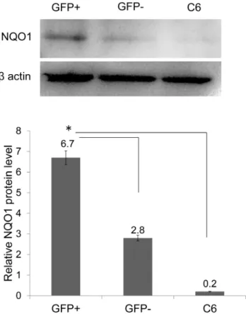
Figure 5. Expression of NQO1 in GFP-positive cells after transfection. Quantification of western blots revealed that GFP-positive cells expressed NQO1 protein prior to treatment with H2O2
when compared with GFP-negative cells and untreated C6 cells (*P,0.05).
doi:10.1371/journal.pone.0086951.g005
Parinaric Acid was added and the incubation was continued for 1 h (total incubation time was 6 h). Cells were collected with PBS and the fluorescence was measured using the BD LSRFortessaTM
apparatus (Becton Dickinson) and analyzed using FlowJo TM Software (Tree Star Inc.) The excitation and emission wavelengths were 320 and 420 nm, respectively.
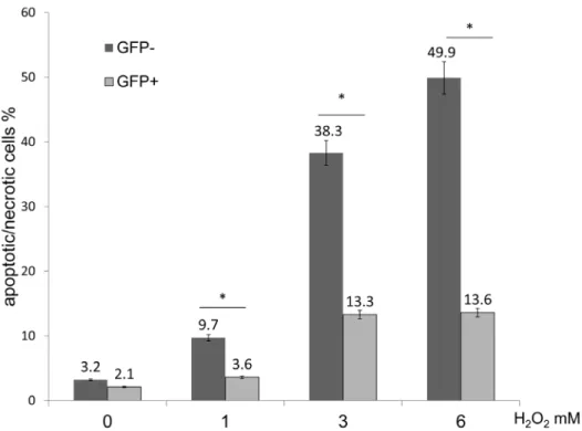
Figure 6. Evaluation of GRP78 overexpressing cells following H2O exposure.FACS analysis of GFP-negative and -positive cells labeling
positive for Annexin V/PI following treatment with H2O2. The percentage of GFP-positive cells that were AP-positive significantly decreased when
compared with GFP-negative cells following treatment with 1, 3 and 6 mM H2O2(*P,0.05).
doi:10.1371/journal.pone.0086951.g006
Figure 7. Evaluation of GRP78 overexpression on cell lipid peroxidation following H2O2 exposure. FACS analysis of the mean
fluorescence intensity of cis-Prinaric acid in GFP-negative and -positive cells following treatment with H2O2. The cis-Parinaric Acid mean fluorescence
intensity of GFP-positive cells was significantly higher when compared with GFP-negative cells following treatment with 1, 3 and 6 mM H2O2
(*P,0.05).
GSH Assay
Cellular GSH levels were monitored and analyzed using the ThiolTracker Violet GSH detection reagentH(Molecular Probes/
Life Technologies) according to the manufacturer’s protocol. Cells were seeded in 6 cm dishes, and treated with 1–6 mM H2O2for
6 h. After designated treatment times, cells were washed with
Figure 8. GSH expression levels in GRP78 overexpressing cells following H2O2treatment. FACS analysis of the mean fluorescence
intensity of GSH in GFP-negative and -positive cells following treatment with H2O2. The GSH mean fluorescence intensity of GFP-positive cells was
significantly higher when compared to GFP-negative cells following treatment with 1, 3 and 6 mM H2O2(*P,0.05).
doi:10.1371/journal.pone.0086951.g008
Figure 9. Evaluation of NQO1 expression levels in GRP78 overexpressing cells following H2O2exposure.FACS analysis of the mean
fluorescence intensity of NQO1 in GFP-negative and -positive cells following treatment with H2O2. The NQO1 mean fluorescence intensity of
GFP-positive cells was significantly higher when compared to GFP-negative cells following treatment with 1, 3 and 6 mM H2O2(*P,0.05).
doi:10.1371/journal.pone.0086951.g009
Dulbecco’s PBS and incubated in Dulbecco’s PBS containing 10mM (final concentration) ThiolTracker Violet for 30 min at 37uC. Cells were measured using the BD LSRFortessaTM apparatus (Becton Dickinson) and analyzed using FlowJo TM Software (Tree Star Inc.). The excitation and emission wave-lengths were 404 and 526 nm, respectively.
Statistical Analysis
Data are expressed as the mean 6 standard deviation (SD). Analysis of variance and the Student’s t-test were used to assess
differences between the means of test samples and controls. Significance was set atP,0.05.
Results
Cell Damage and GRP78 Expression Following H2O2
Treatment
Flow cytometric (FACS) analysis revealed a significant increase in the number of apoptotic/necrotic cells, namely annexin V/PI-positive cells (AP-V/PI-positive cells) following 1–6 mM H2O2treatment
at 6,24 h compared to that in the controls (Fig. 2A). The number
of GRP78-expressing cells following treatment with H2O2did not
significantly increase compared to that in untreated C6 cells at any concentration (Fig. 2A). Similarly, western blot analysis revealed that the expression of the GRP78 protein in the groups treated with 1–6 mM H2O2 for 6 and 24 h did not increase obviously
when compared to untreated C6 cells (n = 3; Fig. 2B).
Evaluation of cis-Parinaric Acid and GSH Following H2O2
Treatment
FACS analysis revealed that the mean fluorescence intensity of cis-Parinaric acid significantly decreased following treatment at all concentrations of H2O2 when compared to untreated C6 cells
(n = 5; P,0.05) (Fig. 3A). This result indicated that H2O2causes
lipid peroxidation of the cell membrane. Similarly, the mean fluorescence intensity of GSH significantly decreased following treatment at all concentrations of H2O2 when compared with
untreated C6 cells (n = 4; P,0.05) (Fig. 3B). These results indicated that intracellular GSH expression levels reduced when cells were subjected to excessive oxidative stress.
Expression of GRP78 Protein and NQO1 in GFP-positive Cells after Transfection
At 36 h post-transfection, an average of 43% of cells was GFP-positive (Fig. 4A). Quantitation of immunoblots of GFP-GFP-positive cell extracts revealed a significant increase in GRP78 protein levels when compared with GFP-negative cells and non-treated C6 cells (n = 3, P,0.05) (Fig. 4B). This result showed that GFP-positive cells overexpress GRP78 protein before exposure to H2O2when
compared with GFP-negative cells and non-treated C6 cells. Moreover, we observed that NQO1 expression significantly increased in GFP-positive cell extracts when compared with GFP-negative cells and non-treated C6 cells (n = 3, P,0.05) (Fig. 5).
Evaluation of GRP78 Overexpressing Cells Following H2O
Exposure
After transfection, a mixture of cells consisting of GFP-positive and -negative cells were incubated for 36 h with 10% (v/v) FBS/ DMEM. Cells were then treated with 1–6 mM H2O2for 6 h. To
evaluate the protective effect of GRP78, apoptotic/necrotic cells were labeled with annexin V and PI, and the ratio of AP-positive cells in GFP-positive and -negative cells were analyzed byFACS.
FACS analysis revealed that the percentage of GFP-positive cells that were AP-positive significantly decreased when compared with GFP-negative cells following exposure to 1,3 and 6 mM of H2O2
(GFP- positive % vs. GFP- negative % at 0 mM = 3.2 vs. 2.1; at 1 mM = 3.6 vs.9.7; at 3 mM = 13.3 vs. 38.3; at 6 mM = 13.6 vs.49.9; at 9 mM = 28.1 vs. 66.3) (n = 5, P,0.05) (Fig. 6). These findings indicated that GRP78 overexpressing cells could survive longer than non-GRP78 overexpressing cells following H2O2
exposure.
Evaluation of GRP78 Overexpression on Cell Lipid Peroxidation Following H2O2Exposure
As described above, a mixture of cells consisting of GFP-positive and -negative cells were treated with H2O21–6 mM for 6 h, and
labeled with cis-Pariaric acid to evaluate lipid peroxidation of cell membranes. FACS revealed that the mean fluorescence intensity of cis-Pariaric acid in GFP-positive cells was significantly higher than that in GFP-negative cells following H2O2exposure at 1, 3
and 6 mM (GFP-positive vs. GFP-negative at 0 mM = 1458 vs. 1287; at 1 mM = 1185 vs.1027; at 3 mM = 1016 vs. 631; at 6 mM = 880 vs. 446) (n = 5, P,0.05) (Fig. 7). These findings indicate that GRP 78 overexpressing cells can suppress lipid peroxidation more than non-GRP78 overexpressing cells following H2O2exposure.
GSH Expression Levels in GRP78 Overexpressing Cells Following H2O2Treatment
We observed GSH expression levels in glutathione using FASC analysis. FACS revealed that the mean fluorescence intensity of GSH in GFP-negative cells significantly decreased when com-pared with GFP-positive cells following exposure to 1, 3 and 6 mM of H2O2(GFP-positive vs. GFP-negative at 0 mM = 62660
vs. 41590; at 1 mM = 56450 vs. 33050; at 3 mM = 60250 vs. 26850; at 6 mM = 29030 vs. 11360) (n = 5; P,0.05) (Fig. 8). Thus, intracellular GSH expression levels in GRP78 overexpressing cells were significantly higher than that of non-GRP78 overexpressing cells.
Evaluation of NQO1 Expression Levels in GRP78 Overexpressing Cells Following H2O2Exposure
The mean fluorescence intensity of NQO1 in GFP-negative cells significantly decreased when compared with GFP-positive cells following 1, 3 and 6 mM of H2O2( positive vs.
GFP-negative at 0 mM = 7190 vs.6530; at 1 mM = 3120 vs. 2050 50; at 3 mM = 3830 vs. 2210; at 6 mM = 2740 vs. 2570) (n = 3, P,0.05) (Fig. 9). These result showed that levels of NQO1 in GRP78 overexpressing cells were higher than non-GRP78 overexpressing cells.
Discussion
involving mainly the maintenance of calcium homeostasis [16]. Meanwhile, it was reported that GRP78 expression did not increase when cells were exposed to H2O2, which suggested that
H2O2exposure may not induce the ER stress pathway [24]. In our
study, an increase in GRP78 expression in C6 cells was not observed after treatment with H2O2, however, the viability of cells
decreased. Considering previous reports, our results suggest that GRP78 itself could play an important role in protecting cells against H2O2 injury regardless of whether the pathways that
mediate GRP78 expression respond to their extracellular stimuli. H2O2 causes cytotoxicity via the formation of more potent
oxidants including OH?, which causes lipid peroxidation of the cell membrane [7,30]. Lipid peroxidation disrupts the normal structure of cellular and subcellular membranes. In addition, the process produces byproducts such as 4-hydroxynonenal (4-HNE) or acrolein, both of which bind to proteins and damage their structure and function [30,31]. The present results show that GRP78 overexpressing cells suppress lipid peroxidation and may contribute to cell survival following H2O2treatment. These data
suggest that GRP78 can promote the expression of some antioxidants and may contribute to the protection of cells against H2O2injury.
While a number of antioxidants are involved in the detoxifica-tion of H2O2, GSH is the primary defense against H2O2 [18].
GSH inhibits lipid peroxidation initiation by scavenging OH?or other ROS. Moreover, GSH also serves as a co-factor for GSH peroxidases that remove H2O2[20,22]. It was reported that GSH
was useful for curtailment of lipid peroxidation damage in acute spinal cord injury [20]. The ratio of GSH reduces when an increase in ROS induces ER stress [31,32]. In our results, when cells were exposed to H2O2, GSH expression in GRP78
overexpressing cells was high when compared with non-GRP78 overexpressing cells. These results suggest that the increase of GRP78 by gene transfection may contribute to the increase in GSH or inhibit GSH consumption, thus leading to cell survival.
We observed the influence of GRP78 on NQO1 in this study. NQO1 catalyzes the electron reduction of quinone and quinoid compounds to hydroquinones, thus limiting the formation of semiquinone radicals, and the subsequent generation of ROS [19,33]. According to reports regarding the role of NQO1, H2O2
-dependent formation of reactive oxygen intermediates was shown to be reduced following treatment with neuroprotective agents that induce NQO1 expression [34,35]. In our results, NQO1 expression levels in GRP78 overexpressing cells was higher than in non-GRP78 overexpressing cells. This phenomenon continued following H2O2 exposure. These findings indicate that GRP78
may be advantageous to the expression of NQO1.
NQO1 has the ability to use both NADPH and NADH equally efficiently, thus NQO1 may contribute to the regulation of the redox balance by modulating reduced/oxidized pyridine nucleo-tide ratios [36]. NADPH is required to reduce oxidized glutathione (GSSG) to GSH [37]. This NQO1 role appears not to contradict our results that GSH expression levels in GRP78 overexpressing cells were higher than non- GRP78 overexpressing cells.
Taken together, this present study suggests that GRP78 plays an important role in protecting glial cells against H2O2 toxicity by
regulating GSH and NQO1 expression. However, there are several pathways and factors related to GRP78 expression in cells and further studies are required to understand the mechanisms involved and the direct relationship between GRP78, GSH and NQO1 in order for molecular/pharmacological treatments of neurotrauma or neurodegenerative diseases to be developed.
Author Contributions
Conceived and designed the experiments: K. Suyama K. Sakabe MW. Performed the experiments: K. Suyama YO. Analyzed the data: K. Suyama YO. Contributed reagents/materials/analysis tools: AO JM TI HT. Wrote the paper: K. Suyama.
References
1. Jia Z, Zhu H, Li J, Wang X, Misra H, et al. (2012) Oxidative stress in spinal cord injury and antioxidant-based intervention. Spinal Cord 50: 264–274. 2. Halliwell B (1992) Reactive oxygen species and the central nervous system.
J Neurochem 59: 1609–1623.
3. Liu D, Liu J, Wen J (1999) Elevation of hydrogen peroxide after spinal cord injury detected by using the Fenton reaction. Free Radic Biol Med 27: 478–482. 4. Schmidley JW (1990) Free radicals in central nervous system ischemia. Stroke
21: 1086–1090.
5. Aragno M, Parola S, Brignardello E, Mauro A, Tamagno E, et al. (2000) Dehydroepiandrosterone prevents oxidative injury induced by transient ischemia/reperfusion in the brain of diabetic rats. Diabetes 49: 1924–1931. 6. Zhu D, Tan KS, Zhang X, Sun AY, Sun GY, et al. (2005) Hydrogen peroxide
alters membrane and cytoskeleton properties and increases intercellular connections in astrocytes. J Cell Sci 118: 3695–3703.
7. Bao F, Liu D (2004) Hydroxyl radicals generated in the rat spinal cord at the level produced by impact injury induce cell death by necrosis and apoptosis: protection by a metalloporphyrin. Neuroscience 126: 285–295.
8. Elmann A, Telerman A, Mordechay S, Erlank H, Ofir R (2012) Antioxidant and astroprotective effects of a Pulicaria incisa infusion. Oxid Med Cell Longev 2012: 157598.
9. Larner SF, Hayes RL, Wang KK (2006) Unfolded protein response after neurotrauma. J Neurotrauma 23: 807–829.
10. Penas C, Guzman MS, Verdu E, Fores J, Navarro X, et al. (2007) Spinal cord injury induces endoplasmic reticulum stress with different cell-type dependent response. J Neurochem 102: 1242–1255.
11. Ni M, Lee AS (2007) ER chaperones in mammalian development and human diseases. FEBS Lett 581: 3641–3651.
12. Kozutsumi Y, Segal M, Normington K, Gething MJ, Sambrook J (1988) The presence of malfolded proteins in the endoplasmic reticulum signals the induction of glucose-regulated proteins. Nature 332: 462–464.
13. Higo T, Hamada K, Hisatsune C, Nukina N, Hashikawa T, et al. (2010) Mechanism of ER stress-induced brain damage by IP(3) receptor. Neuron 68: 865–878.
14. Suyama K, Watanabe M, Sakabe K, Okada Y, Matsuyama D, et al. (2011) Overexpression of GRP78 protects glial cells from endoplasmic reticulum stress. Neurosci Lett 504: 271–276.
15. Gomer CJ, Ferrario A, Rucker N, Wong S, Lee AS (1991) Glucose regulated protein induction and cellular resistance to oxidative stress mediated by porphyrin photosensitization. Cancer Res 51: 6574–6579.
16. Yu Z, Luo H, Fu W, Mattson MP (1999) The endoplasmic reticulum stress-responsive protein GRP78 protects neurons against excitotoxicity and apoptosis: suppression of oxidative stress and stabilization of calcium homeostasis. Exp Neurol 155: 302–314.
17. He S, Yaung J, Kim YH, Barron E, Ryan SJ, et al. (2008) Endoplasmic reticulum stress induced by oxidative stress in retinal pigment epithelial cells. Graefes Arch Clin Exp Ophthalmol 246: 677–683.
18. Jia Z, Zhu H, Misra HP, Li Y (2008) Potent induction of total cellular GSH and NQO1 as well as mitochondrial GSH by 3H-1,2-dithiole-3-thione in SH-SY5Y neuroblastoma cells and primary human neurons: protection against neurocy-totoxicity elicited by dopamine, 6-hydroxydopamine, 4-hydroxy-2-nonenal, or hydrogen peroxide. Brain Res 1197: 159–169.
19. Ross D, Kepa JK, Winski SL, Beall HD, Anwar A, et al. (2000) NAD(P)H:quinone oxidoreductase 1 (NQO1): chemoprotection, bioactivation, gene regulation and genetic polymorphisms. Chem Biol Interact 129: 77–97. 20. Lucas JH, Wheeler DG, Guan Z, Suntres Z, Stokes BT (2002) Effect of
glutathione augmentation on lipid peroxidation after spinal cord injury. J Neurotrauma 19: 763–775.
21. Yen CC, Ho TJ, Wu CC, Chang CF, Su CC, et al. (2011) Inorganic arsenic causes cell apoptosis in mouse cerebrum through an oxidative stress-regulated signaling pathway. Arch Toxicol 85: 565–575.
22. Grant CM, Perrone G, Dawes IW (1998) Glutathione and catalase provide overlapping defenses for protection against hydrogen peroxide in the yeast Saccharomyces cerevisiae. Biochem Biophys Res Commun 253: 893–898. 23. Timblin CR, Janssen YM, Goldberg JL, Mossman BT (1998) GRP78, HSP72/
73, and cJun stress protein levels in lung epithelial cells exposed to asbestos, cadmium, or H2O2. Free Radic Biol Med 24: 632–642.
24. Yokouchi M, Hiramatsu N, Hayakawa K, Okamura M, Du S, et al. (2008) Involvement of selective reactive oxygen species upstream of proapoptotic branches of unfolded protein response. J Biol Chem 283: 4252–4260. 25. Chakravarthi S, Jessop CE, Bulleid NJ (2006) The role of glutathione in
disulphide bond formation and endoplasmic-reticulum-generated oxidative stress. EMBO Rep 7: 271–275.
26. Hedley D, Chow S (1992) Flow cytometric measurement of lipid peroxidation in vital cells using parinaric acid. Cytometry 13: 686–692.
27. Yoshida T, Kondo T, Ogawa R, Feril Jr LB, Zhao QL, et al. (2008) Combination of doxorubicin and low-intensity ultrasound causes a synergistic enhancement in cell killing and an additive enhancement in apoptosis induction in human lymphoma U937 cells. Cancer Chemother Pharmacol 61: 559–567. 28. Oyadomari S, Mori M (2004) Roles of CHOP/GADD153 in endoplasmic
reticulum stress. Cell Death Differ 11: 381–389.
29. Tiffany-Castiglioni E, Qian Y (2012) ER chaperone-metal interactions: links to protein folding disorders. Neurotoxicology 33: 545–557.
30. Hall ED (2011) Antioxidant therapies for acute spinal cord injury. Neurother-apeutics 8: 152–167.
31. Vladykovskaya E, Sithu SD, Haberzettl P, Wickramasinghe NS, Merchant ML, et al. (2012) Lipid peroxidation product 4-hydroxy-trans-2-nonenal causes
endothelial activation by inducing endoplasmic reticulum stress. J Biol Chem 287: 11398–11409.
32. Mohammad MK, Avila D, Zhang J, Barve S, Arteel G, et al. (2012) Acrolein cytotoxicity in hepatocytes involves endoplasmic reticulum stress, mitochondrial dysfunction and oxidative stress. Toxicol Appl Pharmacol 265: 73–82. 33. Dinkova-Kostova AT, Talalay P (2010) NAD(P)H:quinone acceptor
oxidore-ductase 1 (NQO1), a multifunctional antioxidant enzyme and exceptionally versatile cytoprotector. Arch Biochem Biophys 501: 116–123.
34. Bar-Am O, Weinreb O, Amit T, Youdim MB (2009) The novel cholinesterase-monoamine oxidase inhibitor and antioxidant, ladostigil, confers neuroprotec-tion in neuroblastoma cells and aged rats. J Mol Neurosci 37: 135–145. 35. Lim JH, Kim KM, Kim SW, Hwang O, Choi HJ (2008) Bromocriptine activates
NQO1 via Nrf2-PI3K/Akt signaling: novel cytoprotective mechanism against oxidative damage. Pharmacol Res 57: 325–331.
36. Long DJ, 2nd, Gaikwad A, Multani A, Pathak S, Montgomery CA, et al. (2002) Disruption of the NAD(P)H:quinone oxidoreductase 1 (NQO1) gene in mice causes myelogenous hyperplasia. Cancer Res 62: 3030–3036.