J of Evolution of Med and Dent Sci/ eISSN- 2278-4802, pISSN- 2278-4748/ Vol. 3/ Issue 22/June 02, 2014 Page 6053
STUDY OF ANATOMICAL VARIATIONS IN THE SHAPE OF SUPRASCAPULAR
NOTCH IN DRIED HUMAN SCAPULAE AND ITS CLINICAL SIGNIFICANCE
Udayasree L1, Siva Prasad G. V2, Lakshmi V. V. V3
HOW TO CITE THIS ARTICLE:
Udayasree L, Siva Prasad G. V, Lakshmi V. V. V. Study of Anatomical Variations in the Shape of Suprascapular Notch in Dried Human Scapulae and its Clinical Significance . Journal of Evolution of Medical and Dental Sciences 2014; Vol. 3, Issue 22, June 02; Page: 6053-6057, DOI: 10.14260/jemds/2014/2704
ABSTRACT: AIM: The suprascapular nerve arises from the upper trunk of brachial plexus, passes through the suprascapular notch and then through the spinoglenoid notch and supplies the supraspinatus and the infraspinatus muscles. Due to the presence of anatomical variations in the shape of the suprascapular notch, there may be compression of the suprascapular nerve which leads to suprascapular nerve entrapment syndrome. The present study is a simple method to classify the suprascapular notch of dried human scapulae and its clinical significance. MATERIALS & METHOD: 42 dried human scapulae of both sides were examined for variations in the shape of suprascapular notch. RESULT: Mainly three different shapes were observed. They are U, V & J. Some scapulae are having suprascapular foramen instead of notch and some are having absent suprascapular notch. KEYWORDS: Suprascapular notch, suprascapular nerve, Scapula and Anatomical variations. MESHTERMS: Scapula: A02.835.232.087.783
INTRODUCTION: The scapula is a flat bone of shoulder girdle that lies on the posterolateral aspect of the thorax. The lateral part of superior border of scapula contains suprascapular notch which is bridged by superior transverse scapular ligament converting into foramen. This foramen transmits suprascapular nerve to the supraspinous fossa.¹ It supplies motor branches to the supraspinatus, infraspinatus, and sensory branches to rotator cuff muscles and ligamentous structures of the shoulder and acromio-clavicular joints. Complete or partial ossification of superior transverse scapular ligament and anatomical variations in the shape of suprascapular notch leads to nerve compression during movements of shoulder. Suprascapular nerve entrapment is an uncommon cause for shoulder pain and weakness. It was initially described by Thompson and Kopell.²
Many authors proposed different classifications based on measurements and shapes of suprascapular notch. Iqbal et al³ described three types of shapes of suprascapular notch- U, V & J.
N⁷tsis et ⁷l ⁹l⁷ssified the supr⁷s⁹⁷pul⁷r not⁹hes ⁸⁷sed on verti⁹⁷l ⁷nd horizontal diameters of the notch into 5 types. In the present study, we examined the anatomical variations of the shape of suprascapular notch of dried human scapulae which is useful for clinicians to correlate the type of notch causing suprascapular nerve entrapment syndrome.
MATERIALS & METHODS: The present study was conducted on 42 dried human scapulae of both sides from Department of Anatomy, NRIIMS, Sangivalasa and were examined for anatomical variations in the shape of suprascapular notch, absence of notch and partial or complete ossification of superior transverse scapular ligament.
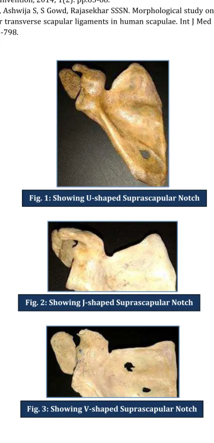
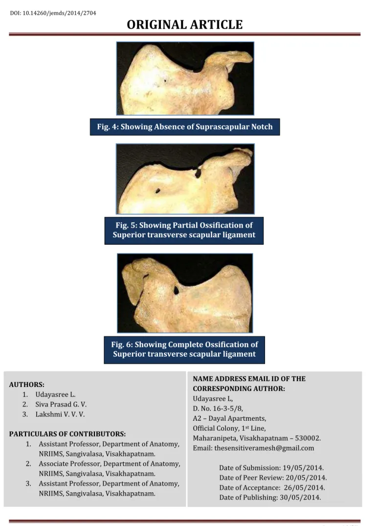
J of Evolution of Med and Dent Sci/ eISSN- 2278-4802, pISSN- 2278-4748/ Vol. 3/ Issue 22/June 02, 2014 Page 6054 superior transverse scapular ligament. Absence of suprascapular notch was also observed. Out of 42 scapulae, 20 scapulae had U-shaped suprascapular notch (Fig. 1), 9 scapulae had J-shaped suprascapular notch (Fig. 2) and 2 scapulae had V-shaped suprascapular notch (Fig. 3). Absence of notch was noted in 5 scapulae (Fig. 4). 2 scapulae had partial ossification of superior transverse scapular ligament (Fig. 5) and 4 scapulae had complete ossification of superior transverse scapular ligament forming suprascapular foramen (Fig. 6). The number and the percentage of different shapes of suprascapular notch and ossification of superior transverse scapular ligament were shown in Table 1.
Shapes Number & Percentage
U 20(47.6%)
J 9(21.4%)
V 2(4.7%)
Absence of Notch 5(11.9%)
Superior Transverse Scapular ligament
Partial Ossification 2(4.7%)
Complete Ossification 4(9.5%)
Table1: Shapes of Suprascapular notch and Ossification of superior transverse scapular ligament
DISCUSSION: The suprascapular notch being a fibro-osseous neural foramen, is the most common site of entrapment of the suprascapular nerve because of an abnormal configuration of the suprascapular notch, traction injury or a ganglion compressing the nerve. ’
In the present study, out of 42 scapulae, 20(47.6%) had U-shaped suprascapular notch with approximately parallel sides and a rounded base. 9(21.4%) scapulae had J-shaped notch with one side short and another side long. 2(4.7%) scapulae had V-shaped notch with convergence of two sides toward a narrow base. According to Iqbal et al, ³ U-shaped notches were 13.2%, V-shaped notches were 20%, J-shaped notches were 22% and 45out of 200 scapulae are without suprascapular notch in the population of Pakistan. In the present study, 5(11.9%) scapulae were not having suprascapular notch. No indentations at the site of notch were found. In a case report by Rekha, occasional absence of suprascapular notch was observed.
In the present study, 2(4.7%) scapulae had partial ossification and 4(9.5%) had complete ossification of superior transverse scapular ligament. Complete ossification leads to the formation of suprascapular foramen. Iqbal et al³ did not mention about the ossification of transverse scapular ligament and foramen. In the study done by Patel et al, 3.75%scapulae showed suprascapular foramen due to complete ossification of transverse scapular ligament. According to the study of Kannan et al, 4% of scapulae had partial and 10% of scapulae had complete ossification of transverse scapular ligament. Most common radiological finding in suprascapular nerve entrapment syndrome is shallowness and deformity of the suprascapular notch.¹⁰
J of Evolution of Med and Dent Sci/ eISSN- 2278-4802, pISSN- 2278-4748/ Vol. 3/ Issue 22/June 02, 2014 Page 6055 to the study done by Vasudha et al¹³ in Indian population, symmetrical U- shaped suprascapular notch was the most common anatomical variation (34.78%) and coexistence of suprascapular notch and suprascapular foramen was least common (0.88%). In the present study also U – shaped notch was most common (47.6%). The narrow V- shaped notch, absence of notch and also the ossification of the superior transverse scapular ligament were commonly associated with suprascapular nerve entrapment syndrome.
CONCLUSION: Knowledge of anatomical variations of suprascapular notch is useful for clinicians for better correlation and early diagnosis of suprascapular nerve entrapment syndrome. Shallow and absence of suprascapular notch and partial or complete ossification of transverse scapular ligament may predispose to entrapment of suprascapular nerve.
REFERENCES:
1. Roger W Soames. Skeletal system: In Williams PL., Bannister LH., Berry MM., Collins P., Dyson M., Dussek JE., Ferguson MWJ: Gr⁷y’s An⁷tomy, 38thed. London, Churchill – Livingstone, 2004;
chapter 6, pp.615-618.
2. Thompson W, Kopell H. Peripheral entrapment neuropathies of the upper extremity. N Engl J Med 1959; 260: pp.1261-1265.
3. Natsis K, Totlis T, Tsikaras P, Appell HJ, Skandalakis K. Proposal for classification of the suprascapular notch: a study on 423 dried scapulas. Clinical Anatomy, 2007; Vol.20: pp.135-139.
4. Iqbal K, Iqbal R, Khan SG. Anatomical variations in the shape of suprascapular notch of scapula. J Morphol Sci, 2010; 27(1): pp.1-2.
5. Clein L J. Suprascapular entrapment neuropathy. J Neurosurg 1975; 43: p.337.
6. Rengachary S, Neff JP, Singer PA, Brackettc F. Suprascapular nerve entrapment neuropathy: a clinical, anatomical and comparative study. Clinical study, Neurosurgery, 1979; vol.5: pp.441-446.
7. Rekha BS. Complete absence of suprascapular notch – A case report. JEMDS.2013; 2(1): pp.19-22.
8. Pragna Patel, SV Patel, SM Patel, Badal Jotania, Sanjay Chavda, Dhara Patel. Study of variations in the shape of the suprascapular notch in Dried Human Scapula. Int J Biol Med Res. 2013; 4(2): pp.3162-3164.
9. Usha Kannan, N. S. Kannan, J. Anbalagan, Sudha Rao. Morphometric study of Suprascapular Notch in Indian dry scapulae with specific reference to the incidence of completely ossified superior transverse scapular ligament. Journal of Clinical and Diagnostic Research, 2014; 8(3): pp.7-10.
10.Edeland HG, Zachrisson BE. Fracture of the scapular notch associated with lesion of the suprascapular nerve. Acta Orthop Scand, 1975; 46: p.758.
J of Evolution of Med and Dent Sci/ eISSN- 2278-4802, pISSN- 2278-4748/ Vol. 3/ Issue 22/June 02, 2014 Page 6056 12.Saritha S. Coexistence of suprascapular notch and suprascapular foramen: A rare anatomical variation and its clinical correlation – A case report. International Journal of Medical Science and Clinical Invention, 2014; 1(2): pp.65-68.
13.Vasudha TK, Ashwija S, S Gowd, Rajasekhar SSSN. Morphological study on suprascapular notch and superior transverse scapular ligaments in human scapulae. Int J Med Res Health Sci. 2013; 2(4): pp.793-798.
Fig. 1: Showing U-shaped Suprascapular Notch
Fig. 2: Showing J-shaped Suprascapular Notch
J of Evolution of Med and Dent Sci/ eISSN- 2278-4802, pISSN- 2278-4748/ Vol. 3/ Issue 22/June 02, 2014 Page 6057 AUTHORS:
1. Udayasree L. 2. Siva Prasad G. V. 3. Lakshmi V. V. V.
PARTICULARS OF CONTRIBUTORS:
1. Assistant Professor, Department of Anatomy, NRIIMS, Sangivalasa, Visakhapatnam.
2. Associate Professor, Department of Anatomy, NRIIMS, Sangivalasa, Visakhapatnam.
3. Assistant Professor, Department of Anatomy, NRIIMS, Sangivalasa, Visakhapatnam.
NAME ADDRESS EMAIL ID OF THE CORRESPONDING AUTHOR: Udayasree L,
D. No. 16-3-5/8, A2 – Dayal Apartments, Official Colony, 1st Line,
Maharanipeta, Visakhapatnam – 530002. Email: thesensitiveramesh@gmail.com
Date of Submission: 19/05/2014. Date of Peer Review: 20/05/2014. Date of Acceptance: 26/05/2014. Date of Publishing: 30/05/2014. Fig. 4: Showing Absence of Suprascapular Notch
Fig. 5: Showing Partial Ossification of Superior transverse scapular ligament