SYNTHESIS, CRYSTAL STRUCTURE, SPECTROSCOPY PROPERTIES AND POTENTIAL ANTIMICROBIAL
POTENTIALITIES OF A NEW SYNTHETIC COMPOUND: AMINO- CHLOROPYRIDINIUM DIAQUA DIOXALATO
IRON(III)
Jawher Abdelhak
1, Essghaier Badiaa
2,3*, Toukebri Nourchene
2, Amani Naouar
2, Rebib Hanene
2, Mohamed Faouzi Zid
1, Najla
Sadfi-Zouaoui
2Address(es): Dr Essghaier badiaa,
1Laboratoire de Matériaux et Cristallochimie, Département de chimie, Faculté des Sciences, 20λ2 El Manar, Tunis, Tunisie.
2Laboratoire Microorganismes et Biomolécul
es Actives, Faculté des Sciences, 20λ2 El Manar, Tunis, Tunisie.
3Département de Biologie, Faculté des Sciences et Techniques Sidi-Bouzid, Tunisie.
*Corresponding author: badiaaessghaier@gmail.com
ABSTRACT
Keywords: Iron (III) complex, antifungal antibacterial activity, spectroscopy studies, single crystal structure
INTRODUCTION
Several years ago, numerous oxalate complexes have been synthesized and investigated due to their extensive application in various fields such as biological (Haikarainen et al., 2001) and even industrial (Ferbinteanu et al., 2005). This is due to the ability of the oxalate ligand to transmit efficiently magnetic interactions through its bridging mode (Jia et al., 2007). His planar shape, its negative charge and its good donor ability due to the presence of four oxygen donors make this ligand very appropriate to build coordination polymers in its interaction with metal ions. The versatility of the oxalate as a ligand is well illustrated by the variety of coordination modes. Further, because of our interest in the magnetic properties of polymeric three-dimensionally linked complexes with chelating oxalate ions as bridging ligand, further studies have been extended to the synthesis transition metal compounds containing oxalate and its derivatives
(Zhang et al., 2007).
A powerful synthetic strategy to design such materials is supramolecular chemistry based on self assembly processes of two different components (Zhang
et al., 2007). In fact, this second ligand can contribute to the cohesion of the
structure by acting as a hydrogen bond donor through the two nitrogen atoms. Additional stability can also be offered by π-π stacking interaction of pyridine rings (Schott et al., 2011). In this context; we quote the protonated 2-amino-5-chloropyridinium as a cationic counter-ion.
The development of multiple antibiotic resistances is a global problem. It is necessary to find new tools whose mechanisms of action differ from those of currently used antibiotics (Spellberg et al., 2004). An active area of research concerns deepening knowledge of chemotherapeutic activity in various pathological conditions, mechanisms of action, development of resistance, kinetics and untoward effects of the available drugs in order to achieve the best utilization (Sensi,1979). Prompted by these observations and in continuation of
the author’s work on the synthesis of new synthetic compounds (Essghaier et al.,
2014), the authors report herein the use of the amino- chloropyridinium diaqua
dioxalato iron (III) to evaluate their antimicrobial activities. Many researches are also directed at identifying the proper use of combinations of antimicrobials to define the synergistic effect or inhibition of resistant strain selection. Finally there are continuous research efforts toward the development of new agents to overcome the drawbacks of the available ones. This problem is approached mainly in two ways, either by modification of the present antimicrobial drugs or by search for completely new entities through various screening sophistications. In the light of data mentioned above, in this work, we aimed at the synthesis of new compounds of amino- chloropyridinium diaqua dioxalato iron (III) and the in vitro estimation of its potential antimicrobial activity against microorganisms such as Gram-positive and Gram-negative bacteria, yeasts and fungi. In this frame, the crystal structure of a shape of sulfanilamide has been widely studied in this work (Scheme 1).
Fe
O O
O O
OH2
OH
C
C O
O C
C O O
-NH+
Cl NH2
.H2O
Scheme 1 The title compound.
We report herein the synthesis and the physicochemical characterization of a new mixed-ligand iron(III) complex of formula (C5H6ClN2)[Fe(C2O4)2(H2O)2].2H2O. This compound has been prepared by slow evaporation at room temperature and characterized by single crystal X-ray diffraction. It has been characterized by IR and UV-VIS spectra and thermal analysis (TG and DTA). In this compound, the iron ion has a slightly distorted square bipyramidal environment, coordinated by two chelating oxalate ion and two water
molecules. Structural cohesion is established essentially by π-π interactions between the rings of pyridine groups and intermolecular
hydrogen bonds connecting the ionic entities and uncoordinated water molecules.
In vitro antimicrobial activities of the amino- chloropyridinium diaqua dioxalato iron (III) against pathogenic fungi, yeast and bacteria were studied in this work. On the whole, our new compound has high antibacterial activities against Pseudomonas aeruginosa, Staphylococcus aureus and Listeria innocua. The amino- chloropyridinium diaqua dioxalato iron (III) used at 200µg m-1, can reduce
Candida albicans survival of about 45.45%, and destruct hyphe mycelial of Trichophyton rubrum. High lysozyme activities were
expressed especially against Listeria innocua with 17 times more than Staphylococcus aureus. The minimal inhibitory concentrations (MIC) are ranging from 16 µg ml-1for bacteria to 256 µg ml-1 for yeast and IC50 values varying from 1.44 to 10.45 µg ml-1 for bacteria and 45.8 for yeast.
ARTICLE INFO
Received 10. 10. 2014 Revised 14. 10. 2014 Accepted 14. 10. 2014 Published 1. 12. 2014
Regular article
MATERIALS AND METHODS
Chemistry
Analytical and physical measurements
All chemicals were commercially available and were used without further purification. TG/DTA 92 SETARAM thermal analyzer was employed for the investigation of the thermal behavior in our atmosphere from room temperature
to 600°C and UV-Vis spectrum was recorded on a Perkin Elmer UV/Vis
spectrometer Lambda 20 in the range 200-700 nm.
The presence of the elements was confirmed by qualitative energy dispersive spectroscopy (EDX) analysis, performed on JEOL-JSM 5400 scanning electron microscope. The infrared (IR) spectrum was recorded within the 4000–400 cm−1region on a FT-IR Paragon 1000 PC spectrometer using KBr pellets.
Synthesis
This compound was prepared by the reaction of iron nitrate Fe(NO3)3.9H2O, 2-amino-5-chloropyridine and oxalic acid dihydrate respectively (1:1:2) in ethanol. The resulting mixture was heated to boiling point and stirred for three hours. A red precipitate formed immediately. After two weeks single red crystals were obtained by slow evaporation from aqueous solution at room temperature. Anal. Found: C,17.12; H,2.53; Fe,11.05; N,4.17; O,35.46; Cl,6.14 Calc. for C9H14FeN2O12Cl (M.W. 433.52): C,17.3; H,2.2; Fe,11.2; N,4.5; O,35.3; Cl,6.2%
Crystal structure determinations and refinements
A prismatic red crystal (0.3×0.27×0.18 mm) is selected for the structural analysis. Diffraction data were collected at 293(2) K with Enraf–Nonius CAD4 automatic four-circle equipped with graphite monochromator using Mo Kα( =0.71073Å) radiation with the w-2θ technique. Unit-cell parameters and orientation matrix of title compoundwere determined by least squares treatment of the setting angles of 25 reflections on the range 10° < θ< 15°.
The structure is solved by standard Patterson methods and refined by the full-matrix least-squares method on F2 for 287 refined parameters. The computations were performed with SHELXS 97 and SHELXL 97 (Sheldrick et al., 1997). All non-hydrogen atoms were treated anisotropically. Except the hydrogen atoms which O12 are calculated, the others were located from a difference synthesis.
IR Spectra
The infrared spectra of compounds exhibit characteristic bands for oxalate ligand. For, the characteristic bands of the oxalate bridging ligand appear in 1684, 1351 and 764 cm-1, corresponding to as(CO),
s (CO) and δ(O-C-O), respectively
(Marinescu et al., 2004). The region of the as(CO) and s (CO) stretching vibrations of the oxalate group often shows slight differences owing to the diverse coordination modes. The split bands are generally characteristic of the bidentate oxalate groups as terminal ligands (Jia et al., 2007). The strong and broad absorption band at 3500–3100 is attributed to the (OH) vibrations of water molecules in the crystal lattice as well as (C-H) and (N-H) (Wrobleski et al., 2004).
The peak located at 493 cm-1are assigned to (Fe–O) (Zheng et al., 1999). Additionally, the bands located in the region 1650 – 1400 cm-1 region, are assignable to C–C and C–N stretching vibration of pyridine groups (Li et al., 2006). Finally, the bands in the region 1250–600 cm-1 can be assigned to the C-Cl and ring deformation absorptions of chloropyridinium cation.
Thermal analysis
Thermal stability of the compound has been studied by differential thermal analysis (DTA) and thermogravimetry (TG) from room temperature to 600°C. Within this interval, several degradation steps were observed. In the interval between 80 and 155°C, the DTA trace shows an endothermic peak. The loss in weight (calculated 16.61%; found 16.20%) suggests that the compound losses four water molecules in two consecutive steps; the first loss corresponds to the weakly coordinated water molecules and the second loss of the coordinated ones. The next large step (ca. 68%) in the decomposition curve, in the temperature range of 220–450 °C, comprises the removal of pyridine cations and oxalate groups as a strongly exothermic process (Czakis-Sulikowska et al., 2000). This technique was used to check the number of water molecules as well as the nature of connections to the network of these molecules. These results are in perfect agreement with the structural study.
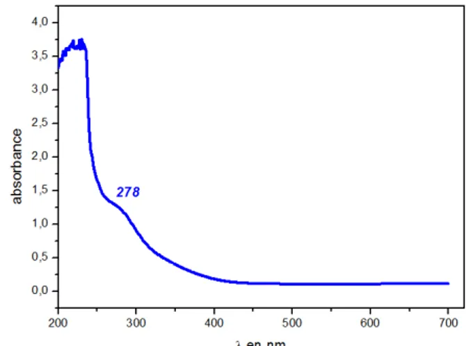
Electronic Spectra
Electronic spectroscope is obtained from ethanol solution. Figure 1 shows the electronic spectrum of compounds. The absorption spectra of the complex show very intense bands in the UV. The spectrum is dominated by one band in UV at 278 nm. This band can be attributed to oxalato-to-FeIII charge transfer
(Pozdnyakov et al., 2008). In the bibliography, many intense bands (not shown) are found between 210 and 250 nm, which can be assigned to pyridine n –π* and π –π* transitions (Snodin et al., 1999).
Figure 1 U.v./vis spectra of title compound in ethanol
Microbiology
Microorganisms and growth conditions
A list of microorganisms was used in this work in order to investigate the antimicrobial activity of the new compound amino-chloropyridinium dark dioxalato iron (III) described here was as follows: gram-negative bacteria
(Pseudomonas aeruginosaν Escherichia. Cole DH5α Acinetobacer spp,
Agrobacteium tumefaciens and Erwinia spp), gram-positive bacteria (Listeria innocua, Staphylococcus aureus), three yeasts (Candida albicans) and three strains of dermatophyte (Trichophyton rubrum); were taken from the culture collection of laboratory of Microorganisms and Biomolecules Actives, Faculty of Sciences in Tunisia. Phytopathogenic fungi: Fusarium oxysporum sp; Botrytis
cinerea, Penicilium, Phytophtora and Alternaria, were also employed. There
have been obtained from the laboratory of Biotechnology applied to the agriculture of the National institute for agronomic research of Tunisia (INRAT). Broth cultures were prepared from the above mentioned bacteria in Tryptone Soy Broth (TSB; 50 ml), inoculated with overnight stationary-phase cultures and held in 100 ml Erlenmeyer flask inside an orbital incubator (110 rpm, 37 °C and 30°C for Listeria innocua). TSB (25 ml) held within 100 ml Erlenmeyer flasks were used to prepare bacterial cultures for the suspension tests.
Chemicals. TSB and TSA were employed throughout the experiments with
bacteria. In the experiments with Candida albicans, yeast malt extract broth (YMB) and agar (YMA) were used. Potato Dextrose Agar (PDA) was used for experiments with fungi.
Agar diffusion method
The in vitro antimicrobial test utilized in the investigation is based on the diffusion method on agar plates (Collins et al., 1989), as previously detailed by us (Essghaier et al., 2014). Before use, the amino- chloropyridinium dark dioxalato iron (III) was diluted in distilled water, sterilized by filtration through a 0.2 m pore size filter and adjusted to the appropriate concentration tested. A 50 l aliquot of filtered compound was placed into paper discs. After overnight pre-diffusion at 4°C, the plates were incubated at appropriate temperature 37°C or 30°C for at least 24h, to develop inhibition zones which diameters were measured in mm.
Antifungal activity on PDA plates
The efficiency of the amino- chloropyridinium dark dioxalato iron (III) on the growth mycelial inhibition of fungi was assayed by applying a dual culture technique, in vitro on PDA plates. A 50µl of spore suspensions of each fungus adjusted at 106spores/ml, were placed in the center of the agar plate, at 2.5 cm apart were placed paper disk containing 50 µl of amino- chloropyridinium dark dioxalato iron (III) at 200, 500 or 1000µg/ml.
The plates with fungi were incubated for 5 days at 25 °C (7days for the dermatophyte Trichophyton rubrum), after overnight pre-diffusion at 4°C, than the diameter zone inhibition were measured. Percentage growth inhibition of fungi was calculated by the following formula as detailed by (Sadfi-Zouaoui et
al., 2008): Growth inhibition (GI %) = (R1-Rβ) ⁄ R1 * 100
Antifungal assay
were added to λ00 l of YM medium containing approximately 105 CFU of yeasts. The culture was then grown at 37°C on a shaker for about 48 h until the A600 of the untreated control was between 0.5 and 1.0. The optical density of the samples was measured at 600nm (A600), and growth inhibition was then determined as followsμ percent yeast survival = 100 × (A600 of test sample) /(A600 of negative control). Each data point was obtained in triplicate.
Determination of MIC and IC50
The MIC ( g/ml) denotes the lowest drug concentration that prevents the visible growth of test microorganisms, and IC50 ( g/ml) is the Concentration at which 50% inhibition of the response is seen. MIC and IC50 value were evaluated by using the serial double dilution method in the appropriate medium which is inoculated with a standardized number of microorganisms. The concentration of amino- chloropyridinium diaqua dioxalato iron (III) incubated with indicator
strain is given in g/ml. Each dilution of amino- chloropyridinium diaqua
dioxalato iron (III) affected in 1000µl of the appropriate medium, was inoculated by 100 µl of 106 UFC/ml of each indicator strain then the different culture tubes were incubated at appropriate temperature. Control tube containing 100 µl of 106 UFC/ml of each indicator strain, added to 1000µl of culture medium without amino- chloropyridinium diaqua dioxalato iron (III). MIC were estimated visually (absence of turbidity) and were determined with 3 independent measurements (Jorgensen et al., 2007).
Determination of bactericidal activity
The antimicrobial activity of the amino-chloropyridinium diaqua dioxalato iron (III) solutions was expressed in arbitrary units per ml (AU/ml) and it was determined by an agar diffusion assay as described by (Graciela et al., 1995). Briefly, a serial twofold dilution in sterile distilled water on the amino- chloropyridinium diaqua dioxalato iron (III) was prepared, and 50 l of each dilution were spotted onto a TSB agar soft plate seeded with about 105 CFU/ml of Staphylococcus aureus. The AU/ml was calculated asμ AU/ml = 1000Χ D/A Where: A is the volume of the amino- chloropyridinium diaqua dioxalato iron (III) aliquot spotted on agar plate (50 l in this case)ν D is the reciprocal of the highest dilution showing a clear inhibition of the indicator strain.
Lysozyme activity
The Lysozyme activity of the amino- chloropyridinium diaqua dioxalato iron (III) was assayed turbidimetrically by measuring the decrease in absorbance at 660 nm of a suspension of Staphylococcus. aureus (Ryazanova et al., 2005).
Mycelial hyphae destruction
Fungal culture was rinsed with distilled sterile water. After centrifugation at 9000rpm for 10min pellet (mycelium) was replaced in an Eppendorf tube containing an appropriate Tris-HCl buffer (0,01M, pH8), in order to obtain the same concentration of mycelial solution (expressed in mg/ml). 200µl of
1000µg/ml of the amino- chloropyridinium diaqua dioxalato iron (III) was added.
The mixture was incubated at 37°C for 14h. After that, optical density was measured at 540nm. Increase of OD compared to control tube (containing only mycelial suspension), make destruction of fungal hyphae by the amino- chloropyridinium diaqua dioxalato iron (III) (Ryazanova et al., 2005).
RESULTS AND DISCUSSION
Description of structure
The different elements (C, O, N, Cl and Fe) in the complex are detected by the EDX on a scanning electron microscope. This compound crystallized in the
triclinic space group P-1. Pertinent details of the structure determination and refinement are listed in Tab 1. The perspective view of the molecular structure is depicted in Figure 2 with atom labeling scheme, selected bond lengths and bond angles are given in Tab 2.
The title compounds contain complex anion [Fe(C2O4)2(H2O)2]-, (C5H6ClN2)+ cations and uncoordinated water molecules. The charge balance of the anion is provided by an uncoordinated 2-amino-5-chloropyridinium cation. The central atom of all anion is hexa-coordinated by two oxygen atoms from cis water molecules and four carboxilate-oxygen atoms from two bidentate oxalato ligands.
Table 1 Crystal data and structure refinement for title compound
Formula C9H14FeN2O12Cl
Formula weight 433.52
Crystal system Triclinic
Space group P-1 a (Å) 7.269(1) b (Å) 7.633(1) c (Å) 14.887(2)
α (°) 99.86(1)
(°) 92.99(1)
(°) 92.34(1)
Volume (Å3) 814.8(2)
Z 2
ρ (g.cm-3) 1.767 μ (mm-1) 1.156
θ Range (°) 2.70 – 26.97
Index ranges -λ ≤ h ≤ 0ν -9 ≤ k ≤ λν -18 ≤ l ≤ 18
Total data collected 3822
Independent reflections 3541
Reflections with I>2σ (I) 3066
Rint 0.0242
Goodness-of-fit on F2 1.077
R [I>2σ(I)] 0.0428
Rw [I>2σ(I)] 0.1248
Largest difference peak and hole (e
Å-3) 0.615 and -0.776
Figure 2 Molecular structure of (C5H6ClN2)[Fe(C2O4)2(H2O)2].2H2O
Table 2 Selected bond lengths (Å)and angles (°) for [Fe(C2O4)2(H2O)2]-
Fe – O1 1.971(2) O1 – Fe – O2 173.9(1) O2 – Fe – O3 83.1(1) Fe – O2 1.968(2) O1 – Fe – O4 82.7(1) O1 – Fe – O3 93.1(1) Fe – O3 1.979(2) O2– Fe – O4 92.5(1) O4 – Fe – O3 90.6(1)
Fe – O4 1.972(2) O2 – Fe – O6 91,5(1) O6 – Fe – O3 173.7(1) Fe – O5 1.994(2) O1 – Fe – O6 92.6(1) O2 – Fe – O5 93.4(1) Fe – O6 1.975(2) O4 – Fe – O6 92.8(1) O1– Fe – O5 91.4(1)
The water oxygen atom O6 and the oxalato oxygen atom O3 occupy an axial position, while the O1, O2, O4 and O5 form the equatorial plane. The three diagonals angles of metal polyhedron [173.7° up to 174.1°] deviates from linearity, therefore the coordination geometry around Fe(III) atom is distorted octahedron. The best equatorial plane is defined by the O1, O2, O4 and O5 atoms (largest deviation from the mean plane 0.07 Å for O1) and the central atoms are 0.03 Å out of this plane that shows a slight distortion. The O-Fe-O (82.7(1)° up to 93.4(1)°) bite angles are far from the ideal one of λ0° because of the usual
small bite size of five-member planar chelate rings formed by the bidentate oxalate ligand (Castillo et al., 2001).
The Fe–oxygen bond distances are in the range from 1.968(2) Å up to 1.994(2) Å. These bonds are comparable with those reported for [Fe(H2O)2(ox)2] (Yu et
al., 2001) where Fe–O bond distances are in the range 1.901(3) Å up to 2.039(2)
In oxalato ligand, the C-O bonds to the chelating O atoms are unexpectedly elongated to 1.283(5) and 1.292(5) Å, substantially larger that 1.219(7) and 1.237(4) Å to the non-coordinating one. The C-C bond distance in the oxalate ligands is as expected for a single C-C bond [between 1.552(λ) and 1.554(3) Å]. The bond length values of the peripheral and inner C-O bonds compare well with those reported for other oxalate complexes, the shorter values being due to the greater double bond character of the free C-O bonds (Abdelhak et al., 2006). The pyridine ligand is planar and the average C-C (1.356 Å) and C-N (1.386 Å) bond lengths, C-Cl distance of the order 1.742 Å, the average angles (120°) within the rings are in good agreement with those currently given in the literature for pyridine non coordinated metal complexes (Karaa et al., 2013).
The structure can be described as segregated positive (C5H6ClN2)+ and negative {[Fe(C2O4)2(H2O)2]- + 2H2O} layers parallel to (001) and interconnected via N-H…O and O-H…O hydrogen bonds. In this compound, the [Fe(C2O4)2(H2O)2]-, (C5H6ClN2)+ cations and uncoordinated water molecules are joined through O–
H….O or N-H…O hydrogen bonds [length d(D…..A) and angle <(D–H…A) are
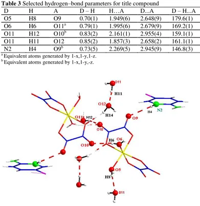
from 2.648(9) up to 2.945(9)Å and from 161.1(1) up to 179.6(1)°, respectively] into 3D supramolecular networks (Figure 3a, Tab 3). In fact, the uncoordinated water molecules (O11) play a role as both acceptors and donors while the coordinated water molecules [O5 and O6] act only as donors. As for the oxalate groups, the peripheral carboxylate-oxygen atoms O9 and O10 are only acceptors. The second kind of hydrogen bond involves N2 atom which acts as a bi-connective node to link two different complex molecules.
Table 3 Selected hydrogen–bond parameters for title compound
D H A D – H H…A D...A D – H...A O5 H8 O9 0.70(1) 1.949(6) 2.648(9) 179.6(1) O6 H6 O11a 0.79(1) 1.995(6) 2.679(9) 169.2(1) O11 H12 O10b 0.83(2) 2.161(1) 2.955(4) 159.1(1) O11 H11 O12 0.85(2) 1.857(3) 2.658(2) 161.1(1) N2 H4 O9b 0.73(5) 2.269(5) 2.945(9) 146.8(3)
a Equivalent atoms generated by 1-x,1-y,1-z. b Equivalent atoms generated by 1-x,1-y,-z.
Figure 3a Fragments of the molecular structure of title compound showing well-directional hydrogen bonding interactions
Figure 3b A view of part of a sheet of cation entities [(C5H6ClN2)]+, linked by π– π stacking interactions (dashed lines) between the neighboring pyridine ligands.
In the crystal structure, the chloropyridinium cations units are stakes by means of face-to-face interactions among the ring system of the pyridine groups to form layers parallel to the bc plane of the unit cell (Figure 3b). The interplanar short distances are of the order of 3.433 and 3.8λ6 Å. A lateral offset of 2.435 Å and
the short interatomic. Obviously, the hydrogen bonds and π- π interactions are responsible for the structural stability of the material.
Antimicrobial activity
The need for new antimicrobial agents is greater than ever because of the emergence of multidrug resistance in common pathogens, the rapid emergence of new infections, and the potential for use of multidrug-resistant agents (Spellberg
et al., 2004). In this context comes the objective of the present work to evaluate
the antimicrobial potentialities of a new synthesized compound described above. For that the antimicrobial activity of the amino- chloropyridinium diaqua dioxalato iron (III), has been evaluated by the filter paper disc method. The effect of the described compound used at 200 µg/ml on the growth inhibition by dual culture technique showed that the results varied with the tested microorganisms; for bacteria the amino-chloropyridinium diaqua dioxalato iron (III) was able to inhibit growth of Pseudomonas aeruginosa with a diameter of inhibition of 18mm, and was able to inhibit two gram-positive bacteria Staphylococcus aureus and Listeria innocua with a diameter inhibition respectively of 20 and 21 mm. The results have shown that the amino-chloropyridinium diaqua dioxalato iron (III) was also able to inhibit Candida albicans with diameter inhibition of 12 mm
when the compound was used at 500 µg/ml. Noteworthy, that the amino
-chloropyridinium diaqua dioxalato iron (III) was unable to inhibit fungi growth of some pathogenic fungi used in this work by means of dual culture technique at the same concentration of 200 µg/ml neither by the application of 500 or 1000µg/ml.
On the whole, the results showed that our compound possesses high antibacterial activity at the used concentration (200 g/ml) compared to others published synthetic compound eg (Nasser et al., 2013). at the highest concentration of
about 500 g/ml of the amino- chloropyridinium diaqua dioxalato iron (III) also
activity against Candida albicans strains has been obtained similar to results obtained by synthetic compound described by the work of Nasser et al. (2013). For antifungal activities numerous compound had shown activity when applied with a concentration more than 500 g/ml. In the present work, in the presence of
the concentration of 1000 g/ml, we have no antifungal activities in solid media.
Compared to the antifungal activity exhibited by others synthetic compound (Nasser et al., 2013; Zani et al., 1995). For example, a number of methyl imidazole derivatives and some of their oxygenated products tested by Zani et al. (1995), were found to exert very low antifungal activity against yeasts and moulds (Zani et al., 1995). As well as the application of Histidine-lysine (HK) polymers against the growth of several species of Candidaalbicans (Zhu et al., 2006).
From the data, it is clear that our compound possesses high activity, against bacteria while it possesses moderate activity against yeast and no activity against fungi when tested on solid medium by the application of the dual culture technique. In light of recently reported data, we decided to analyze the mechanism of action of the compound. For that, we have investigated the antifungal activities in broth medium for yeast and fungi. The results of the antifungal effect of the amino- chloropyridinium diaqua dioxalato iron (III) investigated in broth cultures of Candida albicans was then determined as percent yeast survival by measuring optical density at 600nm. The obtained results showed that the application of amino-chloropyridinium diaqua dioxalato iron (III) at 200µg ml-1, was able to suppress yeast survival by value ranging from 45.45 to 24.53 with various yeasts tested in the present work, after incubation at
37°C for 48h. Previously, we have shown that the sulfanilamide sulphate used at
100µg ml-1, was able to suppress yeast survival with the highest percentage (55.77%) (Essghaier et al., 2014).The values of MIC and IC50 were presented in Tab 4 The results have shown that, the minimal inhibitory concentrations (MIC) ranging from 16 µg ml-1for bacteria to 256 µg ml-1 for yeast and IC50 values
varying from 1.44 to 10.45 µg ml-1
for bacteria and 45.8 for yeast. These results showed that the amino-chloropyridinium diaqua dioxalato iron (III) had especially high activity against bacteria, compared to yeast and fungi. Similar results of the promising antibacterial activities have been reported by the compounds 3j and 3d against B. subtilis with the MIC of 1.12, 3.66 mg/ml as described by Sun et al. (2013).
Table 4 MIC (µg/ml) and IC50 values (µg/ml) of amino-
chloropyridinium diaqua dioxalato iron (III) against pathogenic bacteria and yeast tested.
Microorganisms CMI (in µg/ml) IC50 (in µg/ml)
Bacteria
Listeria innocua 16 1.44
Pseudomonas aeruginosa 16 10.45
Staphylococcus aureus
Yeast 16 1.74
On the whole, our new compound has high antibacterial activities against
Pseudomonas aeruginosa, Staphylococcus aureus and Listeria innocua. In the
same context, we would evaluate the lysozyme as well as the bactericidal activities of the described compound which were related to anti-bacterial activities.
Moreover, in this work, we examine the Bactericide activity of the new synthetic compound described, expressed in arbitrary units per ml (AU ml-1). The results have shown that the amino- chloropyridinium diaqua dioxalato iron (III) tested here had a high bactericidal activity of about 2500 AU ml-1. It should be mentioned that this value was greater than that exhibited by our previous described compound dI (4-sulfamoyl-phenyl-ammonium) sulphate (Essghaier et
al., 2014). The results of the lysozyme activities from the described compound
were presented in Figure 4 .Highlysozyme activities were expressed especially against Listeria innocua with 17 times more than Staphytococcus aureus tested in the present study. Lysozyme activities were directed toward membranes of gram-positive bacteria which are essentially constituted of peptidoglycans.
Figure 4 The Lysozyme activity of the new compound tested at 200 µg/ml by
incubation at 37°C with each pathogenic bacteriaμ Listeria innocua, and
Staphylococcus aureus. Data are the average of three replications and bars present the standard error of the means.
In previous work, we have evaluated the effect of our antifungal compound
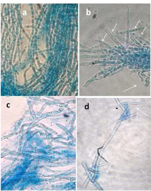
(Essghaier et al., 2014) on spore germination; here we would like to investigate the effect of the present antifungal compound on the direct destruction of mycelial hyphe. The results were presented in Figure 5. The new compound was able to destruct only the hyphe mycelial of the dermatophyte Trichophyton
rubrum with value ranging from 0.018 to 0.69 UA. These results mentioned
above were confirmed by the microscopic observation presented in Figure 6. Where, the effect of our compound was markedly observed compared to untreated mycelium characterized by markedly long hyphe (Figure 6).
Figure 5 The effect of the new compound at 1000µg/ml on mycelial
fragmentation tested by incubation for14h at 37°C with mycelial suspension from three isolates of dermatophyte fungi specie Trichophyton rubrumT1, T2 and T3. AU were expressed by comparison with untreated mycelial suspension (incubation tube without the compound) .Data are the average of three replications ± the standard error of the means.
Figure 6 Microscopic observation of the effect of the new compound at
1000µg/ml on mycelial fragmentation of two strain T2 and T3 of Trichophyton
rubrum respectively (b) and (d) compared to untreated mycelial suspension
(incubation tube without the compound (a) and (c) respectively for strains T2 and
T3. Arrows indicated area fragmentation and destruction hyphea compared to long hyphea in the normal case without treatment.
CONCLUSION
In conclusion, this paper is a study of a new compound mixed ligands containing oxalate bridging ligands. The single-crystal X-ray data show elongated tetragonal-bipyramidal coordination around iron(III) atoms of [Fe(C2O4)2(H2O)2]- anions. In addition to π-π interactions between the rings of pyridine groups, the cations and uncoordinated water molecules are connected through hydrogen bonds into 3D supramolecular frameworks.
In the present study, we describe a new compound as well as its antimicrobial activities. The results had shown that amino- chloropyridinium diaqua dioxalato iron (III) exhibited good activity especially against gram-positive bacteria. Furthermore, it has antifungal activity against Candida albicans as well as it was able to destroy the mycelial hyphe of the dermatophyte Trichophyton rubrum whenculturedin broth media.
SUPPLEMENTARY MATERIAL
Crystallographic data and full lists of bond lengths and angles have been deposited with the Cambridge Crystallographic Data Centre, CCDC No. 968830. Copies of this information may be obtained free of charge from The Director, CCDC, 12 Union Road, CAMBRIDGE CB2 1EZ, UK (fax: +44-1223-336-033; e-mail: deposit@ccdc.cam.ac.uk or http://www.ccdc.cam.ac.uk).
REFERENCES
ABDELHAK, J., CHERNI, S.N., ZID, M.F., DRISS. 2007. Crystal Structure of [Tris(o-phenanthroline-N,N′)iron(II)] dinitrate Oxalic Acid Pentahydrate. Anal. Sci, 23, 65-66.
CASTILLO, O., LUQUE, A., ROMÁN, P. 2001.Synthesis, chemical
characterization and crystal structure of the (oxalato-O,O')bis(1,10-phenanthroline) copper(II) pentahydrate. J. Mol. Struct, 570,181–188.
http://dx.doi.org/10.1107/S1600536801012909
COLLINS, M. D., FACKLAM, R.R., FARROW, J.A.E, WILLIAMSON, R. 1989. Enterococcus raffinosus sp.nov., Enterococcus solitarius sp.nov. and
Enterococcus pseudoavium sp.nov. FEMS Microbiol Lett, 57, 283–288.
http://dx.doi.org/10.1111/j.1574-6968.1989.tb03350.x
CZAKIS-SULIKOWSKA, D., MALINOWSKA, A., RADWAÑSKA -DOCZEKALSKA. 2000. Synthesis, Properties and Thermal Decomposition of Bipyridine–Oxalato Complexes with Mn(II), Co(II), Ni(II) and Cu(II). Polish J. Chem, 74, 607–614.
FERBINTEANU, M., MIYASAKA, H., WERNSDORFER, W., NAKATA, K.,
SUGIURA, K.I., YAMASHITA, M., COULON, C., CLÉRAC, R. 2005.
Single-Chain Magnet (NEt4)[Mn2(5-MeOsalen)2Fe(CN)6] Made of MnIII−FeIII−MnIII Trinuclear Single-Molecule Magnet with an ST = 9/2 Spin Ground State. J. Am. Chem. Soc, 127(9), 3090–3099.
http://dx.doi.org/10.1021/ja0468123
GRACIELA, M., VIGNOLO, M., DE KAIRUZ, N., AIDA, A.P., DE RUIZ H, OILVER, G. 1995. Influence of growth conditions on the production of lactocin 705, a bacteriocin produced by L. casei CRL 705 J Appl Bacteriol, 78, 5-10.
http://dx.doi.org/10.1111/j.1365-2672.1995.tb01665.x
HAIKARAINEN, J., SIPILÄ, J., PIETIKÄINEN, P., PAJUNEN, A., MUTIKAINEN, I. 2001. Synthesis and characterization of bulky salen-type complexes of Co, Cu, Fe, Mn and Ni with amphiphilic solubility properties .J.
Chem. Soc., Dalton Trans, 991-995. http://dx.doi.org/10.1039/b008167l
JIA, H.P., LI, W., JU, Z.F., ZHANG, J. 2008. A series of manganese-carboxylate coordination polymers exhibiting diverse magnetic properties. J. Chem. Soc.,
Dalton Trans, 5350-5357. http://dx.doi.org/10.1039/B808691E
JORGENSEN, J.H., TURNIDGE, J.D. 2007. Antibacterial susceptibility tests: dilution and disk diffusion methods. In: MURRAY PR, BARON EJ, JORGENSEN JH, LANDRY ML, PFALLER M A, editors. Manual of clinical microbiolog. 9th ed. Washington, DC: American
Society for Microbiology, 1152-1172.
KARAA, N., HAMDI, B., BEN SALAH, A., ZOUARI, R. 2013. Synthesis, Infra-red, CP/MAS-NMR characterization, structural study and electrical properties of the bis(4-amino-2-chloropyridinium) tetrachlorozincate (II) monohydrate. J. of Mol. Struc, 1049, 48-58.
LI, L.L., LIN, K.J., HO, C.J., SUNB, C.P., YANG, H.D. 2006. A coordination π– π framework exhibits spontaneous magnetization. Chem. Commun, 1286-1288.
http://dx.doi.org/10.1039/B515681E
MOLLAR, M., CASTRO, I., LLORET, F., JULVE, M., FAUS, J., LATORRE, J. 1991. A solution study of complex formation between iron(III) and oxalate in dimethylsulphoxide Transition Metal Chemistry,16, 31-34 .
MARINESCU, G., VISINESCU, D., CUCOS, A., ANDRUH, M., JOURNAUX, Y., KRAVTSOV, V., SIMONOV, Y.A. 2004. Oxalato-Bridged [CuIICrIII] and [MnIICrIII] Binuclear Complexes: Synthesis, Crystal Structures, Magnetic and EPR Investigations. Euro. J. of Inorg. Chem,14, 2914-2922.
http://dx.doi.org/10.1002/ejic.200400061
ABD EL-SALAM, N.M., MOSTAFA, M.S., AHMED, G.A., ALOTHMAN, O.Y. 2013. Synthesis and Antimicrobial Activities of Some New Heterocyclic Compounds Based on 6-Chloropyridazine-3(2H)-thione. J. of Chemistry; Hind. Pub. Coop. 1-8.
POZDNYAKOV, I.P., KEL, O.V., PLYUSNIN, V.F., GRIVIN, V.P., BAZHIN, N.M. 2008. New insight into photochemistry of ferrioxalate. J. Phys. Chem. A, 112, 8316-8322. http://dx.doi.org/10.1021/jp8040583
RYAZANOVA, L.P., STEPNAYA, O.A., SUZINA, N.E., KULAEV, I.S. 2005.Antifungal action of the lytic enzyme complex from Lysobacter sp. XL.1. Process Biochemistry, 40(2), 557–564.
SADFI-ZOUAOUI. N., ESSGHAIER, B., HAJLAOUI, M.R., FARDEAU, M.L., CAYOL, J.L., OLLIVIER, B., BOUDABOUS, A. 2008. Ability of moderately halophilic bacteria to control grey mould disease on tomato fruits. J
Phytopathol,156,42-52. http://dx.doi.org/10.1111/j.1439-0434.2007.01329.x
SCHOTT, O., FERRANDO-SORIA, J., BENTAMA, A., STIRIBA, S.E.,
PASÁN, J., RUIZ-PÉREZ, C., ANDRUH, M., LLORET, F., JULVE, M. 2011.
Chromium(III) complexes with 2-(2′-pyridyl)imidazole: Synthesis, crystal structure and magnetic properties. Inorg. Chim. Acta, 376, 358-366
http://dx.doi.org/10.1016/j.ica.2011.06.039
SENSI, P. 1979.Trends in the research on antimicrobial agents. Prog Clin Biol Res, 35, 9-15.
SHELDRICK, G.M. 1997. Program for the Refinement of Crystal Structures.
University of Göttingen, Germany
SNODIN, M.D., OUD-MOUSSA, L., WALLMANN, U., LECOMTE, S., BACHLER, V., BILL, E., HUMMEL, H., WEYERMÜLLER, T., HILDEBRANDT, P., WIEGARDT, K. 1999. The Molecular and Electronic Structure of Octahedral Tris(phenolato)iron(III) Complexes and Their Phenoxyl Radical Analoguesμ A Mössbauer and Resonance Raman Spectroscopic Study Chem. Eur. J, 5, 2554-2565 . http://dx.doi.org/10.1002/(SICI)1521-3765(19990903)
SPELLBERG, B., POWERS, J.H., BRASS, E.P., MILLER, L.G., EDWARDS, J.E. JR. 2004.Trends in antimicrobial drug development: implications for the future. Clin Infect Dis. 1;38 (9),1279-86. http://dx.doi.org/10.1086/420937 PMID:15127341. 10.
SUN, J., LV, P-C., YIN, Y., YUAN, R-J., MA, J., et al. Synthesis, Structure and Antibacterial Activity of Potent DNA Gyrase Inhibitors: N9-Benzoyl-3-(4-Bromophenyl)-1H-Pyrazole-5-Carbohydrazide Derivatives. PLoS ONE 20138; (7):69751.http://dx.doi.org/10.1371/journal.pone.0069751
WROBLESKI, J.T., BROWN, D.B. 1980. A study of the variable-temperature magnetic susceptibility of two Ti(III) oxalate complexes J. Inorg. Chem ,38, 227-230.http://dx.doi.org.10.1016/S0020-1693(00)91964-9
YU, J.H., HOU, Q., BI, M.B., LÜ, Z.L., ZHANG, X., QU, X.J., LU, J., XU, J.Q. 2006. Structure characterization of several oxalate-bridged transition-metal
coordination polymers. J. of Mol. Struc, 800, 69-73 .http://dx.doi.org/10.1016/j.molstruc.2006.03.079
ZANI, F.P., MAZZAL, P., BENVENUTIZ, S., SEVERI, F., MALMUSIZ, L., VAMPA, G., ANTOLIN, L. 1995. Synthesis, characterization, crystallographic analysis, antifungal and genotoxic properties of some 1-methyl-1H-imidazoles.
Eur J Med Chem, 729-740. http://dx.doi.org/10.1016/0223-5234(96)88292-4
ZHANG, Y.Y., QI, Y., ZHANG, Y., LIU, Z.Y., ZHAO, Y.F., LIU, Z.M. 2007. Synthesis, structure and magnetic properties of a new iron phosphonate-oxalate with 3D framework. Mat. Res. Bul, 42,1531-1538 .
http://dx.doi.org/10.1016/j.materresbull.2006.10.027
ZHENG, L.M., FANG, X., LII, K.H., SONG, H.H., XIN, X.Q., FUN, H.K., CHINNAKALI, K., ABDUL RAZAK, I.1999. Syntheses, crystal structures and magnetic properties of two novel layered compounds: [Fe3(C2O4)3(4,4′-bpy)4] and [Co(C2O4)(4,4′-bpy)] (4,4′-bpy = 4,4′-bipyridine) J. Chem. Soc. Dalton Trans., 2311-2316.http://dx.doi.org/ 10.1039/A809738K
![Table 2 Selected bond lengths (Å) and angles (°) for [Fe(C 2 O 4 ) 2 (H 2 O) 2 ] -](https://thumb-eu.123doks.com/thumbv2/123dok_br/18161836.328881/3.892.152.742.933.1041/table-selected-bond-lengths-å-angles-fe-c.webp)