Journal of Krishna Institute of Medical Sciences University 132
Ó
Ó
CASE REPORTAbstract:
Adrenal myelolipoma is an unusual, benign and biochemically inactive tumour that is composed of variable mixture of mature adipose tissue and haematopoietic elements. It is usually asymptomatic, unilateral and nonsecreting, though known to be associated with hypertension, obesity, endocrinological disorders and some malignancies. We report a case of adrenal myelolipoma in a 64 year old male, who presented with pain in the left upper abdomen.
Keywords: Adrenal Myelolipoma, Angiomyolipoma, Cholelithiasis
Introduction:
Adrenal myelolipoma is a rare benign endocrine lesion composed of mixture of mature adipose tissue and haematopoietic elements including erythroid, myeloid, megakaryocyte types and often lymphocytes. The tumour has equal sex incidence and commonly found between fifth and seventh decades of life. With an incidence of 0.08-0.4%, myelolipomas account for 3-5% of all primary tumors of the adrenal gland [1, 2]. Incidental detection of myelolipomas are becoming more common, constituting upto 10-15% of incidental adrenal masses, with the widespread use of noninvasive imaging with Ultrasonography (USG), Computed Tomography (CT) and Magnetic Resonance Imaging (MRI). The majority are small, unilateral, and asymptomatic. They are generally nonsecreting. These lesions are often smaller than 4cm in diameter [3].
Adrenal Myelolipoma Associated with Cholelithiasis - A Rare Case Report
1 1*
R.M. Potekar , Shefali Goyal
1
Department of Pathology, BLDEU's Shri B.M.Patil Medical College, Hospital & Research Centre, Vijayapura-586103 (Karnataka) India
Case Report:
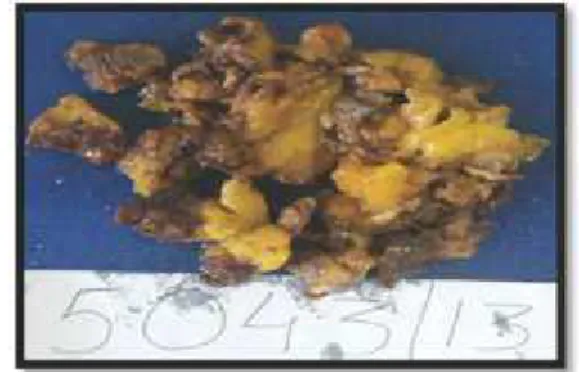
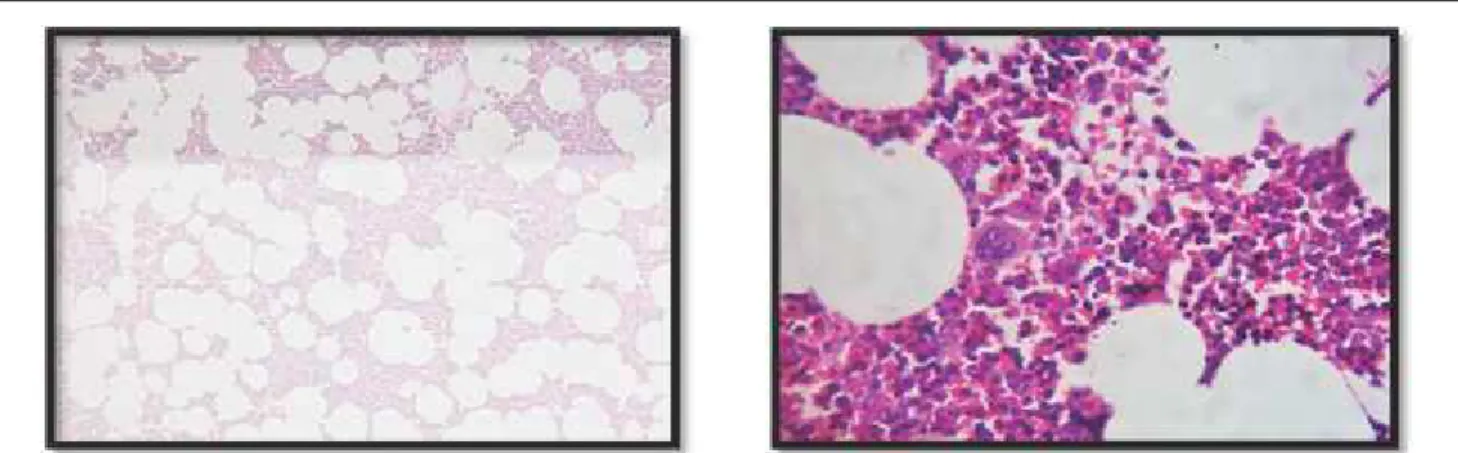
A 64 year old male presented with pain in the left upper abdomen since 3 months that occasionally radiated to the left shoulder. Patient was detected to be hypertensive 1 month earlier and was diabetic for 13 years. Ultrasound abdomen showed presence of large fat density mass lesion in left adrenal gland with small enhancing soft tissue focus within. Gall bladder revealed calculus measuring 15 x 13mm. CT revealed large well-defined rounded 75 x 79 x 61mm sized fat density (-50 to -100 HU) lesion arising from the left adrenal gland. There was evidence of small focus of soft tissue (30-35 HU) density noted within. After a thorough preoperative workup, a surgical left adrenalectomy was performed. Macroscopic examination of the mass revealed fragments of irregular yellow brown tissue measuring 7 x 7 x 6cm (Fig.1). Microscopy revealed mature adipose tissue containing active bone marrow elements admixed with adrenal tissue. (Fig. 2)
ISSN 2231-4261 JKIMSU, Vol. 6, No. 1, January-March 2017
Fig. 1: Gross Specimen of Adrenal Myelolipoma Showing Dark Brown and
Journal of Krishna Institute of Medical Sciences University 133
Ó
Ó
Discussion:Adrenal myelolipomas are rare, benign and clinically silent tumours. The adrenal gland is the most common site but myelolipomas also occur rarely in extra-adrenal locations such as retroperi-toneum, presacral soft tissues, thorax and pelvis [4]. Since their diagnosis is based on autopsy or imaging modalities that are performed for reasons usually unrelated to adrenal diseases, they are also called as 'incidentalomas' [5]. Majority of adrenal incidentalomas are due to pheochromocytoma, adrenal adenoma, adrenal carcinoma and metastatic carcinoma [6]. The histogenesis is uncertain. The tumour may develop from the residual embryonic cells of the bone marrow that, after embolization, reach the adrenal gland. Some authors even consider the development of adrenal myelolipomas as a response to various endocrine stimulations. However, the most widely accepted theory is adrenocortical cell metaplasia that occurs in response to necrosis, infection, inflammation or stress [1, 2]. Four distinct clinico-pathological patterns have been described–isolated adrenal myelolipoma, adrenal myelolipoma with hemor-rhage, extra-adrenal myelolipoma and myelo-lipoma associated with other adrenal diseases like adrenal adenomas or endocrine disorders including Cushing's syndrome, Conn's syndrome
and congenital adrenal hyperplasia [3, 5]. Our patient had an isolated adrenal myelolipoma. A rare feature to consider in our study is the left-sided position of the myelolipoma, because they have been reported in the literature to be prevalent mostly on the right side [2]. The association of adrenal myelolipoma with cholelithiasis has been described in just two case reports [4, 7]. In our patient gallstones were present. These tumors are often quiescent with detection incidental to imaging being >50% [8]. Its association with obesity (25%), diabetes (26%), and hypertension (26%) is also incidental [8]. In our case the patient was both hypertensive and diabetic. Since these tumours are nonfunctional, endocrinological evaluation may not be useful, although there is a report of secreting myelolipoma causing hypertension [1]. These tumours need to be differentiated from benign tumours like renal angiomyolipoma, retroperitoneal lipoma and liposarcoma [1, 2, 8, 9]. Management of adrenal myelolipoma is individualized. Tumours less than 4cm in diameter and asymptomatic, diagnosed incidentally on imaging and/or cytological studies, are best left to long term follow-up. Surgical resection is offered when they are large and symptomatic, exhibits areas of necrosis or R.M. Potekar & Shefali Goyal JKIMSU, Vol. 6, No. 1, January-March 2017
Journal of Krishna Institute of Medical Sciences University 134
Ó
Ó
hemorrhage, or when malignancy is suspected [8]. In the case presented herea left adrenalectomy was performed. Adrenal myelolipoma constitutes a rare entity. The association of obesity with adrenal myelolipoma and cholelithiasis is well documented [7]; however, the occurrence of adrenal myelolipoma and cholelithiasis is extremely rare.
Conclusion:
The co-occurrence of adrenal myelolipoma and cholelithiasis may be an incidental finding or might have common pathophysiologic basis. The 'incidental' diagnosis warrants careful diagnostic study to plan appropriate treatment.
R.M. Potekar & Shefali Goyal JKIMSU, Vol. 6, No. 1, January-March 2017
References 1. Nabi J, Danish R, Authoy FN, Sofi GN. Incidental
detection of adrenal myelolipoma: a case report and review of literature. Case Rep Urol 2013; 2013:1-3. 2. Brogna A, Scalisi G, Ferrara R, Bucceri AM. Giant
secreting adrenal myelolipoma. J Med Case Rep 2011; 5:1-4.
3. Wani NA, Kosar T, Rawa IA, Qayum A. Giant adrenal myelolipoma:Incidentaloma with a rare incidental association. Urol Ann 2010; 2(3):130-3.
4. Kalra R, Dahiya N, Singh G, Modi S, Goel P. Adrenal myelolipoma associated with cholelithiasis – a case report. IJPSR 2011; 2(4): 1031-33.
5. Saha M, Dasgupta S, Chakrabarti S, Chakraborty J. Giant myelolipoma of left adrenal gland simulating a retroperitoneal sarcoma. IJAMHR 2015; 2(2): 122-5.
*
Author for Correspondence: Dr. Shefali Goyal Department of Pathology, BLDEU's Shri B.M. Patil Medical College,
Hospital and Research Centre, Vijayapur-586103 (Karnataka) India
Email: [email protected], [email protected] Cell: 9916481501
6. Puri S, Mardi K, Gupta N, Rao M. Adrenal incidentalomas with review of literature. J Cytol Histol 2016; 7: 1-3.
7. Bano S, Yadav SN, Chaudhary V, Garga UC. Symptomatic giant adrenal myelolipoma associated with cholelithiasis: Two case reports. Urol Ann 2012; 4(1): 55-60.
8. Bhansali A, Dash RJ, Singh SK, Behra A, Singh P, Radotra BD. Adrenal myelolipoma: Profile of six patients with a brief review of literature. Int J Endocrinol Metab 2003; 1: 33-40.