Submitted2 July 2015
Accepted 27 November 2015
Published5 January 2016
Corresponding author
Heng Yin, [email protected]
Academic editor
Joanna Moraczewska
Additional Information and Declarations can be found on page 10
DOI10.7717/peerj.1517 Copyright
2016 Jia et al.
Distributed under
Creative Commons CC-BY 4.0
OPEN ACCESS
A simple and rapid method for measuring
α
-D-phosphohexomutases activity by
using anion-exchange chromatography
coupled with an electrochemical detector
Xiaochen Jia1,2, Jian Kang3and Heng Yin1
1Dalian Institute of Chemical Physics, Chinese Academy of Sciences, Dalian, China 2University of Chinese Academy of Sciences, Beijing, China
3Department of Biochemistry and Molecular Biology, Dalian Medical University, Dalian, China
ABSTRACT
The interconversion of hexose-6-phosphate and hexose-1-phosphate can be directly analyzed by high-performance anion-exchange chromatography coupled with an electrochemical detector (HPAEC-PAD). Thus, this method can be used to measure the activities of N-acetylglucosamine-phosphate mutase (AGM), glucosamine-phosphate mutase (GlmM) and phosphoglucomutase (PGM), which are the members of
α-D-phosphohexomutases superfamily. The detection limits were extremely low as 2.747 pmol, 1.365 pmol, 0.512 pmol, 0.415 pmol, 1.486 pmol and 0.868 pmol for N-acetylglucosamine-1-phosphate (GlcNAc-1-P), N-acetylglucosamine-6-phosphate (GlcNAc-6-P), glucosamine-1-phosphate (GlcN-1-P), glucosamine-6-phosphate (GlcN-6-P), glucose-1-phosphate (Glc-1-P) and glucose-6-phosphate (Glc-6-P), respectively. By employing HPAEC-PAD, activities ofAtAGM (AGM fromArabidopsis thaliana) on these six phosphohexoses can be detected. The Km of AtAGM on
Glc-1-P determined by HPAEC-PAD was 679.18±156.40µM, which is comparable
with theKmof 707.09±170.36µM detected by traditional coupled assay. Moreover,
the activity ofMtGlmM (GlmM fromMycobacterium tuberculosis) on GlcN-6-P tested by HPAEC-PAD was 7493.40±309.12 nmol/min·mg, which is much higher than
288.97±35.28 nmol/min·mg obtained by the traditional coupled assay. Accordingly,
HPAEC-PAD is a more rapid and simple method than the traditional coupled assays given its high specificity and sensitivity, and will certainly bring convenience to further research ofα-D-phosphohexomutases.
SubjectsBiochemistry
Keywords HPAEC-PAD, N-acetylglucosamine-phosphate mutase, N-acetylglucosamine-1-phosphate, N-acetylglucosamine-6-N-acetylglucosamine-1-phosphate,α-D-phosphohexomutases
INTRODUCTION
Figure 1 The hexosamine pathway in eukaryocyte and prokaryocyte.(A) The pathway in eukaryocyte was catalyzed by GlcN-6-P synthase (GFA), GlcN-6-P acetyltransferase (GNA), N-acetylglucosamine-phosphate mutase (AGM) and UDP-GlcNAc pyrophosphorylase (UAP) respectively. (B) The pathway in prokaryocyte was catalyzed by glutamine fructose-6-phosphate transferase (GlmS), phosphoglucosamine mutase (GlmM), glucosamine-1-phosphate acetyltransferase/N-acetylglucosamine-1-phosphate
uridyltransferase (GlmU) respectively. The activity of AGM and GlmM could be detected by HAPEC-PAD method or traditional coupled assay.
hexosamine pathway is a branch of glycolysis. It is associated with posttranslational protein modification by glycosylation and involved in the synthesis of glycolipids, proteoglycans, and glycosylphosphatidylinositol anchors (Vosseller et al., 2002;Wells, Vosseller & Hart, 2001;Zachara & Hart, 2004). The hexosamine pathway is a survival feature of neoplastic cells, plays a prominent role during tumourigenesis, and can be exploited therapeutically to target cancer cells (Vasseur & Manie, 2015). The hexosamine pathway is also essential for the growth of pathogenic bacteria, and can be used as a potential excellent target for anti-bacteria drugs (Li et al., 2012).
In living organisms, there are three variations of hexosamine pathway according to the different reaction sequence and enzymes, which are eukaryotic (Fig. 1A), prokaryotes (Fig. 1B) and mimivirus pathways (Piacente et al., 2014). The hexosamine pathway in mimivirus follows the eukaryotic-like strategy, but also shares some properties with prokaryotic pathway (Piacente et al., 2014). The enzymes in hexosamine pathway have been well studied except N-acetylphosphate mutase (AGM) and glucosamine-phosphate mutase (GlmM) (Fang et al., 2013;Milewski, Gabriel & Olchowy, 2006). The main reason is the limitation of methodology onα-D-phosphohexomutases activity.
Traditionally, the activity of α-D-phosphohexomutase was determined by a coupled enzyme system. PGM catalyze the interconvertion of glucose-1-phosphate (Glc-1-P) and Glc-6-P. The normal way to detect the activity of PGM was using Glc-1-P as substrates, glucose-6-phosphate dehydrogenase (G6PDH) as coupled enzyme, by detect the increasing absorbance values of NADH at 340 nm (Nishinari et al., 2012;Wang & Zhang, 2010). This method was convenience and simple because G6PDH was commercially available and cost-effective. However, the detection of the reverse reaction which uses Glc-6-P as a substrate is still impossible.
6-P was always used as a substrate for detecting the AGM activity, and GlcNAc-1-P uridylyltransferase (UAP), the fourth enzyme in hexosamine pathway, was used as a coupled enzyme (Boles et al., 1994;Greig et al., 2007;Jolly et al., 2000) (Fig. 1A). The detection of GlmM activity was carried out by using GlcN-6-P as substrate, and coupled with the third and forth enzyme GlmU (Li et al., 2012;Li et al., 2011) (Fig. 1B). However, this coupled method was very complex and tedious, and especially UAP and GlmU were commercial unavailable. Furthermore, the most noticeable limitation of the traditional coupled assay is impossible to detect the reverse reaction, which using GlcNAc-1-P or GlcN-1-P as substrate.
In order to overcome the limitations of the traditional coupled assay on the determination of the activity of α-D-phosphohexomutases, a simple and convenient method which can detect the interconversion of hexose-6-P and hexose-1-P directly is urgently needed.
High-performance anion-exchange chromatography (HPAEC) has been used for the quantitation of carbohydrates (Kazlowski, Pan & Ko, 2015;Yang, Hsieh & Lin, 2015) because its strong anion-exchange property allows the highly selective separation of carbohydrates. It has been tried to detect and separate the phosphohexoses directly by HPAEC for decades. In previous studies,Jolly et al. (1999)detected glucosamine phosphates by HPAEC with a post-column derivatization. detected glucose phosphates by HPAEC with14C labeled method.Naught & Tipton, (2005). However, they all needed radiochemical detector or fluorescence detector, and the phosphohexoses pretreatment were complex and expensive. Recently, several phosphohexoses had been successfully separated by HPAEC (Jeong et al., 2007;Marcellin et al., 2009), but the separation of GlcNAc-1-P and GlcNAc-6-P is still unsuccessful. And also, there is no simple and convenient way to determine the activity ofα-D-phosphohexomutases directly.
MATERIALS AND METHODS
Materials
Glc-1-P, Glc-6-P, glucose-1,6-bisphosphate (Glc-1,6-2P) and GlcN-6-P were purchased from Sigma-Aldrich. GlcNAc-6-P, GlcNAc-1-P and GlcN-1-P were purchased from Santa Cruz Biotechnology. Double distilled water was used for HPLC, and all other solvents and chemicals were HPLC grade.
Chromatography
The HPAEC system consists of a Dionex Bio-LC gradient pump with GM-3 (4 mm) gradient mixer, CarboPac PA-100 column (4×250 mm), and an electrochemical detector
with AgCl as reference electrode. The waveform was carbohydrates (standard Quad), the following pulse potentials were used for detection:t=0 s,E=0.10 v;t=0.20 s,E=0.10 v;
t=0.40 s,E=0.10 v;t=0.41 s,E= −2.00 v;t=0.42 s,E= −2.00 v;t=0.43 s,E=0.60 v;t=0.44 s,E= −0.10 v;t=0.50 s,E= −0.10 v. The sample injection volume is 20µL
and column oven temperature is maintained at 30 ◦
C.
Elute conditions
The phosphohexoses should be eluted after 10 min because there were several interference peaks in the enzyme reaction mixture before 10 min on chromatograms. Thus, high pH elution was carried out in 100 mM sodium hydroxide under gradient conditions using a 80–720 mM sodium acetate gradient over 20 min at a flow rate of 0.5 ml/min.
The details of gradient conditions were 0–5 min, 80 mM sodium acetate; 5–15 min, 80–720 mM sodium acetate; 15–18 min, 720 mM sodium acetate; 18–20 min, 80 mM sodium acetate in 100 mM sodium hydroxide, 0.5 ml/min.
Expression and purification of AtAGM andMtGlmM
E. colicells (AtAGM::Amp) were grown exponentially at 37 ◦
C in LB medium with Amp. When the optical density (OD) of the culture reached 0.8, IPTG was added at a final concentration of 1 mM, and growth was continued for 12 h at 16 ◦
C. Harvested cells were disrupted by sonication, centrifugated at 20,000 g for 30 min; the resulting supernatant was loaded onto Ni-NTA column (Qiagen, Hilden, Germany) for purification.
MtGlmM andMtGlmU were expressed and purified according to our previous studies (Li et al., 2012;Li et al., 2011).
AtAGM assay
The activities of AtAGM (N-acetylglucosamine-phosphate mutase, from Arabidopsis thaliana) on different phosphohexoses, were tested in the same reaction mixture as described below. 154.80 pmol (10 µg)AtAGM or boiled enzymes, were incubated in a
substrate buffer (300µl) consisting of 20 mM PBS, pH 7.0, 10 mM MgSO4, and 20µM
Glc-1,6-2P and 0.1 mM different phosphohexose (GlcNAc-1-P, GlcNAc-6-P, GlcN-1-P, GlcN-6-P, Glc-1-P, Glc-6-P) (Fang et al., 2013;Hofmann, Boles & Zimmermann, 1994) for 30 min at 30 ◦C. The reaction was terminated in boiling water and 200µl of 0.2 M NaOH
The Steady-state kinetics of AtAGM was analyzed by two methods using Glc-1-P as substrates. The HPAEC-PAD method was carried out by using 56.66 pmolAtAGM or boiled enzymes, incubated for 10 min at 30 ◦C in a substrate buffer as described above, with varying concentrations of Glc-1-P (60–8,000µM). Then, the assay was terminated
immediately in boiling water and 200 µl of 0.2 M NaOH were added for HPAEC-PAD
analysis.
The traditional coupled assay was carried out in a 200µl reaction volume containing
20 mM PBS, pH 7.0, 10 mM MgSO4, 20µM Glc-1,6-2P, a range of concentrations of Glc-1P
(60–7,000µM), 1 mM NAD+ and 2 units of G6PDH (Anasontzis et al., 2014;Fang et al.,
2013;Kim et al., 2014). The reaction was started by the addition of 56.66 pmol AtAGM and incubated for 10 min at 30 ◦
C. The amount of NADH produced was measured using a micro plate reader at 340 nm (BioTek, Gene 5).
All the experiments had repeated three times, and theKmandVmaxvalues were calculated
by Origin 7.5.
MtGlmM assay
MtGlmM (glucosamine-phosphate mutase, fromMycobacterium tuberculosis) activity was determined by two methods.
The traditional coupled assay was conducted in a coupled enzyme system withMtGlmU, the third- and forth-step enzymes in hexosamine pathway inMycobacterium tuberculosis
(Li et al., 2011) (Fig. 1B). The GlcN-1-P converted from GlcN-6-P by the mutase was quantitatively converted into UDP-GlcNAc in the presence of purifiedMtGlmU in this coupled assay (Li et al., 2012) (Fig. 1B). The 50µl reaction mixture contained 50 mM Tris–
HCl, pH 8.0, 2.5 mM MgCl2, 1 mM GlcN-6-P, 0.6 mM acetyl-CoA, 0.2 mM Glc-1,6-2P,
purifiedMtGlmU (3.5µg) and purifiedMtGlmM (0.18µg). The reaction was incubated
at 37 ◦
C for 20 min and terminated by adding 50µl of stop solution containing 50 mM
Tris–HCl, pH 7.5, 6 M guanidine hydrochloride. The mixture was then incubated for 10 min by the addition of 50µl of Ellman’s reagent solution containing 0.2 mM DTNB in
the buffer with 50 mM Tris–HCl, pH 7.5, and 1 mM EDTA.TNB, the product generated from the reaction of CoA-SH and DTNB, was monitored at 405 nm by Benchmark Plus plate reader (Thermo Fisher Scientific, Waltham, MA, USA).
MtGlmM activity was analyzed using HPAEC-PAD by detecting the interconversion of GlcN-6-P and GlcN-1-P directly. The purifiedMtGlmM (0.18µg) was incubated at 37 ◦C
for 20 min in a substrate buffer (50µl) consisting of 1 mM GlcN-6-P (or GlcN-1-P) as
substrate with 50 mM Tris–HCl, pH 8.0, 2.5 mM MgSO4, and 0.2 mM Glc-1,6-2P. The
reaction was terminated immediately in boiling water and 500µl of 0.2 M NaOH were
added for HPAEC-PAD analysis.
RESULTS AND DISCUSSION
The separation of standards
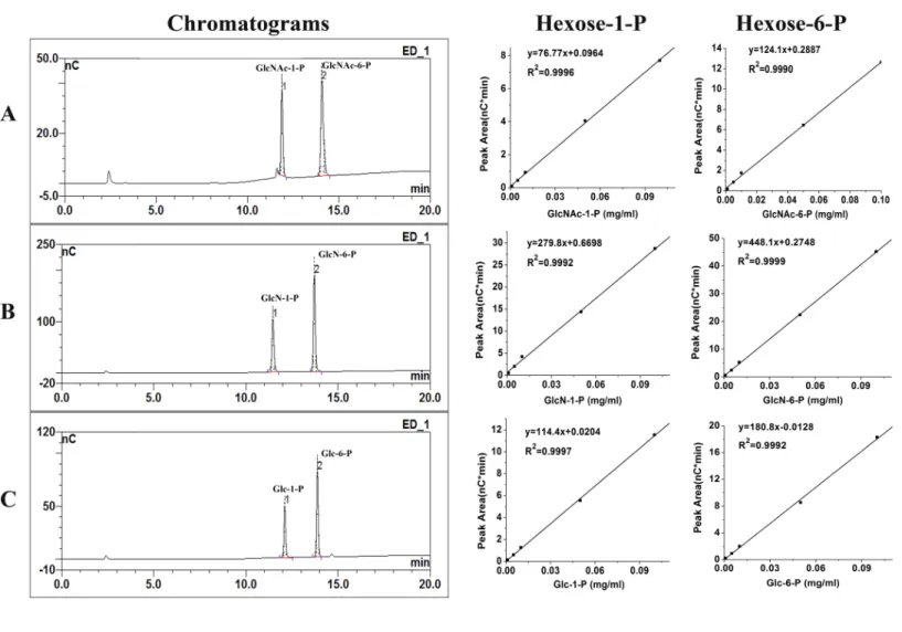
Figure 2 Chromatograms of calibration standards and their standard curves.Under the 80–720 mM sodium acetate gradient over 20 min at a flow rate of 0.5 ml/min, the calibration standards of 0.05 mg/ml GlcNAc-1-P, GlcNAc-6-P (A) ; 0.05 mg/ml GlcN-1-P, GlcN-6-P (B); 0.05 mg/ml Glc-1-P, Glc-6-P (C) were separated excellent on HPAEC-PAD chromatograms. Their standard curves were shown behind their chromatograms re-spectively; ‘‘nC’’ means PAD response.
14.30 min, 11.55 min, 13.75 min, 12.05 min and 13.72 min, respectively (Figs. 2A–2C). Hexose-1-P and hexose-6-P were separated excellent by HPAEC-PAD.
The detection limits are different for different phosphohexoses. The detection limits were 2.747 pmol, 1.365 pmol, 0.512 pmol, 0.415 pmol, 1.486 pmol and 0.868 pmol for GlcNAc-1-P, GlcNAc-6-P, GlcN-1-P, GlcN-6-P, Glc-1-P and Glc-6-P, respectively. The linearity of all the phosphohexoses was excellent (2–400µM,R2>0.999) as shown in
Fig. 2. And HPAEC-PAD response was linearity in the range of concentration 1–15,000µM
(R2>0.99).
Figure 3 AtAGM activity on different substrates was assayed by HPAEC-PAD method.154.80 pmol AtAGM or boiled enzymes, were incubated for 30 min at 30 ◦
C in a substrate buffer (300µl) consisting
of 20 mM PBS, pH 7.0, 10 mM MgSO4, and 20µM glucose-1,6-bisphosphate (Glc-1,6-2P). And using
GlcNAc-1-P (A), GlcN-1-P (B), Glc-1-P (C), GlcNAc-6-P (D), GlcN-6-P (E), and Glc-6-P (F) as substrate respectively (the final concentration is 0.1 mM). The black or red lines means the mixture after reaction, incubated with the boiled enzyme orAtAGM , respectively; ‘‘nC’’ means PAD response.
AGM assay
The chromatograms of the reaction mixtures ofAtAGM incubated with different substrates (GlcNAc-1-P, GlcNAc-6-P, GlcN-1-P, GlcN-6-P, Glc-1-P and Glc-6-P) were shown in
Fig. 3, including the chromatograms before (incubated with boiled enzyme) and after the reaction. The consuming of substrates and the producing of the products could be observed well in the chromatograms. The results showed that AtAGM indeed had the ability to convert these six phosphohexoses.
In order to identify the effectiveness of this novel method, both traditional coupled assay and HPAEC-PAD method were used to detect the steady-state kinetics and kinetic parameters ofAtAGM. The activity ofAspergillus fumigatesAGM on Glc-1-P determined by traditional coupled assay from Fang et al. (2013) was also taken as a comparison. The Steady-state kinetics and kinetic parameters of AtAGM were shown inFig. 4and
Table 1. TheKmofAtAGM detected by HPAEC-PAD was 679.18±156.40µM, which was
comparable with theKmof 707.09±170.36µM detected by traditional coupled assay. And
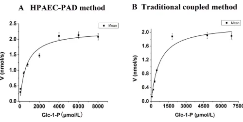
Figure 4 Steady-state kinetics ofAtAGM was analyzed by using Glc-1-P as substrates by two methods.
(A) The HPAEC-PAD method was carried out by using 56.66 pmolAtAGM or boiled enzymes, incubated for 10 min at 30 ◦
C in a substrate buffer (300µl) consisting of 20 mM PBS, pH 7.0, 10 mM MgSO4, and 20µM glucose-1,6-bisphosphate (Glc-1,6-2P), with varying concentrations of Glc-1-P (60–8,000µM).
(B)The traditional coupled assay was carried out in a 200µl reaction volume containing 20 mM PBS, pH
7.0, 10 mM MgSO4, 20µM glucose-1,6-bisphosphate (Glc-1,6-2P), a range of concentrations of
Glc-1-P (60–7,000µM), 1 mM NAD+and 2 units of G6PDH, the reaction was started by the addition of 56.66
pmolAtAGM and incubated for 10 min at 30 ◦
C. The results are the mean±S.D. for three determina-tions.
Table 1 Kinetic parameters ofAtAGM was analyzed by using Glc-1-P as substrates by two methods.The HPAEC-PAD method was carried out by using 56.66 pmolAtAGM or boiled enzymes, incubated for 10 min at 30 ◦C in a substrate buffer (300
µl) consisting of 20 mM PBS, pH 7.0,
10 mM MgSO4, and 20µM glucose-1,6-bisphosphate (Glc-1,6-2P), with varying concentrations of Glc-1-P (60–8,000µM). The traditional coupled
assay was carried out in a 200µl reaction volume containing 20 mM PBS, pH 7.0, 10 mM MgSO4, 20µM glucose-1,6-bisphosphate (Glc-1,6-2P), a range of concentrations of Glc-1-P (60–7,000µM), 1 mM NAD+and 2 units of G6PDH, the reaction was started by the addition of 56.66 pmol
AtAGM and incubated for 10 min at 30 ◦C. The results are the mean±S.D. for three determinations.
Enzyme AtAGM (HPAEC-PAD) AtAGM (Coupled assay) AfAGM (Fang et al., 2013)
Km(µmol L−1) 679.18±156.40 707.09±170.36 400.0±30.0
Vmax(nmol s−1) 2.29±0.13 2.23±0.15 0.41±0.01(µM/s)
Kcat(s−1) 40.43 39.32 41.00
Kcat/Km(µmol L−1s−1) 0.0595 0.0556 0.1025
assay (39.32 s−1). These results were comparable with theK
cat(41.00 s−1) obtained from
AfAGM on the same substrate, which determined by traditional coupled assay (Table 1). The HPAEC-PAD method was simpler, more convenient than the traditional coupled assay (Greig et al., 2007;Hofmann, Boles & Zimmermann, 1994). The results obtained by the HPAEC-PAD method and traditional coupled assay were very similar, further confirmed the effectiveness of this novel method. Another significant advantage of HPAEC-PAD method was the substrate consuming and the product increasing could be detected at the same time, which enabled the detection of reverse reaction.
GlmM assay
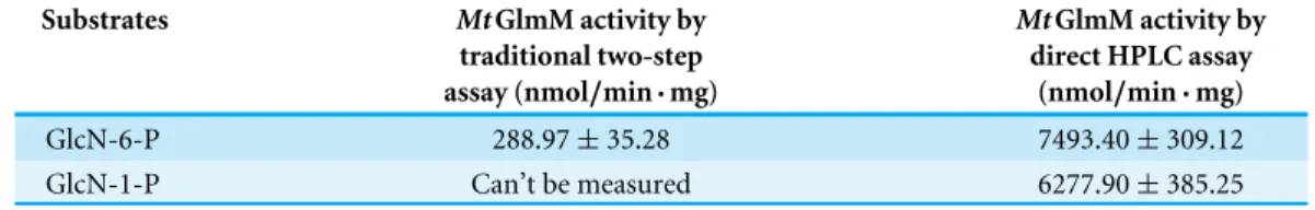
Table 2 The activity ofMtGlmM with GlcN-6-P or GlcN-1-P as substrate was assayed by two meth-ods.The traditional coupled method was assayed in a coupled enzyme system withMtGlmU. The 50µl
reaction mixture contained 50 mM Tris–HCl, pH 8.0, 2.5 mM MgCl2, 1 mM GlcN-6-P, 0.6 mM acetyl-CoA, 0.2 mM Glc-1,6-2P, purifiedMtGlmU (3.5µg) and purifiedMtGlmM (0.18µg). The reaction was
incubated at 37 ◦
C for 20 min. The HPAEC-PAD method was carried out by using the purifiedMtGlmM (0.18µg), incubated at 37 ◦C for 20 min in a substrate buffer (50µl) consisting of 1 mM GlcN-6-P (or
GlcN-1-P) as substrate with 50 mM Tris–HCl, pH 8.0, 2.5 mM MgSO4, and 0.2 mM Glc-1,6-2P. The re-sults are the mean±S.D. for three determinations.
Substrates MtGlmM activity by
traditional two-step assay (nmol/min·mg)
MtGlmM activity by direct HPLC assay
(nmol/min·mg)
GlcN-6-P 288.97±35.28 7493.40±309.12
GlcN-1-P Can’t be measured 6277.90±385.25
catalyzes the interconvertion of GlcN-6-P and GlcN-1-P in prokaryote (Fig. 1B). The well-studiedMtGlmM fromMycobacterium tuberculosiswas chosen to validate this idea.
MtGlmM activity was calculated as nanomoles of GlcN-6-P or GlcN-1-P consumed at per minute for per milligram protein (Table 2). It is noteworthy that the activity ofMtGlmM on GlcN-6-P tested by HPAEC-PAD (7493.40±309.12 nmol/min·mg) was much higher
than the traditional coupled assay (288.97±35.28 nmol/min·mg). One possible reason
was that the accuracy of the traditional coupled assay was mainly dependent on the activity of the coupled enzyme GlmU. GlmU is a bifunctional enzyme (Menginlecreulx & Vanheijenoort, 1993; Menginlecreulx & Vanheijenoort, 1994) (Fig. 1B), catalyzed the third and forth steps in the prokaryotic hexosamine pathway. In this coupled system, the activity of GlmM was measured indirectly by detecting the generation of CoA-SH, which was catalyzed by the acetyl transferase activity of GlmU (third step). But the acetyl transferase activity of GlmU has a very strong product inhibition when GlcN-1-P reached 0.003 mM (Singh, Das & Seshadri, 2012), so the GlmU become the rate-limiting enzyme in the traditional coupled assay, which result in the significantly decrease ofMtGlmM activity. Furthermore, GlmU was non-commercial, the expression and purification of GlmU was tedious, which makes the process complicated and the results unstable.
In contrast, HPAEC-PAD method could detect theMtGlmM activity directly, which made the detection more sensitive, accurate and convenient. Moreover, HPAEC-PAD method could detect the substrate consuming and the product increasing at the same time, making the detection of reverse reaction possible. The activity ofMtGlmM on GlcN-1-P tested by HPAEC-PAD was 6277.90±385.25 nmol/min·mg, which comparable with the
activity on GlcN-6-P.
CONCLUSION
Abbreviations
AGM N-acetylglucosamine-phosphate mutase
GlcNAc N-acetylglucosamine
GlcNAc-1-P N-acetylglucosamine-1-phosphate
GlcNAc-6-P N-acetylglucosamine-6-phosphate
GlcN-1-P Glucosamine-1-phosphate
GlcN-6-P Glucosamine-6-phosphate
Glc-1-P Glucose-1-phosphate
Glc-6-P Glucose-6-phosphate
GlmM Glucosamine-phosphate mutase
HPAEC-PAD High-performance anion-exchange chromatography coupled with electrochemical detector
PGM Phosphoglucomutase
ADDITIONAL INFORMATION AND DECLARATIONS
Funding
This research was supported by the National Natural Science Foundation of China (31370811), the National Key Technology Support Program (2013BAB01B01) and the National High Technology Research and Development Program of China
(2011AA10A205). Dr. Heng Yin was supported by the CAS Youth Innovation Promotion Association (2015144). The funders had no role in study design, data collection and analysis, decision to publish, or preparation of the manuscript.
Grant Disclosures
The following grant information was disclosed by the authors: National Natural Science Foundation of China: 31370811. National Key Technology Support Program: 2013BAB01B01.
National High Technology Research and Development Program of China: 2011AA10A205. CAS Youth Innovation Promotion Association: 2015144.
Competing Interests
The authors declare there are no competing interests.
Author Contributions
• Xiaochen Jia conceived and designed the experiments, performed the experiments,
analyzed the data, wrote the paper, prepared figures and/or tables.
• Jian Kang performed the experiments, analyzed the data.
• Heng Yin conceived and designed the experiments, contributed
reagents/materials/anal-ysis tools, reviewed drafts of the paper.
Data Availability
Supplemental Information
Supplemental information for this article can be found online athttp://dx.doi.org/10.7717/ peerj.1517#supplemental-information.
REFERENCES
Anasontzis GE, Kourtoglou E, Mamma D, Villas-Boas SG, Hatzinikolaou DG, Chris-takopoulos P. 2014.Constitutive homologous expression of phosphoglucomutase and transaldolase increases the metabolic flux of Fusarium oxysporum.Microbial Cell Factories13:43DOI 10.1186/1475-2859-13-43.
Boles E, Liebetrau W, Hofmann M, Zimmermann FK. 1994.A family of hexosephos-phate mutases in saccharomyces-cerevisiae.European Journal of Biochemistry
220:83–96DOI 10.1111/j.1432-1033.1994.tb18601.x.
Fang WX, Du T, Raimi OG, Hurtado-Guerrero R, Marino K, Ibrahim AFM, Albarbarawi O, Ferguson MAJ, Jin C, Van Aalten DMF. 2013.Genetic and structural validation ofAspergillus fumigatusN-acetylphosphoglucosamine mutase as an antifungal target.Bioscience Reports33:689–699 DOI 10.1042/BSR20130053.
Greig KT, Antonchuk J, Metcalf D, Morgan PO, Krebs DL, Zhang JG, Hacking DF, Bode L, Robb L, Kranz C, De Graaf C, Bahlo M, Nicola NA, Nutt SL, Freeze HH, Alexander WS, Hilton DJ, Kile BT. 2007.Agm1/Pgm-3-mediated sugar nucleotide synthesis is essential for hematopoiesis and development.Molecular and Cellular Biology27:5849–5859DOI 10.1128/MCB.00802-07.
Hofmann M, Boles E, Zimmermann FK. 1994.Characterization of the essential yeast gene encoding N-acetylglucosamine-phosphate mutase.European Journal of Biochemistry221:741–747DOI 10.1111/j.1432-1033.1994.tb18787.x.
Jeong JS, Kwon HJ, Lee YM, Yoon HR, Hong SP. 2007.Determination of sugar phosphates by high-performance anion-exchange chromatography coupled with pulsed amperometric detection.Journal of Chromatography A1164:167–173
DOI 10.1016/j.chroma.2007.07.007.
Jolly L, Ferrari P, Blanot D, van Heijenoort J, Fassy F, Mengin-Lecreulx D. 1999.
Reaction mechanism of phosphoglucosamine mutase fromEscherichia coli.European Journal of Biochemistry262:202–210DOI 10.1046/j.1432-1327.1999.00373.x.
Jolly L, Pompeo F, Van Heijenoort J, Fassy F, Mengin-Lecreulx D. 2000. Autophospho-rylation of phosphoglucosamine mutase fromEscherichia coli.Journal of Bacteriology
182:1280–1285DOI 10.1128/JB.182.5.1280-1285.2000.
Kazlowski B, Pan CL, Ko YT. 2015.Monitoring and preparation of neo and agaro-oligosaccharide products by high performance anion exchange chromatography systems.Carbohydrate Polymers122:351–358 DOI 10.1016/j.carbpol.2014.09.003.
Kim HR, Park SY, Kim SB, Jeong H, Choi SK, Park SH. 2014.Inactivation of the phosphoglucomutase gene pgm in Paenibacillus polymyxa leads to overproduction of fusaricidin.Journal of Industrial Microbiology & Biotechnology 41:1405–1414
Li S, Kang J, Yu WD, Zhou Y, Zhang WL, Xin Y, Ma YF. 2012.Identification of M-tuberculosisRv3441c and smegmatis MSMEG_1556 and essentiality of M-smegmatis MSMEG_1556.PLoS ONE7(8):pe42769
DOI 10.1371/journal.pone.0042769.
Li YM, Zhou Y, Ma YF, Li XB. 2011.Design and synthesis of novel cell wall in-hibitors ofMycobacterium tuberculosisGlmM and GlmU.Carbohydrate Research
346:1714–1720DOI 10.1016/j.carres.2011.05.024.
Marcellin E, Nielsen LK, Abeydeera P, Kromer JO. 2009.Quantitative analysis of intracellular sugar phosphates and sugar nucleotides in encapsulated streptococci using HPAEC-PAD.Biotechnology Journal4:58–63DOI 10.1002/biot.200800197.
Menginlecreulx D, Vanheijenoort J. 1993.Identification of the Glmu gene encoding N-acetylglucosamine-1-phosphate uridyltransferase inEscherichia-coli.Journal of Bacteriology175:6150–6157.
Menginlecreulx D, Vanheijenoort J. 1994.Copurification of glucosamine-1-phosphate acetyltransferase and N-acetylglucosamine-1-phosphate uridyltransferase activities ofEscherichia-Coli—characterization of the Glmu gene-product as a bifunctional enzyme catalyzing 2 subsequent steps in the pathway for Udp-N-acetylglucosamine synthesis.Journal of Bacteriology176:5788–5795.
Milewski S, Gabriel L, Olchowy J. 2006.Enzymes of UDP-GlcNAc biosynthesis in yeast.
Yeast 23:1–14DOI 10.1002/yea.1337.
Naught LE, Tipton PA. 2005.Formation and reorientation of glucose 1,6-bisphosphate in the PMM/PGM reaction: transient-state kinetic studies.Biochemistry44:6831–6836
DOI 10.1021/bi0501380.
Nishinari M, Aoyama N, Ogawa Z, Yukino S, Oka S, Yano K, Kurosaki Y, Takeuchi I, Imaki R, Tojo T, Shimohama T, Takehana H, Izumi T. 2012.Phosphoglucomutase activity as a novel biomarker in patients with acute myocardial infarction.Circulation Journal 76:2197–2203DOI 10.1253/circj.CJ-11-1444.
Piacente F, Bernardi C, Marin M, Blanc G, Abergel C, Tonetti MG. 2014. Characteriza-tion of a UDP-N-acetylglucosamine biosynthetic pathway encoded by the giant DNA virus Mimivirus.Glycobiology 24:51–61DOI 10.1093/glycob/cwt089.
Shackelford GS, Regni CA, Beamer LJ. 2004.Evolutionary trace analysis of the alpha-D-phosphohexomutase superfamily.Protein Science13:2130–2138
DOI 10.1110/ps.04801104.
Singh VK, Das K, Seshadri K. 2012.Kinetic modelling of GlmU reactions— prioritization of reaction for therapeutic application.PLoS ONE7(8):e43969
DOI 10.1371/journal.pone.0043969.
Vasseur S, Manie SN. 2015.ER stress and hexosamine pathway during tumourigenesis: a pas de deux?Seminars in Cancer Biology33:34–39
DOI 10.1016/j.semcancer.2015.04.001.
Vosseller K, Sakabe K, Wells L, Hart GW. 2002.Diverse regulation of protein function by O-GlcNAc: a nuclear and cytoplasmic carbohydrate post-translational modifica-tion.Current Opinion in Chemical Biology6:851–857
Wang Y, Zhang YHP. 2010.A highly active phosphoglucomutase from clostridium thermocellum: cloning, purification, characterization and enhanced thermostability.
Journal of Applied Microbiology108:39–46DOI 10.1111/j.1365-2672.2009.04396.x.
Wells L, Vosseller K, Hart GW. 2001.Glycosylation of nucleocytoplasmic proteins: signal transduction and O-GlcNAc.Science291:2376–2378
DOI 10.1126/science.1058714.
Yang LC, Hsieh CC, Lin WC. 2015.Characterization and immunomodulatory activity of rice hull polysaccharides.Carbohydrate Polymers124:150–156
DOI 10.1016/j.carbpol.2015.02.025.
Zachara NE, Hart GW. 2004.O-GlcNAc a sensor of cellular state: the role of nucleo-cytoplasmic glycosylation in modulating cellular function in response to nutrition and stress.Biochimica et Biophysica Acta (BBA)—General Subjects1673:13–28