Alireza Manafi Rasi, MD; Gholamhossein Kazemian, MD; Mohamad M Omidian, MD; Ali Nemati, MD Research performed at Department of Orthopedic Surgery, Imam Hosein Hospital, Shahid Beheshti University of Medical Sciences, Tehran,Iran
Introduction
T
ibiofibular syndesmosis is an anatomical term that describes the connection between the distal tibia and the fibula which forms ankle mortise (1). This structure is composed of four ligaments including the an -terior tibiofibular ligament (AITFL), the pos-terior inferior tibiofibular ligament (PITFL), the transverse tibiofibular ligament, and the interosseous tibiofibular ligament (2). Ankle fractures, one of the most common orthopedic in -juries, can be associated with syndesmosis injury and diastasis, especially when resulting from an external ro -tation mechanism (3-8). It is reported that syndesmosis injury occurs in more than 13% of patients with ankle fracture and in 20% of patients requiring internal fixation (2, 9-11). Injury to the syndesmosis results in severe an -kle instability and requires long-term treatment (12, 13). There is consensus that anatomical reduction of ankle fractures and preservation of the stability and congruity of the mortise is an important prerequisite for achievinggood long-term results and can decrease the incidence of posttraumatic osteoarthritis, but the appropriate method for evaluating syndesmosis stability in ankle fractures re -mains controversial (1, 14-22).
There are several methods used to evaluate the syndes -mosis stability including plain radiography, magnetic res -onance imaging (MRI), CT scanning and arthroscopy (4-6, 13). HR Chissell et al believed that CT imaging is more sen -sitive than radiography in the diagnosis of syndesmosis injuries; however, diagnosing a very small (1 mm) diasta -sis is difficult using this method (22).
Based on the necessity of ankle anatomical reduction in patient function and prevention of late complications, it is obvious that we need to find an accurate method to assess the quality of syndesmosis reduction after ankle surger -ies. Some believe that plain radiography is not a sensitive and accurate method for the diagnosis of syndesmosis in -juries and orthopedic literature suggests that postopera -tive CT scanning is useful to assess mortise reduction (5,
Corresponding Author:Gholamhossein Kazemian, Department of
Orthopedic Surgery, Imam Hossein Hospital, Shahid Madani St, Tehran, Iran.
Email: Ali_nemati802002@yahoo.com
Arch Bone Joint Surg. 2013; 1(2): 98-102. http://abjs.mums.ac.ir
the online version of this article abjs.mums.ac.ir
Received: 16 July 2013 Accepted: 14 December 2013
Syndesmotic Malreduction after Ankle ORIF;
Is Radiography Sufficient?
Abstract
Background: Ankle fractures, especially those resulting from external rotation mechanisms are associated with injury to
the distal tibioibular syndesmosis. Some authors have recommended performing CT scanning after open ankle surgery to evaluate the reduction of syndesmosis. In this current study, we aimed to investigate the sensitivity of plain radiography in diagnosing syndesmosis malreduction after open reduction and internal ixation (ORIF) in patients with ankle fractures.
Methods: Thirty patients with ankle fractures participated in this prospective study. ORIFs were performed with respect
to all of the technical guidelines shown in orthopedic literature for exact syndesmosis reduction, such as ibular length and proper settings. In the operating room, plain radiography was performed in anteroposterior, mortise and lateral views to as
-sess whether syndesmosis was malreduced. If malreduction was detected, the patient was revised. As the gold standard, patients underwent postoperative bilateral CT scanning to investigate the syndesmosis reduction which was then compared to the healthy side. Finally, the sensitivity of plain radiography in the diagnosis of syndesmosis malreduction was determined by comparing this method to CT scanning.
Results: In both of the methods we did not ind any patient with syndesmosis malreduction. Hence, the sensitivity of plain radiography was determined 100%.
Conclusion: Based on our indings, there is no need to perform CT scanning to evaluate syndesmosis reduction after ankle
ORIF in patients with ankle fractures. Plain radiography is suficient and has satisfactory sensitivity in these patients.
23, 24). However, plain radiography is inexpensive, rapid and available and most of the previous studies have inves -tigated the adequacy of plain radiography in diagnosis of syndesmosis injuries preoperatively. To our knowledge, only one study has evaluated the adequacy of standard postoperative radiographic measurements in syndesmo -sis reduction compared to CT scanning (23). In this cur -rent study we have aimed to investigate the diagnostic value of plain radiography in diagnosing postoperative syndesmosis malreduction in patients with ankle fracture and syndesmosis diastasis compared to CT scanning.
Materials and Methods
In this prospective study, inclusion criteria are patients with closed ankle fractures diagnosed by a physical ex -amination (squeeze test and external rotation stress test) and plain radiography in the anteroposterior (AP), lateral and mortise, who were older than 18 and needed open reduction and internal fixation (ORIF). Before the study, patients signed informed consent forms. Radiographic criteria for the diagnosis of syndesmosis disruption were: the tibiofibular clear space >5 mm and tibiofibular over -lap <10 mm in the AP view; tibiofibular over-lap <10 mm in the mortise view; and posterior or anterior subluxation in the lateral view. The AP radiograph was taken when the leg was in neutral rotation and the foot was plantigrade. The x-ray beam was placed anterior to the ankle. For the mortise view, the technique was similar to the AP view except that the leg position was in 15° internal rotation.
However, the definite syndesmosis disruption was deter -mined based on intraoperative examinations such as the Cotton test. If the patient required syndesmosis reduction, a cortical screw was placed 1.5-2 cm distance from the joint line. The screw was placed parallel to the joint line.
Immediately after surgery, the syndesmosis reduction was assessed in the operating room by plain radiography in the AP, lateral and mortise views and if malreduction was detected, the cortical syndesmosis screw was removed and it was again reduced. If, based on the radiographic pa -rameters, syndesmosis reduction was acceptable, patient was taken out of the operating room and bilateral ankle CT scanning was performed within the first postoperative day. In this study, based on the findings of Dikos et al, we
used CT imaging of the healthy side to judge the quality of syndesmosis reduction on the operated side (25). Lastly, the presence of incoherency between the findings of CT scanning and plain radiography was investigated and the adequacy of postoperative plain radiography in the diag -nosis of syndesmosis malreduction was determined by comparing it to CT scanning.
Results
Thirty (30) patients enrolled in this study and the mean age was 26.7±5.5 years (range: 20-37 years). Of them, 22 patients were male (73.3%) and eight patients were fe -male (26.7%). Ankle fractures were the result of direct trauma in 25 patients (83.3%) and ankle sprain in five pa -tients (16.7%). Based on the Lauge-Hansen classification,
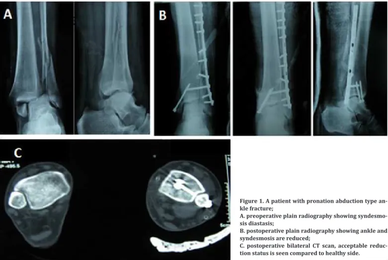
Figure 1. A patient with pronation abduction type an-kle fracture;
A. preoperative plain radiography showing syndesmo-sis diastasyndesmo-sis;
B. postoperative plain radiography showing ankle and syndesmosis are reduced;
there were six (20%), 15 (50%) and nine (30%) fractures that resulted from supination-external rotation, prona -tion-external rotation and pronation-abduction mecha -nisms, respectively. There was no patient with supination-adduction fracture in our study.
After the operation, we found that adequate syndesmosis reduction, as shown by plain radiography, was approved by CT scanning (Figure 1). In this study, we found no pa -tient with syndesmosis malreduction in CT imaging. The accuracy of plain radiography for diagnosing syndesmosis malreduction after ankle ORIF was determined 100%.
Discussion
This study showed that the sensitivity of plain radiog -raphy was high in diagnosing syndesmosis malreduction after ankle ORIF and based on this finding, it seems that postoperative CT scanning to evaluate syndesmosis re -duction is not necessary.
Stability of the syndesmosis, which is the anatomical relationship between the tibia and fibula and plays an important role in the mortise architecture, is a vital pre -requisite for normal ankle function and lack of this, is associated with pain and ankle instability and weakness (12, 13). Also, lack of treatment or improper treatment of syndesmosis injuries can result in latent diastasis, chronic instability, further injury, arthritic changes, chronic pain, osteochondral lesions, and other sequelae (26). Sagi et al examined the correlation between syndesmosis mal -reduction and functional outcome after at least two years of follow-up and found that patients with malreduction have worse functional outcomes based on the Short Form Musculoskeletal Assessment and Olerud-Molander ques -tionnaires. Moreover, they observed that open reduction of the syndesmosis results in a substantially lower rate of malreduction (27). For this reason, examining syndesmo -sis stability and reduction is crucial in assessing patients with ankle traumas. However, there is an important draw -back with examining syndesmosis reduction in patients with ankle fracture and it is that the appropriate method for the assessment of syndesmosis reduction in ankle fractures remains controversial (1, 14-22). Currently, we have no definite quantitative parameters for examining and diagnosing syndesmosis injuries and most clinical tri -als with greater sample sizes are required (14, 25, 28).
CT scanning is the main method for assessing syndesmo -sis reduction and the advantages of this method in diag -nosing syndesmosis injuries are shown in several studies (29-31). However, due to limited studies efforts to find some quantitative criteria for normal syndesmosis on CT imaging, the diagnostic efficacy of this method for detec -tion of syndesmosis malreduc-tion is to some extent con -troversial (25, 32). Also, in spite of several advantages of CT scanning, plain radiography is rapid and inexpensive and in the case of acceptable diagnostic efficacy for diag -nosing syndesmosis malreduction, can be a proper option to assess syndesmosis. Several authors have investigated the adequacy of plain radiography in diagnosing syndes -mosis injuries compared to other methods.
Jenkinson et al compared the intraoperative fluoroscop -ic stress testing, stat-ic radiographs, and biomechan-ical criteria for the diagnosis of syndesmosis instability in 38 patients with external rotation type ankle fractures and found that preoperative radiographs and biomechanical criteria are not appropriate methods to detect syndesmo -sis instability. Moreover, they advised intraoperative stress
fluoroscopy as a valuable tool to detect unstable syndes -mosis injuries (33). Takao et al compared the accuracy of
standard anteroposterior (AP) radiography, mortise radi -ography and MRI with arthroscopy of the ankle to diagno -sis syndesmo-sis disruption in 52 patients. They found that in comparison with arthroscopy, the sensitivity, specificity and accuracy were 44.1%, 100% and 63.5% for standard AP radiography and 58.3%, 100% and 71.2% for mortise radiography. For MRI they were 100%, 93.1% and 96.2% for a tear of the anterior inferior tibiofibular ligament and 100%, 100% and 100% for a tear of the posterior inferior tibiofibular ligament. They concluded that AP and mortise radiography did not provide a correct diagnosis, contraty to MRI imaging. However, Takao et al demonstrated that
ankle arthroscopy is crucial for the accurate diagnosis of a tear of the tibiofibular syndesmosis (6). Also, Lui et al
compared the intraoperative stress radiographic and ar -throscopic diagnosis of syndesmosis disruption in 53 pa -tients with acute ankle fractures (type B or C based on the Weber classification) and found that ankle arthroscopy is superior to intraoperative stress radiography in diagno -sising syndesmosis disruption (5). In another study, Oae
et al compared the diagnostic value of MRI and arthros -copy in syndesmosis injuries in 58 patients with ankle sprain or distal fibular fracture. They considered ligament disruption when MRI showed ligament discontinuity or nonvisualization of the ligament and concluded that MRI using the both criteria is highly accurate to diagnose syn -desmosis injury (4). Also, Paredes-Vázquez et al compared
the diagnostic efficacy of plain radiography and CT scan -ning in syndesmosis diastasis and showed that the abil -ity of plain radiography is questionable to define the syn -desmosis diastasis and the CT scanning should be used in cases of a doubtful diagnosis (34).
As mentioned above, most of the studies have shown that plain radiography is not an appropriate diagnostic method for syndesmosis disruption. However, it is of importance that most of these studies have evaluated the diagnostic value of radiography before operating and postoperative diagnostic value has been limitedly investigated. Gardner
et al determined the adequacy of standard postoperative
radiographic measurements in assessing syndesmosis reduction compared to CT in 25 patients with ankle frac -tures and syndesmosis instability, who underwent open reduction and syndesmosis fixation. Postoperative stand -ard radiography and CT scanning were performed for all patients. In the CT scan, if the distance between the fibula and the anterior and posterior facets of the incisura was >2 mm, the syndesmosis was considered as incongruous. The sensitivity and specificity of the radiographs were 31% and 83%, respectively. Gardner et al concluded that
radiographic measurements were not accurate in diag -nosing syndesmosis malreduction and postoperative ra -diographic measurements were not accurate to assess the quality of the reduction (23). Sagi et al found that after
the operation, 39% of the patients had syndesmosis mal -reduction and they recommended that surgeons obtain a postoperative CT scan in order to compare it to the healthy side (27). In addition to these findings, important ortho -pedic literature recommends obtaining a postoperative CT scan to assess the quality of syndesmosis reduction (24). Also, nowadays there are portable CT scanners used for intraoperative evaluations in some medical centers.
1. Bartoní�cek J. Anatomy of the tibiofibular syndesmosis and its clinical relevance. Surg Radiol Anat. 2003;25(5-6):379-86.
2. Pettrone FA, Gail M, Pee D, Fitzpatrick T, Van Herpe LB. Quantitative criteria for prediction of the results after displaced fracture of the ankle. J Bone Joint Surg Am. 1983; 65:666-77.
3. van den Bekerom MP, Haverkamp D, Kerkhoffs GM, van Dijk CN. Syndesmosis stabilization in pronation exter -nal rotation ankle fractures. Clin Orthop Relat Res. 2010;468(4):991-5.
4. Oae K, Takao M, Naito K, Uchio Y, Kono T, Ishida J, Ochi M. Injury of the tibiofibular syndesmosis: value of MR imaging for diagnosis. Radiology. 2003;227(1):155-61.
5. Lui TH, Ip K, Chow HT. Comparison of radiologic and arthroscopic diagnoses of distal tibiofibular syndes -mosis disruption in acute ankle fracture. Arthroscopy. 2005;21(11):1370.
6. Takao M, Ochi M, Oae K, Naito K, Uchio Y. Diagno -sis of a tear of the tibiofibular syndesmo-sis. The role of arthroscopy of the ankle.J Bone Joint Surg Br. 2003;85(3):324-9.
7. Hermans JJ, Beumer A, Hop WC, Moonen AF, Ginai AZ. Tibiofibular syndesmosis in acute ankle fractures: ad -ditional value of an oblique MR image plane. Skeletal Radiol. 2012;41(2):193-202.
8. Hermans JJ, Beumer A, Hop WC, Moonen AF, Ginai AZ. Tibiofibular syndesmosis in acute ankle fractures: ad -ditional value of an oblique MR image plane.Skeletal Radiol. 2012;41(2):193-202.
9. Kennedy JG, Johnson SM, Collins AL, DalloVedova P, McManus WF, Hynes DM, et al. An evaluation of the Weber classification of ankle fractures. Injury. 1998;29(8):577-80.
10. Court-Brown CM, McBirnie J, Wilson G. Adult ankle fractures-an increasing problem? Acta Orthop Scand. 1998;69(1):43-7.
11. Lindsjo U. Operative treatment of ankle fractures. Acta Orthop Scand. 1981;189:1-131.
12. Wagener ML, Beumer A, Swierstra BA. Chronicin -stability of the anterior tibiofibular syndesmosis of the ankle. Arthroscopic findings and results of ana -tomical reconstruction. BMC Musculoskelet Disord. 2011;12:212.
13. Taser F, Shafiq Q, Ebraheim NA. Three-dimension -al volume rendering of tibiofibular joint space and quantitative analysis of change in volume due to tibiofibular syndesmosis diastases. Skeletal Radiol. 2006;35(12):935-41.
14. Liu X, Yu G.Progress in diagnosis and treatment of dis -tal tibiofibular syndesmosis injury.Zhongguo Xiu Fu Chong Jian Wai Ke Za Zhi. 2012;26(5):612-6.
15. Yamaguchi K, Martin CH, Boden SD, Labropoulos PA. Operative treatment of syndesmosis disruptions with -out use of a syndesmosis screw: a prospective clinical study. Foot Ankle Int. 1994;1(8):407-14.
16. Weening B, Bhandari M. Predictors of functional out -come following transsyndesmosis screw fixation of ankle fractures. J Orthop Trauma. 2005;19(2):102-8. 17. Ramsey PL, Hamilton W. Changes in tibiotalar area of
contact caused by lateral talar shift. J Bone Joint Surg
References
Alireza Manafi Rasi MD Gholamhossein Kazemian MD Mohamad M Omidian MD Ali Nemati MD
Department of Orthopedic Surgery Imam Hossein Hospital
Shahid Beheshti University of Medical Sciences Tehran, Iran
other words, we have tried to find out whether postop -erative CT scanning is necessary to assess the quality of syndesmosis reduction in patients with ankle fracture and syndesmosis disruption. However, in contrast to previous studies, we have found that plain radiography is an accu -rate and appropriate tool that can reflect the status of syn -desmosis after the operation and mainly due to that we reoperated on those patients who were diagnosed with had syndesmosis malreduction in the postoperative plain radiography.
In this study, we used the contralateral ankle as the standard to evaluate the quality of reduction and congru -ity of the fractured side on CT imaging. Based on previ -ous studies there is no quantitative parameter available to accurately evaluate the status of the syndesmosis and the authors have recommended using contralateral ankle CT images as the norm. Dikos et al. investigated the charac -teristics of normal tibiofibular relationships on CT images and have shown that due to significant anatomic varia -tions between individuals, comparing a patient’s ankles together provides a precise definition of normal tibiofibu -lar relationships (25). Also, Mukhopadhyay et al. have shown the need for an individual assessment method for
Am. 1976;58(3):356-7.
18. Cedell CA. Total rupture of the syndesmosis in the an -kle joint. Lakartidningen. 1969;66(23):2427-33. 19. Nelson OA. Examination and repair of the AIT
-FL in transmalleolar fractures. J Orthop Trauma. 2006;20(9):637-43.
20. Hovis WD, aiser BW, Watson JT, Bucholz RW. Treat -ment of syndesmosis disruptions of the ankle with bioabsorbable screw fixation. J Bone Joint Surg Am. 2002;84(1):26-31.
21. Kennedy JG, Soffe KE, Dalla Vedova P, Stephens MM, O’Brien T, Walsh MG, et al. Evaluation of the syndes -mosis screw in low Weber C ankle fractures. J Orthop Trauma. 2000;14(5):359-66.
22. Chissell HR, Jones J. The influence of a diastasis screw on the outcome of Weber type-C ankle fractures. J Bone Joint Surg Br. 1995;77(3):435-8.
23. Gardner MJ, Demetrakopoulos D, Briggs SM, Helfet DL, Lorich DG. Malreduction of the tibiofibular syndesmo -sis in ankle fractures.Foot Ankle Int. 2006;27(10):788-92.
24. Davidovitch RI, Egol KA. Ankle fractures. In: Bucholz RW, Court-Brown CM, Heckman JD, Tornetta III P. Rockwood and Green’s fractures in adults. 7th ed. Phil -adelphia: Lippincott Williams & Wilkins; 2010: 1974-2021.
25. Dikos GD, Heisler J, Choplin RH, Weber TG. Normal tib -iofibular relationships at the syndesmosis on axial CT imaging.J Orthop Trauma. 2012;26(7):433-8.
26. Porter DA. Evaluation and treatment of ankle syndes
-mosis injuries. Instr Course Lect. 2009;58:575-81. 27. Sagi HC, Shah AR, Sanders RW. The functional con
-sequence of syndesmosis joint malreduction at a minimum 2-year follow-up. J Orthop Trauma. 2012; 26(7):439-43.
28. Mukhopadhyay S, Metcalfe A, Guha AR, Mohanty K, Hemmadi S, Lyons K, O’Doherty D. Malreduction of syndesmosis--are we considering the anatomical vari -ation? Injury. 2011;42(10):1073-6.
29. Ebraheim NA, Lu J, Yang H, Mekhail AO, Yeasting RA. Radiographic and CT evaluation of tibiofibular syn -desmosis diastasis: a cadaver study. Foot Ankle. 1997;18(11):693-8.
30. Sclafani SJ. Ligamentous injury of the lower tibiofibu -lar syndesmosis: radiographic evidence. Radiology. 1985;156:21-7.
31. Harper MC. An anatomic and radiologic investiga -tion of the tibiofibular clear space. Foot Ankle Int. 1993;14:455-8.
32. Elgafy H, Semaan HB, Blessinger B, Wassef A, Ebraheim NA. Computed tomography of normal distal tibiofibu -lar syndesmosis.Skeletal Radiol. 2010;39(6):559-64. 33. Jenkinson RJ, Sanders DW, Macleod MD, Domonkos A,
Lydestadt J. Intraoperative diagnosis of syndesmosis injuries in external rotation ankle fractures. J Orthop Trauma. 2005;19(9):604-9.