ORIGIN
AL RESEAR
CH
Lung analysis in children mechanically ventilated
with atelectasis after cardiac surgery
Análise pulmonar em crianças mecanicamente ventiladas
com atelectasia após cirurgia cardíaca
Análisis pulmonar en niños mecánicamente ventilados
con atelectasia después de cirugía cardiaca
Adriane Muller Nakato1, Eddy Krueger2, Adriana Maria Trevisan Domingues3,
Cássio Fon Ben Sum4, Bertoldo Schneider Junior1,2
Mailing address: Adriane Muller Nakato – General Carneiro, 181 – CEP 80060-900 – Curitiba (PR), Brazil. E-mail: [email protected] – Phone: +55 41 9901 2692
Presentation: July 2014 – Accepted for publication: Apr. 2015 – Financing sources: Financial support: Coordination of Improvement of Higher Education Personnel (Capes), National Counsel of Technological and Scientiic Development (CNPq) and Postgraduate Program in Electrical Engineering and Industrial Informatics (CPGEI) – Conlicts of interest: none – Approved by the Ethics and Research committee, under registration number 1034-11.– Protocol no. 1034-11.
Study developed at Hospital Pequeno Príncipe l, Department of Cardiac Intensive Care – Curitiba (PR), Brazil.
1 Graduate School of Electrical Engineering, Biomedical Engineering Program, Universidade Tecnológica do Paraná (UTFPR) – Curitiba
(PR), Brazil.
2 Graduate Program in Biomedical Engineering, Universidade Tecnológica do Paraná (UTFPR) – Curitiba (PR), Brazil. 3 Pediatric Intensive Care Unit, Hospital Pequeno Príncipe – Curitiba (PR), Brazil.
4 Pediatric Cardiologist of Cardiac Pediatric Intensive Care Unit, Hospital Pequeno Príncipe – Curitiba (PR), Brazil.
ABSTRACT | The main objective of this study is to associate the healthy area of the lung (evaluated by radiography) with the data of respiratory mechanics in children with atelectasis after cardiac surgery, under mechanical ventilation in the assisted controlled mode. Altogether, 46 children were selected, but 16 were excluded due to irregular respiratory waves or lack of the data on arterial blood gases. A group of 30 children under assisted controlled mode were analyzed, and 10 from this group developed atelectasis. The data were analyzed before and after the onset of atelectasis, and respiratory mechanics was correlated to radiography. We also analyzed the data related to arterial blood gas of these children – who initially had no pulmonary complication – to verify possible changes due to assisted controlled cycles. Atelectasis may modify some parameters of respiratory mechanics. In the association of the healthy area of the lung with the respiratory mechanics, the Spearman correlation results showed statistical signiicance of the lung area with airway resistance (ρ=−0.648 and p=0.043). Our results show that it is possible to analyze respiratory mechanics waves by selecting controlled cycles in the assisted controlled mode, since we found insigniicant changes in potential ionic hydrogen. The analysis of respiratory mechanics allows checking lung function and undesired lung injuries; the analysis of respiratory mechanics can be daily performed in these children to have important information on the pulmonary function. Our research also showed that under
268
the assisted controlled mode is also possible to evaluate respiratory mechanics.
Keywords | Pulmonary Atelectasis; Pediatric Thoracic
Surgery; Respiratory Mechanics; Blood Gas Analysis; X-Rays.
269
dos ciclos controlados, uma vez que não houve alteração signiicativa no potencial de hidrogênio iônico. A análise da mecânica respiratória permite veriicar a função pulmonar e as possíveis lesões pulmonares. A análise da mecânica respiratória pode ser usada diariamente nessas crianças, permitindo obter informações importantes da função pulmonar. O estudo também mostrou que no modo ventilatório assistido controlado também é possível avaliar a mecânica respiratória.
Descritores | Atelectasia Pulmonar; Cirurgia Torácica; Mecânica
Respiratória; Gasometria; Raios X.
RESUMEN | El objetivo de este estudio es asociar la área sana del pulmón (evaluado mediante radiografía) a los datos de mecánica respiratoria en niños en el postoperatorio de cirugía cardiaca, con atelectasia en ventilación mecánica en el modo asistido controlado. En el total, fueron seleccionados 46 niños, de los cuales 16 fueron excluidos debido a las ondas respiratorias irregulares o a la falta de datos de la gasometria arterial. Se analizó un grupo de 30 niños en el modo asistido controlado, de los cuales 10 han desarrollado atelectasia. Los datos fueron analizados antes y después del inicio de la atelectasia, y la
mecánica respiratoria se correlacionó a las medidas del área del pulmón. En los 30 niños inicialmente sin complicación pulmonar fueron analizados los datos de la gasometria arterial para comprobar posibles cambios debido a los ciclos asistidos. La atelectasia puede modiicar algunos parámetros de la mecánica respiratoria. En la asociación del área saludable del pulmón con la mecánica respiratoria, los resultados de correlación de Spearman demostraron signiicancia estadística entre el área del pulmón con la resistencia de las vías aéreas (ρ=-0,648 y p=0,043). Los resultados mostraron que es posible el análisis de las ondas de mecánica respiratoria a través de la selección de los ciclos controlados, puesto que no hubo alteración signiicativa en el potencial de hidrógeno iónico. El análisis de la mecánica respiratoria permite comprobar la función pulmonar y las posibles lesiones pulmonares. Es posible utilizar el análisis de la mecánica respiratoria diariamente en estos niños, permitiendo obtener informaciones importantes de la función pulmonar. El estudio también mostró que en el modo de ventilación controlada es posible evaluar la mecánica respiratoria.
Palabras clave | Atelectasia Pulmonar; Cirugía Torácica; Mecánica
Respiratoria; Análisis de los Gases de la Sangre; Rayos X.
INTRODUCTION
Congenital heart defects are the leading causes of newborn mortality. In the United States, around 35,000 babies are born alive with congenital heart disease each year. Of which, 10,000 need heart surgery before one year old1. In Brazil the number is 8 to 10 for every 1,000 born alive2,3. hese babies usually do not reach
adulthood without any surgical procedure2,4-5, whereas
only 20% of them have spontaneous healing3, which
requires attention in the postoperative cardiac surgery and Intensive Care Unit (ICU).
Pulmonary complications are common in the postoperative cardiac surgery, being the causes of morbidity and mortality in these patients6,7. he most
common pulmonary complications in the postoperative period after congenital cardiac surgery are atelectasis and microatelectasis, pleural efusion, bronchospasm, pneumonia and pneumothorax8. hese complications
should be avoided to remove the child from Invasive Mechanical Ventilation (IMV) as soon as possible.
Many of the studies involving analysis of respiratory mechanic devices are performed in patients who are under the efect of sedation and controlled mode in
the mechanical ventilation to exclude the interference of the patient in the ventilation and evaluation9-12.
Although these children generally remain alive for 1 to 2 days under sedatives, after that, ventilation mode is changed to assisted controlled mode to allow the active inluence of the child on respiratory cycle.
he X-ray examination should be avoided, but in clinical practice it is often performed. Ionizing radiation performed by X-ray apparatuses may cause complications and somatic efects later on children since they are more sensitive and vulnerable to radiation13-14.
Ensuring safety and respecting the minimum doses required for this exam without adversely afecting the information obtained is extremely important for good results13. However, we hypothesize that respiratory parameters can reduce the X-ray evaluation rate.
mechanics in assisted controlled mode in comparison with changes in arterial blood gases, analyzing the possible changes that assisted cycles can inluence and change the blood gases analysis.
METHODOLOGY
he study was approved by the Ethics and Research committee of the Hospital Pequeno Príncipe, Curitiba, Brazil, under registration number 1034-11. he parents signed a free and enlightened consent form and an authorization for the research that was carried out in the hospital’s cardiac critical care unit (from January to August, 2012).
In the period analyzed we selected all children in cardiac ICU within the inclusion criteria: age between 0-12 months in a postoperative cardiac surgery situation in the IMV; the exclusion criteria were children with neurological problems, children that were not in a controlled or assisted controlled mode, and children with great hemodynamic instability. During the study period we selected 46 children of both gender, aged 0-12 months, and 16 of them were excluded due to irregular respiratory waves or lack of arterial blood gases data. Groups of 30 children (20 boys and 10 girls) were in assisted controlled mode, not having any pulmonary complication in the initial period in ICU. Ten out of these children developed atelectasis during the ICU period (age 2.25±1.62 months), and 20 children who had other pulmonary complications were excluded – such as pneumothorax and pleural efusion, irregular respiratory signals or lack of arterial blood gases data. Samples for arterial blood gas analysis were obtained from a catheter in the radial artery with a heparinized syringe (Monovette® LH; Sarstedt, Nümbrecht, Germany). he equipment used for analysis was Cobas B121 system (Roche Diagnostics®, Mannheim, Germany) that was daily calibrated.
Respiratory mechanics data were acquired by mechanical ventilator Inter® 5 plus and the Inter® GMX graphic monitor, both from the Intermed, through the use of a low sensor to capture and storage the data, related later to the pressure variation. For low, pressure and volume we used Wintracer® software (Intermed). Respiratory mechanics data were obtained almost simultaneously with radiographic examination and arterial blood analysis. hese procedures were daily performed for each child during their staying at
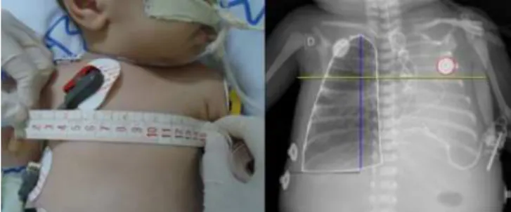
ICU. At the end of child’s period in the ICU, most representative radiographs and respiratory waves were chosen. Children who developed atelectasis were analyzed separately, and their radiographs were examined before and after lesions. For the analysis of the lung area, we used AutoCad® 2012, in which the projection of parts without atelectasis was visually deined, being classiied as functional areas of the lung. he axillary distance and height of the child’s lung was used to establish a calibration of the measures in AutoCad® 2012 (Figure 1).
Figure 1. Analysis of the lung area with the AutoCad® 2012 program
Statistical analysis was performed using the SPSS®
(version 18). he normality test Shapiro-Wilk was used, and presented non-normal distribution in the groups. It was applied non-parametric statistics, Mann-Whitney test to non-paired group, Wilcoxon test to paired group and Spearman to correlation coeicient.
RESULTS
All children were analyzed in the assisted controlled mode. he characteristics of the group without lung injury are shown in Table 1. he pulmonary complication most frequent during Invasive Mechanical ventilation (IMV) was atelectasis, followed by pleural efusion and pneumothorax.
Before injury, we veriied a signiicant change compared with normal arterial blood gases and no changes in the blood’s potential ionic hydrogen (pH). In the arterial partial pressures of carbon dioxide (PaCO2), we found a mild hypercapnia, but this small increase in PaCO2 did not signiicantly altered the pH (signiicance threshold used was p=0.05). he bicarbonate (HCO3-)
271
Table 1. Descriptive characteristics of the group without lung injury (n=30)
Variables Mean±sd Minimum – Median – Maximum
Age (months) 3.23±2.52 0.5–2–9
Weight (kg) 4.29±1.46 2.25–3.94–7.33
Time in IMV (days) 2.43±1.97 1–2–9
VT (ml) 50.22±23.27 3.9–50.22–97.9
PPEAK (cmH2O) 25.39±4.93 17.6–25.05–35.5
PPLAT (cmH2O) 21.16±4.61 14.4–22.11–31.2
PEEP (cmH2O) 6.02±1.26 4.6–6–10.1
Cst (ml/cmH2O) 3.54±2.10 0.22–3.25–9.02
Cdyn (ml/cmH2O) 2.64±1.45 0.02–2.53–5.98
Surface Lung Area (cm2) 84.98±23.18 30.92–88.12–135.3
Raw (cmH2O/L/seg) 41.61±16.74 16.64–38.87–77.33
RR (bpm) 25.43±6.18 18–23.5–41
pH 7.36±0.06 7.2–7.39–7.5
PaCO2 (mmHg) 43.87±8.26 31.4–40.1–64.3
PaO2 (mmHg) 124.99±60.78 33.4–141.9–255.4
HCO3- (mEq/L) 24.12±4.71 11.7–23.2–39.1
SaO2 (%) 93.17±10.01 64.1–99.15–99.9
Abbreviations:VT – tidal volume; RR – respiratory rate; PPEAK – peak pressure; PPLAT – plateau
pressure; PEEP – positive end-expiratory pressure; Cdyn – dynamic compliance; Cst – static
compliance; Raw – airway resistance; pH – potential hydrogen; PaCO2 – partial pressure of arterial
carbon dioxide; PaO2 – partial pressure of arterial oxygen; HCO3–bicarbonate; SpO2 – oxygen
saturation; mmHg – millimeters of mercury; ml – milliliter; mL/kg – milliliter per kilogram; bpm – breaths per minute; s – seconds; cmH2O – water centimeter; l/min – liters per minute; ml/cmH2O
– milliliter per water centimeter; cm2 – square centimeter; mEq/L – milliequivalents per liter
*Signiicant diference at a signiicance level of p<0.05
Table 2. Comparison of the group without lung injury with the normal values (n=30) using the Wilcoxon (W) test (n=30)
Variables Reference value p
pH 7.4 (7.35-7.45mmHg) 0.36
PaCO2 40 (35-45mmHg) 0,0449*
PaO2 90 (80-100mmHg) 0,006*
PaO2 (1-6months) 72.5 (60-85mmHg) 0.001*
HCO3- 24 (22-26mEq/L) 0.8936
*Signiicant diference at a signiicance level of p<0.05
he data show no signiicant pH change in these children without lung injury in IMV, showing that the assisted-controlled ventilation mode did not inluenced the blood gas analysis, which presented only mild hypercapnia (PaCO2) – the accumulation of carbon dioxide (CO2) can occur due to hypoventilation. hus, the children’s Respiratory Rate (RR) decreased and they could not eliminate this CO2. It is probable that assisted breath was not enough to alter the data on the blood gas analysis because few inspiratory intentions were captured.
Regarding RR in children without lung injury, the average was 25.43bpm, which takes into account that this estimated RR for the age group is below the expectance for the average during 3.23 months (30-35 bpm for children with 3 months of age). his could lead to a slight increase in CO2 levels.
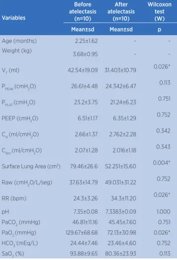
Among the groups with atelectasis, the reduction of parameters statistically signiicant was the Tidal Volume (TV), surface lung area and PaO2, and the increase of RR and Time in IMV. In the group with atelectasis, RR had a signiicant increase (p=0.025), with higher respiratory efort. he results that had statistical signiicance using the Wilcoxon test to compare the groups without/with atelectasis are presented in Figures 2 and 3, and the descriptive characteristics of the atelectasis group are presented in Table 3.
Without atelectasis 40 20 25 30 35 40 45 50 55 60 65 60 80 100 120 140 Without atelectasis With atelectasis R espir a tory Ra te (bpm) Surf ac
e Lung Ar
ea ( cm 2) p=0.004 (A) (B) p=0.026 With atelectasis
Figure 2. Signiicant reduction in (A) surface lung area and signiicant increase in (B) respiratory rate in the groups with and without atelectasis Without atelectasis 50 10 20 30 40 50 60 70 80 90 100 100 150 200 250 300 P aO2 (mmHg) T idal V olume (ml) (A) (B) p=0.026 p=0.026
With atelectasis Without atelectasis With atelectasis
Figure 3. Signiicant reduction in (A) PaO2 and in the (B) tidal volume in the group with and without atelectasis
with atelectasis (37.63 to 49.03cmH2O/L/s). he group had higher values compared with the normal values for the age (10 to 20cmH2O/L/s). In the group without lesion, the Raw also increased(41,61±16,74cmH2O/L/s). he results of the Spearman coeicient (ρ) showed statistical signiicance between the surface lung area and the Raw parameters (ρ=−0.648 with p=0.043) for the group with atelectasis point, whereas the group with atelectasis had a negative moderate correlation. his means that the smaller the area of the lung, the greater the value of Raw.
Table 3. Comparison between before and after the appearance of the atelectasis
Variables
Before atelectasis
(n=10)
After atelectasis
(n=10)
Wilcoxon
test
(W)
Mean±sd Mean±sd p
Age (months) 2.25±1.62 -
-Weight (kg)
3.68±0.95 -
-VT (ml) 42.54±19.09 31.403±10.79 0.026*
PPEAK (cmH2O) 26.61±4.48 24.342±6.47 0.113
PPLAT (cmH2O) 23.2±3.75 21.24±6.23 0.751
PEEP (cmH2O) 6.51±1.17 6.35±1.29 0.752
Cst (ml/cmH2O) 2.66±1.37 2.762±2.28 0.342
Cdyn (ml/cmH2O) 2.07±1.28 2.016±1.18 0.343
Surface Lung Area (cm2) 79.46±26.6 52.251±15.60 0.004*
Raw (cmH2O/L/seg) 37.63±14.79 49.031±31.22 0.752
RR (bpm) 24.3±3.26 34.3±11.20 0.026*
pH 7.35±0.08 7.3383±0.09 1.000
PaCO2 (mmHg) 46.81±11.16 45.45±7.60 0.751
PaO2 (mmHg) 129.67±68.68 72.13±30.98 0.026*
HCO3-(mEq/L) 24.44±7.46 23.46±4.60 0.752
SaO2 (%) 93.88±9.65 80.36±23.93 0.113
*Signiicant diference at a signiicance level of p<0.05
DC S
his study showed that some parameters of respiratory mechanics decreased and other increased due to atelectasis, but this also may be related to the cardiac surgical procedure itself. he lung function is altered by pulmonary complications as well as by the surgical
procedure and mechanical ventilation itself15. Static
compliance may be reduced compared with normal values for each age group due to a cardiac surgery16.
he Raw can increase due to injury and pulmonary edema, presence of secretions and loss of lung volumes17,18. his increase in airway resistance is
common during surgery and return to normal values after the procedure19.
Another parameter of respiratory mechanics showed that change was VT. As a protective measure, the VT should be below 6ml/kg10, therefore the V
T between
these values was not associated with increased mortality, being used as margin trust20. Dividing the value of the
VT by weight, both groups (with/without atelectasis) and the group without lung injury had values above recommended for VT – thus, this parameter should be considered a protective measure in an ICU.
Regarding the main objective of this study, which was to correlate the healthy area of the lung through X-ray to pulmonary function by respiratory mechanics, it had statistical signiicance between the surface lung area and the respiratory parameters for the atelectasis group, which were the RAW parameters (ρ=−0.648 and p=0.043). We observed that the smaller the area of the lung, the greater the value of RAW. his correlation of the physical with the functional aspect is normally not checked, and we found few studies about the subject. herefore, this study brings new data that can improve pulmonary analysis in children in IMV, since many factors can afect respiratory mechanics (e.g., X-ray). A single study consisting of high-resolution images from computed tomography compared the respiratory mechanics of newborns with very low weight, aiming at to ind an association between lung mechanics and structural changes of the lung parenchyma, and proving thus the relationship between the lung morphological structure and functional alterations in asymptomatic infants of very low birth weight21.
7 3 Although the data presented changes before and after
the appearance of atelectasis, many had no statistical power, even with changes in all parameters – probably due to the sample size. A study with a larger number of subjects would be interesting to check if changes occur before and after the onset of pulmonary complications and with the correlation between pulmonary function and physical changes of the lung.
CO U O
he daily analysis of respiratory mechanics allowed us to verify the functional changes of the lung, analyzing changes both due to pulmonary complications and the surgical procedure.
Only one parameter was related to physical changes in the lung, making room for other studies in this area, since the number of this sample was small and this factor should be considered. Although the health area evaluated for X-ray shows correlation with only one mechanical parameter – airway resistance (ρ=−0.648 and p=0.043) –, it is not viable to reduce the X-ray acquisition rate to perform pulmonary assess and reassessment.
Most studies involving respiratory mechanics suggest the controlled mode and sedation as the best options, but the assisted controlled mode is also possible to perform this analysis, since the children of our study showed no considerable changes in relation to arterial blood gases. Moreover, this procedure can be daily performed with the monitoring of the child on an ICU bed.
ACKNOWLEDGMENTS
he research was carried out in the Department of Cardiac Pediatric Intensive Care, Hospital Pequeno Príncipe. We thank the team of the Hospital and professor PhD Hugo Vieira Neto for statistics support and assistance.
REFERENCES
1. Karamichalis JM. Barachd PR, Nathana M, Henainee R, Nidoa PJ, Bacha EA. Assessment of technical competency in pediatric cardiac surgery. Progress in Pediatric Cardiol. 2012; 33:15-20.
2. Silva ZM, Perez A, Pinzon AD, Ricachinewsky CP, Rech DR, Lukrafka JL, Rovedder PME. Factors associated with failure in ventilatory weaning of children undergone pediatric cardiac surgery [in Portuguese]. Rev Bras Cir Cardiovasc. 2008;23(4):501-6.
3. Pinto-Jr VC, Daher CV, Sallum FS, Jatene MB, Croti UA. Situação das cirurgias cardíacas congênitas no Brasil [in Portuguese]. Rev Bras Cir Cardiovasc. 2004;19(2):3-6. 4. Tchervenkov CI, Jacobs JP, Bernier PL, et al. The
improvement of care for pediatric and congenital cardiac disease across the World: a challenge for the World Society for Pediatric and Congenital Heart Surgery. Cardiol Young. 2008;18(2):63-9.
5. Hofman, JIE, Kaplan S. The Incidence of Congenital Heart Disease. J Am College Cardiol. 2002;39(12):1890-900. 6. Johnson D, Hurst T, Thomson D, Mycyk T, Burbridge B, To
T, Mayers I. Respiratory function after cardiac surgery. J Cardiothoracic Vasc Anesth. 1996;10:571-7.
7. Gergely Albu, Barna Babik, Klára Késmárky, Mariann Balázs, Zoltán Hantos, Ferenc Peták. Changes in airway and respiratory tissue mechanics after cardiac surgery. Ann Thorac Surg. 2010;89:1218-26.
8. Hulzebos EH, Helders PJ, Favié NJ, De Bie RA, Brutel de la Riviere A, Van Meeteren NL. Preoperative intensive inspiratory muscle training to prevent postoperative pulmonary complications in high-risk patients undergoing CABG surgery: a randomized clinical trial. JAMA. 2006; 296:1851-7.
9. Kissoon N, Rimensberger PC, Bohn D. Ventilation strategies and adjunctive therapy in severe lung disease. Pediat Clin North Am. 2008;55:709-33.
10. Amato MBP, Barbas CSV, Medeiros DM, Magaldi RB, Schettino GPP, Lorenzi-Filho G, et al. Efect of a protective-ventilation strategy on mortality in the acute respiratory distress syndrome. New Engl J Med. 1998;338(6):347-54. 11. Tantucci C, Eissa NT, Ranieri VM, Corbeil C, Milic-Emili J.
Respiratory Mechanics in Ventilated Patients. J Crit Care. 1992;7(4):251-5.
12. Faustino EA. Concepts and Monitoring of Pulmonary Mechanic in Patients under Ventilatory Support in Intensive Care Unit [in Portugese]. Rev Bras Ter Inten. 2007;19(2):161-9. 13. Carvalho E, Grilo R, Matela N, Pereira P. Evaluation of the
dose standards in pediatric radiology: comparison between conventional ilm systems and image digitalization systems in infants between 0-5 years old, in the x-ray of the chest in antero-posterior projection [in Portuguese]. Rev. Lusófona Ciênc Tecnol Saúde. 2007;4(21):37-46.
14. Bartley K, Metayer C, Selvin S, Ducore J and Buler P. Diagnostic X-rays and risk of childhood Leukaemia. Int J Epidemiol. 2010;39:1628-37.
15. Caséca MB, Andrade LB, Britto MCA. Pulmonary function assessment in children and teenagers before and after surgical treatment for rheumatic valve disease [in Portuguese]. J Pediat. 2006;82(2):144-50.
Mecânica Prolongada após Cirurgia Cardíaca [in Portuguese]. Arq Bras Cardiol. 2003;80 (3):301-5.
17. D’Angelo E, Pecchiari M, Baraggia P, Saetta M, Balestro E, Milic-emili J. Low-volume ventilation causes peripheral airway injury and increased airway resistance in normal rabbits. J Appl Physiol. 2002;92:949-56.
18. Westaby S. Organ dysfunction after cardiopulmonary bypass. A systemic inlammatory reaction initiated by the extracorporeal circuit. Int Care Med. 1987;13(2):89-95. 19. Lanteri CJ, Kano S, Duncan AW, Sly PD. Changes in respiratory
mechanics in children undergoing cardiopulmonary bypass. Am J Respir Crit Care Med. 1995;152(Pt 1):1893-900.
20. Khemani RG, Conti D, Alonzo TA, Bart III RD, Newth CJL. Efect of tidal volume in children with acute hypoxemic respiratory failure. Intensive Care Med. 2009; 35:1428-37.
21. Mello RR, Dutra MVP, Ramos JR, Daltro P, Boechat M, Lopes JMA. Lung mechanics and high-resolution computed tomography of the chest in very low birth weight premature infants. Sao Paulo. Med J. 2003;121(4):167-72.