Secondary caries is the most common cause of dental restoration failures. This study aimed to compare the diagnostic accuracy of conventional and digital intraoral radiography and cone beam computed tomography (CBCT) for detection of recurrent caries around composite restorations. mesio-occluso-distal (MOD) cavities were prepared using bur on 45 extracted sound human molar teeth. The teeth were divided into 3 groups. In the control group, cavities were restored with composite resin after etching and bonding (n=15). In Group 2, 500-μm thick wax was placed over the buccal, lingual and gingival walls and the cavities were restored with composite resin. Group 3 specimens were subjected to pH cycling and artificial caries were created on the buccal, lingual and gingival walls. The cavities were restored with composite. Conventional and digital photo-stimulable phosphor (PSP; Optime) radiographs and two CBCTs images (NewTom 3G and Cranex 3D) were obtained from them. Presence or absence of caries in the cavity walls was assessed on these images. Data were analyzed using Kappa statistic. The diagnostic accuracy of CBCT was significantly higher than that of digital and conventional intraoral radiography (p<0.05). The accuracy was 0.83, 0.78, 0.55 and 0.49 for CBCT Cranex 3D, CBCT NewTom 3G, conventional and digital intraoral radiography, respectively. CBCT has a higher diagnostic accuracy than digital and conventional intraoral radiography for detection of secondary caries around composite restorations.
C o m p a r i s o n o f C o n e - B e a m
Computed Tomography and Intraoral
Radiography in Detection of Recurrent
Caries under Composite Restorations
Shahin Kasraei1, Abbas Shokri2, Jalal Poorolajal3, Samira Khajeh4, Hamid Rahmani5
1Dental Research Center, Department
of Restorative Dentistry, Dental School, Hamadan University of Medical Sciences, Hamadan, Iran 2Dental Research Center, Department
of Oral and Maxillofacial Radiology, Dental School, Hamadan University of Medical Sciences, Hamadan, Iran 3Modeling of Noncommunicable
Diseases Research Center, Department of Epidemiology, School of Public Health, Hamadan University of Medical Sciences, Hamadan, Iran 4Department of Oral and
Maxillofacial Radiology, Dental School, Kurdistan University of Medical Sciences, Sanandaj, Iran 5Dental School, Hamadan University
of Medical Sciences Hamadan, Iran
Correspondence: Abbas Shokri, Shahid Fahmideh Blvd, in front of Mardom Park, Hamadan, Zip code: 6516647447, Iran. Tel: +98-813-825-1885. e-mail: [email protected]
Key Words: dental caries, composite resins, cone beam computed tomography, dental digital radiography.
Introduction
Despite the advances in composite restorative materials and dentin bonding systems, secondary caries is still a main cause for failure of resin restorations (1,2). Accurate, early detection of recurrent caries is the key for success and longevity of dental restorations. Radiography is among the most important techniques for detection of caries, particularly in the posterior teeth (3). Secondary caries are defined as carious lesions developed at the margins of existing restorations (3,4). These carious lesions are formed as extrinsic lesions. Histopathologically, they resemble the primary caries; however, the walls of the secondary carious lesions are thinner than those of the primary caries that develop within ftthe enamel or dentin (5). The primary diagnosis of recurrent caries around composite restorations is especially important because these restorations lack self-sealing and antibacterial properties . Resin restorations of the posterior teeth must be radiopaque because detection of marginal defects, overhangs and secondary caries around these restorations highly depends on the radiopacity of these restorations (6). Diagnosis of secondary caries at the interproximal surfaces is extremely difficult unless the carious lesion is large enough or has resulted in loss of tooth structure (7-9). Considering the high prevalence
of secondary caries and the importance of preserving tooth vitality, early detection of these lesions is of utmost importance to prevent further extension.
CBCT is a high-quality radiography for diagnosis and treatment planning. This imaging modality provides three-dimensional (3D) images of axial, coronal and sagittal planes with excellent submillimeter resolution (10). CBCT has been suggested as a suitable tool for detection of small carious lesions (11). Intraoral radiography provides 2D images of the teeth and thus, caries on the buccal and lingual walls cannot be detected using this technique; late or no diagnosis of these lesions may lead to pulp involvement.
In the literature, studies comparing the detection of recurrent caries around composite restorations using CBCT and intraoral radiography are scarce. The current study aimed to compare the diagnostic accuracy of different CBCT systems with analog and digital intraoral radiography for detection of secondary caries around composite restorations.
Material and Methods
S. Kasraei et al.
Hamadan University of Medical Sciences (Protocol No:2014-16p 142). Forty-five extracted human molar teeth were collected and disinfected with 5.25% sodium hypochlorite and stored in saline solution. Teeth with caries, fracture, previous restorations or cracks were excluded.
Using an 008 fissure bur (Tizkavan, Tehran, Iran), MOD cavities with 2-mm occlusal width, 3-mm gingival width and gingival floor 1 mm below the cementoenamel junction were prepared in all teeth. After cavity preparation, the teeth were divided into three groups of 15 each. Group 1 was considered as the control group (n=15) and the cavities were etched with 36% phosphoric acid (3M ESPE Dental Products, St. Paul, MN, USA) for 15 s, rinsed and dried. Bonding resin (Single Bond; 3M ESPE) was applied according to the manufacturer’s instructions and light
cured for 10 s using Demi LED light curing unit (Kerr Corp., Orange, CA, USA). Dialog composite (Dialog Schutz Dental GmbH, Rosbach, Germany) was incrementally (horizontally) applied in 4 1.5-mm thick layers and cured for 40 s using the light-curing unit. After restoring the cavities, the teeth were stored in water at 37 °C. In Group 2, cavities were prepared and according to previous studies (1,12), a wax sheet 500 μm thick measuring 2 x2 mm was randomly placed over the walls to simulate secondary caries. The cavities were then restored as in the control group. In Group 3, secondary caries was artificially created on the walls using pH cycling method with a cariogenic solution (13). In this group, all tooth surfaces except for the selected walls were coated with nail varnish for the cariogenic solution to exert its effect only on the selected walls. Thus, carious lesions were only induced on the respective walls. The cariogenic solution has two demineralizing and remineralizing solutions. The demineralizing solution contained 2.2 mM CaCl2, 2.2 mM KH2PO4 and 0.05 M acetic acid and the pH was adjusted to 4.4 with 1 M KOH. The remineralizing solution contained 1.5 mM CaCl2, 0.9 mM NaH2PO4 and 0.15 M KCl with a 7.0 pH. For pH cycling, the specimens in Group 3 were stored for 18 h per day in the demineralizing solution and 6 h per
Figure 1. Digital intraoral radiograph of specimens.
Figure 2. Analog (conventional) intraoral radiograph of specimens.
CBCT versus intraoral radiography
day in the remineralizing solution at 37 °C for two weeks, induce carious lesions on the respective walls. Based on a previous study, pH cycling causes 500-μm deep caries in dentin (13); this depth of caries was equal to the thickness of wax used in Group 2.
After pH cycling in Group 3, nail varnish on the remaining surfaces was gently removed with bur and the cavities were restored as in other groups.
Specimens in the Groups 2 and 3 were divided into three subgroups (n=5) to induce caries. Carious lesions were induced in the buccal and lingual walls in the first 5 specimens, in the buccal and gingival walls in the second 5 specimens, and in all gingival, buccal and lingual walls in the final 5 specimens.
Finally, all teeth were mounted in 6 red wax blocks in the form of mandible in separate rows (7 or 8 specimens
in each row). To recognize the teeth on the radiographs, each wax block was coded with a metal wire.
Digital intraoral radiographs (Fig.1) were obtained using PSP system (Digora Optime; Soredex, Tsuula, Finland). After fixing the sensor, the tube was adjusted to a 30 cm focal distance and the exposure was done using Minray dental X ray unit (Soredex) operating at 0.06 s, 6 mA and 60 kVp. Analog intraoral radiographs were obtained using E-speed film (Kodak, NY, USA) and the same dental X-ray unit and adjustments, except for the exposure time (0.25 s). To match the film processing conditions, all films were processed using the same processor (HOPE Dentemax, Warminster, PA, USA) and fresh solutions (Fig. 2).
CBCT systems have a minimum exposure setting for imaging (i.e. 90 kVp for Cranex 3D). Also, in this study, the teeth were mounted in wax and there was no hard or soft
Table 2. Accuracy and reliability of various imaging techniques versus the gold standard in detection of recurrent caries
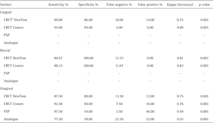
Surface Sensitivity % Specificity % False negative % False positive % Kappa (Accuracy) p value Lingual
CBCT* NewTom 90.00 86.00 10.00 14.00 0.75 0.001 CBCT Cranex 95.00 94.00 5.00 6.00 0.89 0.001
PSP - - -
Analogue - - -
-Buccal
CBCT NewTom 86.67 100.00 13.33 0.00 0.81 0.001 CBCT Cranex 88.33 100.00 11.67 0.00 0.83 0.001
PSP - - -
Analogue - - -
-Gingival
CBCT NewTom 87.50 88.00 12.50 12.00 0.75 0.001 CBCT Cranex 92.50 84.00 7.50 16.00 0.76 0.001
PSP 97.50 54.00 2.50 46.00 0.49 0.001
Analogue 77.50 78.00 22.50 22.00 0.55 0.001 CBCT: Cone beam computed tomography. PSP: photostimulable phosphor.
Table 1. Accuracy and reliability of various imaging techniques versus the gold standard in detection of recurrent caries Imaging
technique Sensitivity % Specificity % False negative % False positive % Kappa (Accuracy) p value CBCT* NewTom 87.86 90.00 12.14 10.00 0.78 0.001 CBCT Cranex 91.43 91.54 8.57 8.46 0.83 0.001
PSP 97.50 54.00 2.50 46.00 0.49 0.001
S. Kasraei et al.
(Tizkavan, Tehran, Iran) in different dimensions and the sections were evaluated under a stereomicroscope (SZ240, Olympus, Tokyo, Japan) to ensure presence of caries.
The collected data were entered into the STATA software (version 11.2, Stata Corp, College Station, TX, USA) and analyzed.
Results
The inter-observer agreement between the first and the second observer was calculated to be 0.79 according to Kappa statistic for different radiographic techniques; which indicates excellent agreement between the observers.
Sensitivity, specificity, positive and negative predictive values and the overall diagnostic accuracy for each radiographic technique are shown in Table 1. As seen in Table 1, the highest and the lowest diagnostic accuracy were for CBCT Cranex and the PSP digital radiography, respectively.
Table 2 shows the sensitivity, specificity, positive and negative predictive values and the overall diagnostic accuracy for each cavity wall. In analog and PSP digital intraoral radiographs, only the gingival wall carious lesions were evaluated due to their 2D nature. No significant difference was found in the diagnostic accuracy of CBCT Cranex and CBCT NewTom for the assessment and detection tissue around teeth (like in the clinical setting) to attenuate
the beam. Thus, in order to simulate the in-vivo conditions, specimens mounted in each mandible were placed in 1 L water container and then the exposure was made. CBCT images were then obtained of all teeth using New Tom 3G CBCT system (Verona, Italy) at the exposure settings of T=0.6 s, 1.80 mA and 110 kVp and also the Cranex 3D CBCT system (Soredex) at the exposure settings of T=14 s, 6 mA=and 90 kVp.
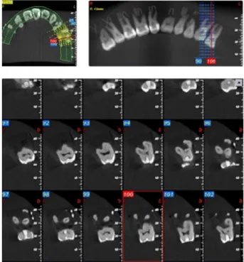
Radiographs obtained with New Tom 3G were entered into NNT Viewer software (Newtom, Verona, Italy) (Fig. 3), while radiographs obtained with Cranex 3D were entered into OnDemand 3D Dental software (Cybermed, Seoul, Korea) (Fig.4) and were evaluated by two observers in sagittal, axial and coronal planes. Data were recorded in a check list.
Since the buccal and lingual caries could not be observed on analog and digital intraoral radiographs, only the gingival wall carious lesions were assessed; while on CBCT images obtained with both systems, carious lesions on all three walls were evaluated.
After taking the radiographs, specimens with artificial caries induced by the cariogenic solution (Group 3) were sectioned at the restoration site using a diamond disc
Table 3: Accuracy and reliability of various imaging techniques versus the gold standard in detection of recurrent caries by type of caries (groups 1, 2 and 3)
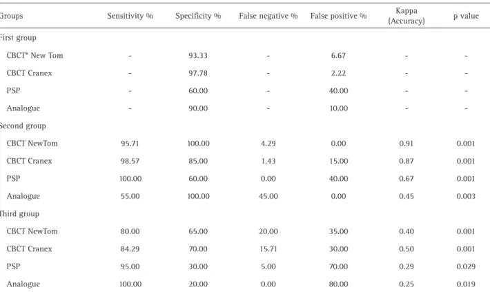
Groups Sensitivity % Specificity % False negative % False positive % Kappa
(Accuracy) p value First group
CBCT* New Tom - 93.33 - 6.67 -
CBCT Cranex - 97.78 - 2.22 -
PSP - 60.00 - 40.00 -
Analogue - 90.00 - 10.00 -
-Second group
CBCT NewTom 95.71 100.00 4.29 0.00 0.91 0.001 CBCT Cranex 98.57 85.00 1.43 15.00 0.87 0.001
PSP 100.00 60.00 0.00 40.00 0.67 0.001
Analogue 55.00 100.00 45.00 0.00 0.45 0.003 Third group
CBCT NewTom 80.00 65.00 20.00 35.00 0.40 0.001 CBCT Cranex 84.29 70.00 15.71 30.00 0.50 0.001
PSP 95.00 30.00 5.00 70.00 0.29 0.029
CBCT versus intraoral radiography
of recurrent caries in buccal, lingual and gingival surfaces (p=0.190 and 0.217, respectively).
Table 3 summarizes the sensitivity, specificity, positive and negative predictive values and the overall diagnostic accuracy for each type of caries.
Based on the results, the diagnostic accuracy of all imaging techniques in Group 2 was higher than the one in Group 3. In Group 2, the highest diagnostic accuracy was for CBCT NewTom while in Group 3, the highest diagnostic accuracy was for CBCT Cranex.
Discussion
Observation and detection of recurrent caries in interproximal surfaces is clinically difficult; these carious lesions are often detected radiographically. Intraoral bitewing radiography is the most commonly used radiographic technique for detection of these lesions; however, recent evidence shows that this technique has relatively low sensitivity and specificity for this purpose (1,14,15).
In previous studies (12), wax has been used to simulate secondary caries around restorations for the assessment of sensitivity and specificity of different radiographic systems. In the current study, in order to better simulate the clinical setting, pH cycling was used to induce artificial caries on the cavity walls. Previous studies have demonstrated that pH cycling for two weeks can cause 500 μm deep caries in the enamel and dentin (13).
In the present study, CBCT Cranex 3D exhibited the highest diagnostic accuracy, while the PSP digital radiography had the lowest diagnostic accuracy. Although the PSP digital radiography has high diagnostic sensitivity, it has high frequency of false positive results, attributed to the defects in digital systems and application of different enhancement filters such as sharpness, which cause high contrast at the restoration-tooth interface, leading to the formation of pseudo caries on radiographs of the respective areas (10).The high sensitivity of this system is due to the software features allowing adjustment of the contrast and density of images. Increasing the contrast enhances the detection of caries. However, such image manipulation may cause pseudo-caries and increase the frequency of false positive results (10).
In comparison between the two CBCT systems evaluated in the present study, Cranex 3D had higher diagnostic accuracy than New Tom 3G CBCT; the reason could be the different voxel sizes and type of detectors in the two systems. In Cranex 3D system, the voxel size is 200 μm in a 6x8 cm field of view (FOV) and 0.133 μm in a 4x6 cm FOV. But in New Tom 3G system, the voxel size is 210 μm; this factor affects the spatial resolution and the quality of the reconstructed images. Smaller voxel size results in higher
resolution of the final image (16). Moreover, the detector in the Cranex 3D system is CMOS and in the New Tom 3G CBCT the detector system is Image Intensifier plus Charge Coupled-Device (II/CCD). In systems using intensifier plates, image resolution decreases due to light emission (11).
Different sensor types, FOVs and exposure settings are used in CBCT systems. However, beam hardening and metal artifacts in CBCT scans complicate the detection of recurrent caries around restorations (12,16).
In the current study, similar to the study by Haak et al, no statistically significant difference was noted between PSP digital radiography and analog systems in detection of recurrent caries around composite restorations. They concluded that the accuracy of detecting marginal defects around composite restorations was slightly influenced by the type of radiographic system (analogue or digital) (1).
Qu et al. (17) reported no significant difference was found among different CBCT systems for detection of proximal caries; this finding is in contrast to the present study results. In the current study, Cranex 3D system had a higher diagnostic accuracy than NewTom. The difference between our results and those of Qu et al (17). may be attributed to the different CBCT systems used (New Tom 9000, Accuitomo 3DX, Kodak 9000, Promax 3D DCT PRO) and the fact that in this study, secondary carious lesions were evaluated while in their study, the primary proximal carious lesions were assessed (17).
The present study accepted the hypothesis stating the superior accuracy of CBCT over that of other radiographic techniques for detection of secondary caries. Different authors (12,17-19) also reported the high accuracy of CBCT for detection of carious lesions compared with intraoral radiographic techniques.
In line with our findings, Abesi et al. (20) also demonstrated the low accuracy of conventional and PSP digital intraoral radiography for detection of caries.
The 2D nature of intraoral radiography limits the detection of carious lesions due to superposition. This explains the low sensitivity of intraoral radiography for detection of these caries.
Haiter et al.(11) indicated the low sensitivity of CBCT for detection of dental caries (44% sensitivity); this result is most probably due to the used CBCT system. They used a CBCT system with a large FOV (16 inches); which decreases the sensitivity of CBCT system (15). Larger FOV of CBCT system results in lower image resolution (16).
S. Kasraei et al.
gingival surfaces were 0.83, 0.89 and 0.76 respectively; the reason may be the different sections required for the assessment of these walls. Buccal and lingual carious lesions are better observed in coronal sections while gingival carious lesions are better detected in sagittal sections. Although in this study tooth surfaces in all sagittal, axial and coronal sections were evaluated, most cross-sectional slices are obtained coronally; this issue per se can affect the diagnostic accuracy at different tooth surfaces. In analog and PSP systems, only the gingival carious lesions were evaluated because these systems are 2D and do not allow evaluation of caries in the buccal and lingual surfaces. Nair et al. (21) reported that lesions in the gingival wall are detected much easier and more accurately by PSP digital radiography and CBCT systems.
In PSP digital radiography, although the sensitivity for detection of recurrent gingival caries is very high (even higher than that of CBCT systems), the specificity is low due to the high number of false positive results; the reason may be the enhancement filters in digital systems that may cause pseudo-caries (10). The sensitivity of analog intraoral systems is lower than that of PSP; because in the analog systems the contrast or density of the image cannot be adjusted for better observation of details. However, they have a higher specificity than PSP due to less false positive results.
The diagnostic accuracy of all imaging techniques evaluated in the current study was higher in Group 2 (caries simulation with wax) than in Group 3 (caries induction with cariogenic solution); the reason is the different wax structure from that of actual caries. Also, the wax sheets applied had well defined borders that made them easily recognizable, whereas actual caries do not have well defined margins and the penetration depth of lesions into the tooth structure may vary at different points. Moreover, the composition of carious lesion is totally different from that of wax. Some previous studies have also used wax to create a lucent area beneath the restorations. In the mentioned studies, after cavity preparation, a hole is created by a bur (in dimensions similar to those of bur) at the pulpal floor of the cavity, which is then filled with wax. Using a bur for this purpose also causes a well defined simulated carious lesion (12). These studies also confirmed that the lucency caused by wax (due to high contrast) was 2.92 times clearer than the lucency due to actual caries (22).
In the current study, cariogenic solution was used to induce artificial caries on cavity walls. The ability to observe radiographically these caries was exactly the same as that of lucencies due to actual caries in the clinical setting; this issue makes our results more reliable than those of the wax technique used in previous studies.
In Group 2 (wax group) in the present study, the highest accuracy was by New Tom CBCT while in the pH cycling
group (Group 3), the highest diagnostic accuracy was by Cranex 3D CBCT. Thus, it may be concluded that under
in-vivo conditions, Cranex 3D has greater efficacy for detection of recurrent caries due to its higher resolution.
The high diagnostic accuracy of CBCT systems is due to the fact that they enable easy observation of all tooth surfaces in sagittal, coronal and axial planes and any gap or caries around the restorations are seen as lucent areas at the tooth-restoration interface. However, in metal restorations, metal artifacts on the cavity walls compromise accurate assessment of the walls for caries. Titanium and zirconia restorations as well as composite resins containing a minimum of 20% AlSiO2 cause clear artifacts on CBCT scans. The intensity of artifacts increases by an increase in the radiopacity of composite resins (23). In the present study, the cavity walls were easily assessed due to the absence of artifacts.
Although CBCT does not have many of the disadvantages of intraoral radiography, it should be noted that patient radiation dose is higher in CBCT compared to intraoral and panoramic radiography (24). Therefore, available CBCT images taken for various purposes should be used only if conventional methods do not have sufficiently high diagnostic accuracy.
In conclusion, it may be stated that CBCT has a higher diagnostic accuracy than digital and conventional intraoral radiography for detection of secondary caries around composite resin restorations.
Resumo
A causa mais comum de falha das cáries dentais são as cáries secundárias. Este estudo objetivou comparar a acurácia diagnóstica da radiografia intraoral digital e convencional com a tomografia computadorizada de feixe cônico (TCFC) para a detecção de cáries recorrentes em torno restaurações com compósitos. Cavidades mésio-oclusais-distais (MOD) foram produzidas com broca em 45 molares hígidos humanos extraídos. Os dentes foram dividdos em 3 grupos. No grupo controle, as cavidades foram restauradas com compósito após condicionamento e colagem (n=15). No Grupo 2, foi aplicada uma camada de cera de 500 µm de espessura sobre as paredes bucais, linguais e gengivais; as cavidades foram então restauradas com resina composta. No Grupos 3, as amostras foram submetidas a ciclagem de pH e criação artificial de cáries sobre as paredes bucais, linguais e gengivais; as cavidades foram então restauradas com resina composta. Radiografias convencional e digital com fósforo foto-estimulável (PSP, Optima) e duas imagens em TCFC (NewTom 3G e Granex 3D) foram obtidas de cada amostra. A presença ou não de cáries foi avaliada nestas imagens. Os dados foram analisados por estatística Kappa. A acurácia diagnóstica da TCFC foi significantemente (p<0,05) maior que na radiografia intraoral convencional e digital. A acurácia foi de 0,83, 0,78, 0,55 e 0,49, respectivamente para TCFC Cranex 3D, TCFC NewTom 3G, a radiografia intraoral convencional e digital. A TCFC tem maior acurácia diagnóstica que a radiografia intraoral convencional e digital para detecção de cáries secundárias em torno de restaurações com compósito.
Acknowledgements
CBCT versus intraoral radiography
Chancellor of Research, Hamadan University of Medical Sciences, for supporting this study.
References
1. Haak R, Wicht MJ, Hellmich M, Noack MJ. Detection of marginal defects of composite restorations with conventional and digital radiographs. Eur J Oral Sci 2002;110:282-286.
2. Levin L, Coval M, Geiger SB. Cross-sectional radiographic survey of amalgam and resin-based composite posterior restorations. Quintessence Int 2007;38:511-514.
3. Newman B, Seow WK, Kazoullis S, Ford D, Holcombe T. Clinical detection of caries in the primary dentition with and without bitewing radiography. Aust Dent J 2009;54:23-30.
4. Mjor IA, Toffenetti F. Secondary caries: a literature review with case reports. Quintessence Int 2000;31:165-179.
5. Kuper NK, Opdam NJ, Ruben JL, de Soet JJ, Cenci MS, Bronkhorst EM, et al.. Gap size and wall lesion development next to composite. J Dent Res 2014;93:108S-113S.
6. Goshima T, Goshima Y. Optimum radiopacity of composite inlay materials and cements. Oral Surg Oral Med Oral Pathol 1991;72:257-260.
7. Boston DW. Initial in vitro evaluation of DIAGNOdent for detecting secondary carious lesions associated with resin composite restorations. Quintessence Int 2003;34:109-116.
8. Bamzahim M, Shi XQ, Angmar-Mansson B. Secondary caries detection by DIAGNOdent and radiography: a comparative in vitro study. Acta Odontol Scand 2004;62:61-64.
9. Brouwer F, Askar H, Paris S, Schwendicke F. Detecting secondary caries lesions: A systematic review and meta-analysis. J Dent Res 2016;95:143-151.
10. White S, Pharoah M. Oral radiology, principle and interpretation. 6 ed. St. Louis: Mosby; 2009. Chapter 7,14.
11. Haiter-Neto F, Wenzel A, Gotfredsen E. Diagnostic accuracy of cone beam computed tomography scans compared with intraoral image modalities for detection of caries lesions. Dentomaxillofac Radiol 2008;37:18-22.
12. Charuakkra A, Prapayasatok S, Janhom A, Pongsiriwet S, Verochana K, Mahasantipiya P. Diagnostic performance of cone-beam computed tomography on detection of mechanically-created artificial secondary caries. Imaging Sci Dent 2011;41:143-150.
13. Mukai Y, Ten Cate JM. Remineralization of advanced root dentin lesions in vitro. Caries Res 2002;36:275-280.
14. Ludlow JB, Platin E, Delano EO, Clifton L. The efficacy of caries detection using three intraoral films under different processing conditions. J Am Dent Assoc 1997;128:1401-1408.
15. Wenzel A. Bitewing and digital bitewing radiography for detection of caries lesions. J Dent Res 2004;83 Spec No C:C72-75.
16. Cheng JG, Zhang ZL, Wang XY, Zhang ZY, Ma XC, Li G. Detection accuracy of proximal caries by phosphor plate and cone-beam computerized tomography images scanned with different resolutions. Clin Oral Investig 2012;16:1015-1021.
17. Qu X, Li G, Zhang Z, Ma X. Detection accuracy of in vitro approximal caries by cone beam computed tomography images. Eur J Radiol 2011;79:e24-27.
18. Young SM, Lee JT, Hodges RJ, Chang TL, Elashoff DA, White SC. A comparative study of high-resolution cone beam computed tomography and charge-coupled device sensors for detecting caries. Dentomaxillofac Radiol 2009;38:445-451.
19. Murat S, Kamburoğlu K, Isayev A, Kurşun S, Yüksel S. Visibility of artificial buccal recurrent caries under restorations using different radiographic techniques. Oper Dent 2013;38:197-207.
20. Abesi F, Mirshekar A, Moudi E, Seyedmajidi M, Haghanifar S, Haghighat N, et al.. Diagnostic accuracy of digital and conventional radiography in the detection of non-cavitated approximal dental caries. Iran J Radiol 2012;9:17-21.
21. Nair MK, Tyndall DA, Ludlow JB, May K, Ye F. The effects of restorative material and location on the detection of simulated recurrent caries. A comparison of dental film, direct digital radiography and tuned aperture computed tomography. Dentomaxillofac Radiol 1998;27:80-84.
22. Kang BC, Farman AG, Scarfe WC, Goldsmith LJ. Mechanical defects in dental enamel vs. natural dental caries: observer differentiation using Ektaspeed Plus film. Caries Res 1996;30:156-162.
23. Kuusisto N, Vallittu PK, Lassila LV, Huumonen S. Evaluation of intensity of artefacts in CBCT by radio-opacity of composite simulation models of implants in vitro. Dentomaxillofac Radiol 2015;44:20140157. 24. Ludlow JB, Davies-Ludlow LE, White SC. Patient risk related to common
dental radiographic examinations: The impact of 2007 International Commission on Radiological Protection recommendations regarding dose calculation. J Am Dent Assoc. 2008;139:1237-1243.