International Journal of Advanced Biotechnology and Research (IJBR) ISSN 0976-2612, Online ISSN 2278–599X,
Vol-7, Special Issue-Number4-June, 2016, pp988-993 http://www.bipublication.com
Research Report
Antibacterial properties of Artemisia khorassanica on food pathogens
Sepideh Tabrizie1 and Ali Mohammadi Sani2*
1
Department of Food Science and Technology, Quchan Branch, Islamic Azad University, Quchan, Iran
2
Young Researchers and Elite Club,
Quchan Branch, Islamic Azad University, Quchan, Iran
*Corresponding Author: Ali Mohamadi Sani , mohamadisani@yahoo.com
ABSTRACT:
The present study aimed at evaluating the in antibacterial activity of methanolic extract of Artemisia Khorassanica against different pathogenic microorganisms. The agar disk diffusion method was used to study the antibacterial activity of Artemisia Khorassanica three extracts against 2 gram-positive and 2 gram-negative bacteria at concentration 300 and 400 mg/ml.The results revealed that the methanol extract of Artemisia Khorassanica presented the highest zone of inhibition against tested pathogens (8 mm inhibition zones). Other plants did not show significant zone inhibition .Minimum inhibitory concentrations (MIC) and minimum bactericidal concentrations (MBC) were quantified by micro-dilution method. The leaf extract of artemisa against Staphylococcus aureus (PTCC 1431) and Listeria monocytogenes (PTCC1221) and Esherichia coli (PTCC1399) and Salmonella typhimurium (PTCC1709) strains showed the best activities, with the lowest minimal inhibitory concentration (MIC) of 12.5 mg ml-1, 12.5 mg ml-1, 25mg ml-1 and 25 mg ml-1 and MBC was 50,>100,>100 and >100 mg ml-1 respectively for Staphylococcus aureus (PTTC 1431) and and Listeria monocytogenes (PTCC1221) and Esherichia coli (PTCC1399) and Salmonella typhimurium (PTCC1709). The results showed that the methanol extract of the Artemisia Khorassanica has antibacterial activity and therefore it could be used as a natural preservative ingredient in food and/or pharmaceutical industries.
Keyword: Artemisia Khorassanica, Patogenic, Listeria, Escherichia, methanolic extracts, Antibacterial activity.
INTRODUCTION
Food processors, food safety researchers, and regulatory agencies have been increasingly concerned with the growing number of food-borne illness outbreaks caused by pathogens like Staphylococcus aureus, Salmonella sp., Clostridium perfringens, Campylobacter, Listeria monocytogenes, Vibrio parahaemolyticus, Bacillus cereus, and entero-pathogenic Escherichia coli. These bacteria cause over 90% of all cases of food poisoning (Sokovic et al., 2010). The contamination of raw and/or processed foods with micro flora can take place at various stages from the production to the sale and distribution. Thus, food industry at present uses chemical preservatives to prevent the growth of food spoiling microbes. Due to the economical impacts of spoiled foods and the consumers concerns over the safety of foods
plants and essences are rich sources in antibacterial compounds which can be an alternative to combat bacterial diseases.These bioactive compounds are actually combinations of secondary products present in the plant. They have been used as food preservatives, pharmaceuticals, alternative medicines and natural therapies for centuries.
These compounds are mostly alkaloids, steroids, tannins, phenolic compounds, flavonoids, resins and fatty acids. These compounds are odorous, complex, volatile compounds produced by special cells or groups of cells and concentrated in one particular region of plant such as the leaves, bark and stems (Ahmad et al., 2013). Artemisia Khorassanica occurs in large parts of mainly continental temperate Eurasia (and including northwestern Africa, Iran and Turkmenistan, excluding South East Asia).Its distribution is often scattered and mainly concentrated in the mountains of the meridionaland step and Wilderness zones, while having a clear center of diversity in Iran and the Desert Jajarm. (Poudledge, 2007).
Artemisia khorassanica consists of approximately 400 species in temperate regions of the northern hemisphere of which 19 occur in most regions of Iran, but its main distribution range includes Alborz Mts. and elevations in northwest Iran (Azerbaijan province) and northeast Iran ( Khorasan ) (Heravi et al., 2013).Artemisia khorassanica is belonging to family Compositae that has a variety of medicinal properties (Singh et al., 2009).The stem is fairly stout, erect or spreading, 30 to 40 cm tall, branched, covered sparsely to heavily with ash-colored soft hairs. Artemisia khorassanica is native to western Asia, including Iran, and Eastern Europe and is an invasive species in North America.The aims of the present study were to evaluate the potential antimicrobial activities of methanol extracts of Artemisia khorasanica on typical food-borne pathogens.
MATERIALS AND METHODS Chemicals and Plant materials
Gentamicin (Sinadaroo, Iran), methanol and Dimethyl Sulfoxide (DMSO) (Merck, Germany)
were purchased. The aerial part (leaves) of Artemisia Khorassanica was collected in May 2016 and aerial parts of Artemisia Khorassanica was collected in May 2016 and also all organ of Artemisia Khorassanica were collected in Avril 2016 from the mountains of North Khorasan Province in Iran. The plants were identified by the Research Center of Natural Products Health (NPH), North Khorasan University of Medical Sciences (Iran).
Extraction
The plant samples were dried at room temperature under shade (Umer et al., 2013), finely ground with a hammer mill, and the powdered sample from each plant was extracted with methanol (1.5 L) (Merck, Germany) for 48 hrs at room temperature (Seukep et al., 2013). The extracts were filtered through filter paper, afterwards extracts dried in vacuum at 40ºC (Salvat et al., 2004) and were kept at 4ºC until further uses (E Djeussi et al., 2013).
Organisms and Inoculation Conditions
Authentic pure cultures of bacteria were obtained from Persian Type Culture Collection (PTCC). They included gram positive bacteria; Staphylococcus aureus (PTCC 1431),Listeria monocytogenes (PTCC1221) and gram-negative bacteria; Salmonella typhimurium (PTCC1709), Escherichia coli (PTCC 1399).
They were maintained on agar slant at 4°C and sub cultured on a fresh appropriate agar plates 24 hrs prior to any antimicrobial test. Mueller Hinton Agar (MHA) was used for the activation of bacteria and the Mueller Hinton Broth (MHB) was used for the MIC determinations (Seukep et al., 2013). Finally, suspensions were adjusted to 0.5 McFarland standard turbidity. Bacterial suspensions were standardized to concentrations of 1.5×10⁸ CFU ml-1 (Library of Congress Cataloging-in-Publication Data, 2005).
Antimicrobial assay
Kirby-Bauer disc diffusion method (Selim et al., 2014).Methanolic extract were dissolved in dimethyl Sulfoxide (DMSO) to a final concentration of 100, 50, 25, 12.5 25,3/125,1/562, 0/781, 0.390and 0.195 mg ml-1 as stock solution and sterilized by filtration
through 0.45 µm Millipore filters. The discs (6
mm in diameter) were (Ahmad et al., 2013; Rishikesh et al 2012) immediately placed on the surface (Thompson et al., 2013) plates (Petri dishes, 80 mm diameters) containing a suitable medium (MHA) seeded with the test organisms
(1.5×10⁸). The amount of 15 µl of methanolic
extract was poured onto discs. These plates were kept at low temperature (4oC) for 15 min to allow maximum diffusion (Rahman and Sultana, 2011). Negative controls were prepared using the same solvent employed to dissolve the
extract (DMSO) (10 µl). Gentamycin used as standard antibiotic (positive control) (10 µl)
(Assam et al., 2010). The test plates were incubated at 37oC for 24 hrs (Mhaske et al., 2011; Billah et al., 2013; Rishikesh et al., 2012). The test materials having antibacterial activity inhibited microorganism growth, and a clear, distinct zone of inhibition surrounding the discs was visualized (Billah et al., 2013). Antimicrobial activity was evaluated by measuring the zone of inhibition (Billah et al.,
2013; Selim et al., 2014) ruler to an accuracy of 0.5 mm (Thompson et al., 2013) against the test organisms (Selim et al., 2014).
Minimum Inhibitory Concentration (MIC) Test
The antibacterial activity of extracts were tested using the micro-dilution antibacterial assay for the minimum inhibitory concentration (MIC) values (Fawole et al., 2012) and MBC (Haobin et al., 2009). The studied microorganisms included strains of (Mbveng et al., 2012) Staphylococcusaureus (PTCC 1431) and Listeria monocytogenes (PTCC1221) and Esherichia coli (PTCC1399) and Salmonella typhimurium (PTCC1709).MIC were determined by the broth micro-dilution method (Coccia et al., 2012) in a 96-wells micro-plate (Mbveng et al., 2012). All tests were performed in Mueller Hinton broth (MHB) (Haobin et al., 2009). The microorganism inoculum was standardized with appropriate culture medium (MHB) to a final concentration of (Coccia et al., 2012) 1.5×10⁶ CFU ml-1 (standardized at 1.5×10⁶ CFU ml-1 by adjusting the optical density to 0.1 at 600 nm by Shimadzu UV-120-01 spectrophotometer) (Kuete et al., 2011). Each extract was dissolved in dimethyl Sulfoxide (DMSO) and added to MHB (Boussaada et al., 2008).
Microorganism methanolic extract (mg/ml)
Positive control Negative control Gentamicin DMSO
Staphylococcus aureus 8 27 6
Salmonella typhimurium 6 27 6
Escherichia coli 6 29 6
Listeria monocytogenes 8 28 6
Table 1: inhibition zone in diameter (mm) for methanol extract of Artemisia Khorassanica
The final concentration of DMSO was lower than 2.5% and does not affect the microbial growth (Mbveng et al., 2012). The extracts were serially diluted to give a concentration of 100, 50, 25, 12.5,3/125,1/562, 0/781, 0.390 and 0.195mg ml-1 (Dhiman et al., 2011). Then, 100
µl of each concentration was added in a well
(96-well micro plate) containing 95 µl of MHB
and 5 µl of inoculum (1.5×10⁶ CFU ml-1) (Kuete et al., 2011). The micro plate was incubated at 37°C ± 1°C for 24 hrs. Dilution of the extract corresponding to respective test organism showing no visible growth was
considered as MIC (Umer et al., 2013). To
determine MBC, 10 µl broth was taken from
each well and inoculated in MHB for 24 hrs at 30 or 37ºC for bacteria. The MBC is defined as the lowest concentration the methanol extracts at which inoculated microorganism was completely killed (99.99%) (Haobin et al., 2009).
RESULTS AND DISCUSSION Results of disc-diffusion test
zones are presented in Table1. These results indicated that the diameters of inhibition zones varied from 8 mm and 27–29 mm for the various concentration of extracts and gentamycin respectively.The methanolic extract from aerial parts (leaves) of Artemisia Khorassanica had substantial of antimicrobial activity against 4 bacteria (Staphylococcus aureus, Salmonella typhimurium, Escherichia coli, Listeria monocytogenes) species tested. On the other hand, the methanolic extracts from aerial parts of Artemisia Khorassanica and all organ of it
showed no antibacterial activity and inhibition zone against Staphylococcus aureus and Listeria monocytogenes diameter and did not show antibacterial activity against the Esherichia and Salmonella all the tested bacterial strains et the 10 concentrations of 100, 50, 25, 12.5, 6.25, 3.125, 1.562, , 0/781, 0.390 and 0.195 mg ml-1 . The maximal inhibition zones for bacterial strains, which were sensitive to the methanolic extract of Artemisia Khorassanica in the range of 8-12 mm respectively.
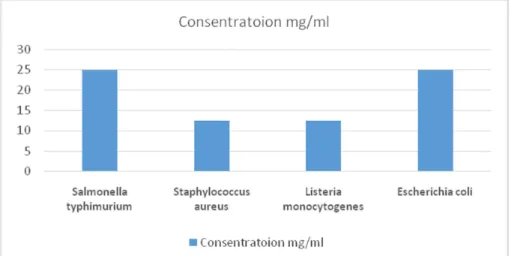
Microorganism MIC MBC
Salmonella typhimurium 25 >100
Staphylococcus aureus 12.5 50
Listeria monocytogenes 12.5 >100
Escherichia coli 25 >100
Table 2: MIC and MBC for methanolic extract of Artemisia Khorassanica (mg/ml)
DISCUSSION
Since pre-historic times, man has gone in different ways to search for cures and relief from various diseases by using numerous plants, plant products and plant-derived products. Recently, there is a scientific interest and a certain popularity with regard to screening essential oils and extracts from plants used medicinally all over the world. Historically, many plants essential oils and crude extracts have been used as topical antiseptics, or have been reported to have antimicrobial properties (Hossain et al., 2012). The gram-positive bacteria were found to be more sensitive towards the plants methanol extracts than gram-negative bacteria. Antibacterial activity of MeOH extracts and its polar fractions could also be attributed to the presence of several types of compounds such as flavonoids and phenolic acids (Rahman et al., 2011). Generally, the higher resistance among Gram-negative bacteria could be ascribed to the presence of their outer phospholipidic membrane, almost mpermeable to lipophilic compounds.
Figure 1: MIC for methanolic extract of Artemisia Khorassnica(mg ml-1) against different bacteria
The absence of this barrier in Gram-positive bacteria allows the direct contact of the essential oils hydrophobic constituents with the
leakage of vital intracellular constituents, or impairment of the bacteria enzyme (Selim et al., 2014; Delamare et al., 2007).The results of the antibacterial screening showed that MeOH extracts of this plants had potential activity against some of the representative food-borne pathogens. Antibacterial activity of MeOH extract and its fractions could also be attributed to the presence of several types of compounds such as flavonoids and phenolic acids (Rahman et al., 2011).
CONCLUSION
Among methanolic extracts of Artemisia Khorassanica, the extract obtained from Artemisia Khorassanica showed stronger results. The extract from this plant was showed antimicrobial activity against Staphylococcus aureus, Salmonella typhimurium, Escherichia coli, Listeriamonocytogenes food borne pathogen. Therefore it can be concluded methanolic extracts of this plants especially Artemisia Khorassanica in appropriate combination, can act as an effective food preservative. Of course, this was the first study to compare the antimicrobial properties of methanolic extracts three plants on food-borne pathogens.
REFERENCES
1. Ahmad M, Pin Lim C, AkyiremAkowuah G, Ismail N.N, Hashim M.A, Yee Hor S, Fung Ang L, Fei Yam M. 2013. Safety assessment of standardised methanol extract of
Cinnamonumburmannii. Phytomedicine. 20,
1124-1130.
2. Assam JP A, Dzoyem J, Pieme C, Penlap V. 2010. In vitro antibacterial activity and acute toxicity studies of aqueous-methanol extract of SidarhombifoliaLinn. (Malvaceae). BMC Complementary and Alternative Medicine.10 (40), 1-7.
3. Delamare A.P.L, Moschen-Pistorello I.T, Artico L, Atti-Serafini L, Echeverrigaray S. 2007. Antibacterial activity of the essential oils of Salvia officinalisL. and Salvia trilobaL. cultivated in South Brazil. Food Chemistry. 100, 603-608.
4. Dhiman A, Nanda A, Ahmad A, Narasimhan B. 2011. In vitro antimicrobial activity of methanolic leaf extract of
(PsidiumguajiavaL.).Journal of Pharmacy and Bioallied Science. 3 (2), 226-229.
5. Dickore W.B, Kasperek G. 2010. Species of
Cotoneaster (Rosaceae, Maloideae)
indigenous to, naturalising or commonly cultivated in Central Europe. Willdenowia. 40, 13-45.
6. Heravi M.M, Rodi S, Ardalan P. 2013. Study of Antioxidant and Free Radical Scavenging Activities of Cotoneaster medicus and
Glycyrrhizaglabra Plants. Journal of
Chemical Health Risks. 3 (2), 27-34.
7. Hossain M.A, Dawood Shah M, Vun Sang S, Sakari M. 2012. Chemical composition and antibacterial properties of the essential oils and crude extracts of Merremiaborneensis. Journal of King Saud University–Science.24, 243-249.
8. Jarald E.E, Joshi S.B, Jain D.C. 2008. Antidiabetic activity of aqueous extract and non‐polysaccharide fraction of
CynodondactylonPers. Ind J Exp Bio. 46, 660‐667.
9. Kuete V., Kamga J, P Sandjo L, Ngameni B, MP Poumale H, Ambassa P, T Ngadjui B. 2011. Antimicrobial activities of the methanol extract, fractions and compounds from FicuspolitaVahl. (Moraceae). BMC Complementary and Alternative Medicine. 11 (6), 1-6.
10.Library of Congress Cataloging-in-Publication Data. 2005. Manual of antimicrobial susceptibility testing. 39-41. 11.Mbaveng A.T, Kuete V, Ngameni B, Beng
V.P, Ngadjui B.T, Marion Meyer J.J, Lall N. 2012. Antimicrobial activities of the methanol extract and compounds from the twigs of Dorsteniamannii (Moraceae). BMC Complementary and Alternative Medicine.12 (83), 1-6.
12.Selim S.A, AdamM.E, Hassan S.M, Albalawi A.R. 2014. Chemical composition, antimicrobial and antibiofilm activity of the essential oil and methanol extract of the
Mediterranean cypress
Complementary and Alternative Medicine. 14 (179), 1-8.
13.Seukep J.A, Fankam A.G, Djeussi D.E, Voukeng I.K, Tankeo S.B, Noumdem J.AK, HLN Kuete A, Kuete V. 2013. Antibacterial activities of the methanol extracts of seven Cameroonian dietary plants against bacteria expressing MDR phenotypes. Springer Plus. 2 (363), 1-8.
14.Singh SK, Rai PK, Mehta S, Gupta RK, Watal G. 2009. Curative effect of
Cynodondactylonagainst STZ induced
hepatic injury in diabetic rats. Ind J ClinBiochem. 24, 410‐413.
15.Sokovic M, Glamoclija J, Marin P.D, Brkic D, JLD.VanGriensven L. 2010. Antibacterial Effects of the Essential Oils of Commonly Consumed Medicinal Herbs Using an In Vitro Model. Molecules.15, 7532-7546.
16.Thompson A, Meah D, Ahmed N, Conniff-Jenkins R, Chileshe E, O Phillips C, C Claypole T, W Forman D, E Row P. 2013. Comparison of the antibacterial activity of essential oils and extracts of medicinal and culinary herbs to investigate potential new treatments for irritable bowel syndrome. BMC Complementary and Alternative Medicine. 13 (338), 1-19.
17.Teke G.N, Elisee K.N, Roger K.J. 2013. Chemical composition, antimicrobial properties and toxicity evaluation of the essentioal oil of CupressusLusitanica Mill.