Antimicrobial activity of Tachyplesin 1
against
Burkholderia pseudomallei
:
an in vitro and in silico approach
Lyn-Fay Lee1, Vanitha Mariappan1, Kumutha Malar Vellasamy1, Vannajan Sanghiran Lee2and Jamuna Vadivelu1
1Department of Medical Microbiology, Faculty of Medicine, University of Malaya, Kuala Lumpur, Malaysia
2Department of Chemistry, Faculty of Science, University of Malaya, Kuala Lumpur, Malaysia
ABSTRACT
Burkholderia pseudomallei, the causative agent of melioidosis, is intrinsically resistant to many conventional antibiotics. Therefore, alternative antimicrobial agents such as antimicrobial peptides (AMPs) are extensively studied to combat this issue. Our study aims to identify and understand the mode of action of the potential AMP(s) that are effective against B. pseudomalleiin both planktonic and biofilm state as well as to predict the possible binding targets on using in vitro and in silico approaches. In the in vitro study, 11 AMPs were tested against 100B. pseudomallei isolates for planktonic cell susceptibility, where LL-37, and PG1, demonstrated 100.0% susceptibility and TP1 demonstrated 83% susceptibility. Since the
B. pseudomalleiactivity was reported on LL-37 and PG1, TP1 was selected for further investigation. TP1 inhibitedB. pseudomalleicells at 61.69mM, and membrane blebbing was observed using scanning electron microscopy. Moreover, TP1 inhibited B. pseudomalleicell growth, reaching bactericidal endpoint within 2 h post exposure as compared to ceftazidime (CAZ) (8 h). Furthermore, TP1 was shown to suppress the growth ofB. pseudomalleicells in biofilm state at concentrations above 221mM. However, TP1 was cytotoxic to the mammalian cell lines tested. In the in silico study, molecular docking revealed that TP1 demonstrated a strong interaction to the common peptide or inhibitor binding targets for lipopolysaccharide ofEscherichia coli, as well as autolysin, pneumolysin, and pneumococcal surface protein A (PspA) of Streptococcus pneumoniae. Homology modelledB. pseudomalleiPspA protein (YDP) also showed a favourable binding with a strong electrostatic contribution and nine hydrogen bonds. In conclusion, TP1 demonstrated a good potential as an anti-B. pseudomalleiagent.
Subjects Microbiology, Infectious Diseases
Keywords Burkholderia pseudomallei, Melioidosis, Antimicrobial peptides, Molecular docking, Tachyplesin 1
INTRODUCTION
Burkholderia pseudomallei, the causative agent for melioidosis, is commonly found in the soil and water of Southeast Asia, and Northern Australia (Cheng & Currie, 2005). Due to the ease of dissemination where infection can be acquired either through an open wound, inhalation, or ingestion (Puthucheary, 2009), coupled with the genomic Submitted4 December 2015
Accepted21 August 2016 Published25 October 2016
Corresponding author
Jamuna Vadivelu, jamuna@um.edu.my
Academic editor
Siouxsie Wiles
Additional Information and Declarations can be found on page 23
DOI10.7717/peerj.2468
Copyright
2016 Lee et al.
Distributed under
plasticity of the bacteria (Holden et al., 2004;Schweizer, 2012), it is labelled as a potential biological warfare agent and classified as a category B bio threat agent by the Centers for Disease Control and Prevention where specific enhancements are required for diagnostic capacity and disease surveillance (Sto¨ckel et al., 2015). Currently, melioidosis is treated using parenteral therapy of ceftazidime (CAZ) followed by prophylactic therapy of co-trimoxizole (Dance, 2014). Over the years,B. pseudomalleihas been reported to resist the commonly used antibiotics (increased usage of CAZ and amoxicillin/clavulanic acid in treatment), and also to the ability to form biofilm in vitro and in vivo (Schweizer, 2012). To exacerbate the problem, incomplete treatment resulted in a high rate of melioidosis relapse. Worst of all, despite appropriate antimicrobial therapy, mortality from
melioidosis septic shock remained high (Wiersinga et al., 2006). As such, there is a need to consider alternative antimicrobial agents, one of which is the antimicrobial peptides (AMPs) (Rotem & Mor, 2009).
AMPs, both naturally occurring and synthetic, have considerable advantages for therapeutic applications, including broad-spectrum activity, rapid onset of activity and relatively low possibility of emergence of resistance (Seo et al., 2012). Furthermore, they act on slow-growing or even non-growing bacteria due to the ability to permeablise and form pores within the cytoplasmic membrane (Batoni et al., 2011). Groups of AMP’s i.e., defensins, cathelicidins, and dermicins have previously been reported to show potential against various pathogens (Wiesner & Vilcinskas, 2010). However, to date, only a few cathelicidin AMPs have been reported to be effective againstB. pseudomallei, including LL-37 (Kanthawong et al., 2012), protegrin 1 (PG1) (Sim et al., 2011), bovine lactoferrin (Puknun et al., 2013), phospholipase A2 inhibitors (Samy et al., 2015), and sheep cathelicidin (SMAP-29) (Blower, Barksdale & van Hoek, 2015).
activity as well as to determine the orientation of the 2,5-dichloro thienyl substituted thiazole derivatives (4a–4d) bound in the active site of 3-deoxy-D-glucosamine (GlcN)-6-P synthase (target for antifungals). Similar use of in silico molecular docking to compliment the findings of the in vitro/in vivo experiments and elucidate the antimicrobial-protein interaction was also been reported in a study by inAlves et al. (2013)on the antimicrobial properties of mushroom phenolic compounds andAl-Sohaibani & Murugan (2012)on the inhibition effects ofSalvadora persicaonStreptococcus mutans.
Taking into account the effectiveness of AMPs to inhibit potentially antibiotic resistant bacteria, our study aimed to identify the potential AMP(s) that are effective against B. pseudomalleiin both planktonic and biofilm state and to predict the possible binding targets onB. pseudomalleiprotein structures. Overall, there were two approaches in this study; in vitro and in silico. Initially, the in vitro experiments were executed to identify the potential AMP(s) that are effective againstB. pseudomalleiin both planktonic and biofilm state. Based on those observations, the in silico experiments were then carried out on the selected AMP to predict the possible binding targets on the available B. pseudomalleiprotein structures. Both of these approaches will assist in elucidating the action of the AMP onB. pseudomalleiand contribute to the development of AMPs as an alternative anti-B. pseudomalleiagent.
MATERIALS AND METHODS
Bacteria strains and growth conditions
A total of 90B. pseudomalleiclinical isolates from melioidosis patients (archive collection of University of Malaya Medical Centre (UMMC), Kuala Lumpur and Hospital Tengku Ampuan Afzan, Pahang) and 10 non-clinical isolates (one environmental and nine animal) were included in this study. All isolates were identified asB. pseudomalleiusing routine phenotypic characterization including selective growth on Ashdown, biochemical profiles on API 20 NE tests (BioMe´rieux, France) and in-house polymerase chain reaction (PCR) analysis (Suppiah et al., 2010). Prior to the study, theB. pseudomallei isolates were cultured aerobically in Luria Bertani (LB) broth at 37C and agitated at 180 rpm for 24 h according to Al-Maleki et al. (2014).
Peptide storage and handling
A total of 11 AMPs used in this study were selected based on the potential to inhibit other Gram-negative organisms; LL-37, Magainin 2, Tachyplesin 1 (TP1), PG1, Sushi 1, Sushi 2, 1037, 1018, DJK5, V2D, and ornithine. Among these LL-37, Magainin 2, TP1, and PG1 were synthesized by SBS Genetech, China; Sushi 1 and Sushi 2 were synthesised by First Base Laboratories, Singapore; Peptide 1037, 1018, and DJK5 were kindly provided by our collaborators from University of British Columbia, Canada; while V2D and ornithine were provided by our collaborators in National University of Singapore (Table 1).
The AMPs used in this study were stored in lyophilized form at-20C (Travis et al.,
a concentration of 10 mg/ml. The AMPs were diluted in 0.01% acetic acid containing 0.2% bovine serum albumin (BSA) for the preliminary screening whereas the dilution of AMPs with serum free Roswell Park Memorial Institute 1640 media (RPMI 1640; Life Technologies) was performed to minimize the dilution of the RPMI used in subsequent experiments. In addition, the microtiter plates used in this study were made of
polypropylene while the storage vials were made of polypropylene (Hancock, 1999).
Preliminary screening of AMPs antimicrobial activity on in
B. pseudomalleiisolates from melioidosis patients in Malaysia
The 100 strains mentioned were screened for AMP susceptibility by colony counting (Sieuwerts et al., 2008). The 11 AMPs were diluted to 1 mg/ml before use. Screening was performed as described previously (Sheafor et al., 2008) with slight modifications. Briefly, 80ml of 24-hB. pseudomalleiculture in LB broth was diluted with 1 X phosphate buffer saline (PBS; pH 7.4) to a bacteria density of 105CFU/ml and mixed with 20ml AMPs prior to incubation. The mixture was then plated on fresh nutrient agar (NA) and incubated at 37C for 24 h to determine viability of the bacteria. Isolates were
categorized as “sensitive” when no growth was observed and categorized as “resistant” if there one or more colonies grown after 24-h incubation on NA. Three biological replicates were performed.
Minimum inhibitory concentration (MIC) and minimum bactericidal concentration (MBC) of AMPs onB. pseudomalleicells
in planktonic state
Based on the preliminary screening results TP1 that inhibitedB. pseudomallei,was selected for further study with LL-37 and PG1. In this study,B. pseudomalleistrain K96243 and randomly selected B. pseudomalleiclinical strains UMC031, UMC080, and UMC089, andEscherichia coli(ATCC 29522; control), were exposed to TP1, LL-37, and PG1 concentrations ranging from 0 to 200mM, with two-fold increase. The exponential phase
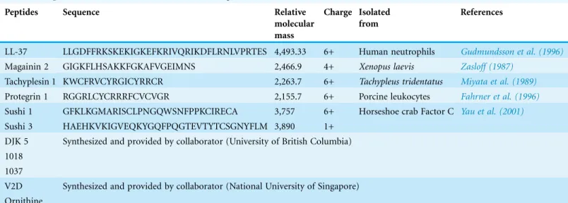
Table 1 Sequences and characteristics of AMPs investigated.
Peptides Sequence Relative
molecular mass
Charge Isolated from
References
LL-37 LLGDFFRKSKEKIGKEFKRIVQRIKDFLRNLVPRTES 4,493.33 6+ Human neutrophils Gudmundsson et al. (1996)
Magainin 2 GIGKFLHSAKKFGKAFVGEIMNS 2,466.9 4+ Xenopus laevis Zasloff (1987)
Tachyplesin 1 KWCFRVCYRGICYRRCR 2,263.7 6+ Tachypleus tridentatus Miyata et al. (1989)
Protegrin 1 RGGRLCYCRRRFCVCVGR 2,155.7 6+ Porcine leukocytes Fahrner et al. (1996)
Sushi 1 GFKLKGMARISCLPNGQWSNFPPKCIRECA 3,757 6+ Horseshoe crab Factor C Yau et al. (2001)
Sushi 3 HAEHKVKIGVEQKYGQFPQGTEVTYTCSGNYFLM 3,890 1+ DJK 5 Synthesized and provided by collaborator (University of British Columbia) 1018
1037
V2D Synthesized and provided by collaborator (National University of Singapore)
culture of planktonicB. pseudomalleiwas centrifuged at 10,000 rpm for 15 min to pellet the bacteria. The resulting pellet was washed three times with PBS and the bacterial density was adjusted to a 105CFU/ml using serum free RPMI 1640 medium; as
recommended bySchwab et al. (1999)in order to obtain the highest AMP activity. A total of 20ml of each AMPs were added to 180ml of the bacterial suspension in 96 well U-bottom microtiter plates (Eppendorf). Following incubation at 37C for 24 h, the plates were subjected to optical density (OD) measurement at 570 nm using a microplate absorbance reader (Tecan Sunrise, Ma¨nnedorf, Switzerland) using the settings for U-bottom microtiter plates. Each well was subjected to a 10 times serial dilution and plated on NA to determine the viability of the bacteria (Sieuwerts et al., 2008). The MIC was read as the lowest concentration of AMP that inhibited visible growth of the bacteria (Schwab et al., 1999) compared to the untreated after 24-h incubation.
Additionally, the MBC was determined when no growth was observed on NA following 24-h incubation. Three technical replicates were performed on three different occasions.
Time-kill kinetic assay ofB. pseudomallei
The time-kill kinetic assay was carried out with a similar set up as the MIC and MBC studies usingB. pseudomalleistrain K96243. Briefly, 20ml of AMP (TP1 at 55, 110, and 221mM; approximately the MIC and MBC ofB. pseudomalleistrains tested) was added to 180 ml of the bacterial suspension in a 96 well U-bottom microtiter plates. The plates were incubated 37C for 24 h and the absorbance was taken at OD 570 nm using the settings for U-bottom microtiter plates at 60-min interval. At every two hours, the bacterial suspension in each well was subjected to a 10 times serial dilution and plated on NA to determine bacteria viability (Sieuwerts et al., 2008). In addition, the inhibition patterns of TP1 was also compared with 54mM of CAZ (30mg/ml; equivalent to the concentration on the antibiotic disk; Oxoid, UK), and also the untreated bacteria. Three technical replicates were performed on three different occasions, and the data obtained were analyzed with one-way ANOVA followed by Dunnett’s test to determine the significance relative to the untreated bacteria (P < 0.05).
Inhibition activity of AMPs againstB. pseudomallei
in biofilm state
containing the antimicrobial agents tested in serum-free RPMI (antimicrobial challenge plates) before incubation at 37C for 24 h. Following these, the biofilms were disrupted using a water bath sonicator (Thermoline Scientific, Wetherill Park NSW, Australia) for 30 min in PBS and the viability of the bacteria from the biofilm (CFU/biofilm) was determined by colony counting (Sieuwerts et al., 2008). Three biological replicates were performed, and the data obtained were analysed with one-way ANOVA followed by Dunnett’s test to determine the significance relative to the untreated bacteria (P < 0.05).
Scanning electron microscopy (SEM)
SEM was performed according toSong et al. (2012)with slight modifications. Briefly, B. pseudomalleiK96243 culture was incubated with 62mM of TP1 at 37C for 24 h. Following centrifugation (10 min at 2,000g, 4C), the bacterial pellet was washed twice with PBS, and resuspended in 3.0% glutaraldehyde (incubated overnight at ambient temperature). Subsequently, the solution was rinsed three times with double distilled water and dehydrated in a graded series of ethanol solutions. After critical-point drying and layering with 20 nm gold coating (Leica EM SCD005, Leica Microsystems,
Singapore), the microscopic analysis was performed using Quanta 650 FEG Scanning Electron Microscope (FEI, Oregon, USA). BothB. pseudomalleiK96243 cell suspension without the AMPs andE. coliwere treated as the control.
Cytotoxicity of AMPs on mammalian cell lines
In this study, human lung epithelial (A549; ATCCÒ
CCL185TM), human gastric adenocarcinoma (AGS; ATCCÒ
CRL-1739TM) and human hepatocellular carcinoma (HepG2; ATCCÒ
HB8065TM) cell lines were cultured in RPMI 1640 containing foetal bovine serum (FBS) (10%) at 37C with 5% CO2and 95% relative humidity). Cytotoxicity was determined based on the reduction of 3-[4, 5-dimethylthiazol-2-yl]-2, 5 diphenyl tetrazolium bromide (MTT; Sigma, St. Louis, MO, USA) by cellular oxidoreductases of viable cells, which yields a crystalline blue formazan product (Hansen et al., 2012). Briefly, cells (approximately 2104cells/well) were seeded in tissue culture treated 96-well plates (Corning, Corning, NY, USA). After 24 h, the cells were rinsed with PBS and 200mg/ ml of the TP1 (110mM), PG1 (115mM) and LL-37 (55mM) were added as a solution in fresh serum free medium to a final volume of 100ml/well, respectively. Following 2 h of incubation, the MTT reagent (20ml) was added to obtain a final concentration of 500 mg/l and further incubated for 3 h. After which DMSO (solubilizing solution) was added to lyse the cells and to dissolve the formazan crystals and the absorbance was recorded at OD 570 nm. The percentage of viable cells was calculated as followed:
Absorbance of peptide treated cells
ð Þ
ðAbsorbance of untreated cellsÞ 100
Possible TP1 interactions with protein targets from in silico molecular docking study
Preparation of TP1 structure from PDB
Solution NMR structures of TP1 (PDB ID: 2RTV) were obtained from Protein Data Bank (PDB) (http://www.rcsb.org/pdb). The first model of 20 peptide configuration models available in the PDB file was used. Prior to docking, AutoDock tools 4.2.6 (ADT) software was used to prepare the ligand before proceeding to energy minimization using
Accelrys Discovery Studio 2.5.5 (DS) software (Accelrys Software Inc., San Diego, CA, USA). The backbone of TP1 was kept rigid whereas most of the side chains were defined as flexible in molecular docking experiment.
Interaction of TP1 on selected bacteria structures
A total of four bacteria structures were selected to visualize TP1 interaction;
lipopolysaccharide (LPS) of E. coli,andStreptococcus pneumoniaeproteins; autolysin, pneumolysin, and pneumococcal surface protein A (PspA).
TP1 has been reported to bind to the LPS ofE. coli(Kushibiki et al., 2014). However, the LPS structure ofB. pseudomalleiis yet to be reported. Thus, the LPS model from E. coliwas used to predict the interaction of TP1 withB. pseudomalleias both are Gram-negative bacteria andE. coliis often used as a representative for Gram negative bacteria (Fernandez-Recio et al., 2004;Galdiero et al., 2012). The molecular docking study was carried out with the prepared TP1 structure and the LPS structure ofE. coli
(PDB ID: 1QFG) based onKushibiki et al. (2014)with a slight modification to add the missing atoms (e.g. hydrogens) in the model. The initial structure was modified according to the CHARMm force field with partial charge Momany-Rone (Momany & Rone, 1992) and minimizations of the structures were performed, satisfying the root mean squared gradient tolerance of 0.1000 kcal/(molAngstrom) before docking was carried out using AutodockVina (ADV) (Trott & Olson, 2010) instead of Autodock 4.2 as published. A 70 8080-point grid box of the structure was then generated with a grid spacing of 0.375 A˚, and centred on GlcN II in lipid A between TP1 and the LPS structure.
TP1 was reported to bind to both Gram negative and Gram positive bacteria
stable system and thus likely to bind. In addition, the software was also used to detect intermolecular hydrogen (H+) bonds and hydrophobic interactions which play a role in stabilizing the docked complex.
Interaction of TP1 on homology modelled B. pseudomallei protein structure
At this stage, all the three S.pneumoniaeproteins demonstrated negative IE with TP1 which indicated a strong interaction. When we searched for the protein sequences (BLAST search; blastp; non-redundant proteins with 95% sequence similarity) for all the three protein structures againstB. pseudomalleiprotein sequence database, only the PspA sequence secured a hit in the search; YD repeat-containing protein (accession:
CFU00865). This may indicate that there are high similarities in the proteins of the Gram positive and Gram negative bacteria. Similarly,Alloing, Trombe & Claverys (1990) have also demonstrated that the Ami proteins ofS. pneumoniaeexhibited homology with components of the oligopeptide permeases of the Gram-negativeSalmonella typhimurium andE. coli. In addition, autolysin exists in the peptidoglycan bacterial cell walls, which applies to both Gram positive and Gram negative bacteria (Beveridge, 1999). Thus, it is possible to execute homology modelling using the protein sequences of either Gram-positive or Gram-negative due to the similarities between both groups of bacteria. Taking into consideration of the above points, we have carried out homology modelling of the B. pseudomalleiprotein using PspA as a template.
Using DS, multiple sequence alignment was performed and the model was built using MODELLER function. The overall quality of the minimized model was evaluated by utilizing PROCHECK (Laskowski et al., 1993) for evaluation of Ramachandran plot quality, ERRAT (Colovos & Yeates, 1993) to verify patterns of non-bonded atomic interactions, and VERIFY3D (Lu¨thy, Bowie & Eisenberg, 1992) for assessing the
compatibility of each amino acid residue. Subsequently with ADV, TP1 was docked onto the YDP model using the binding site of PspA as a reference in order to predict the intermolecular interaction.
RESULTS
Preliminary screening of AMPs antimicrobial activity on
B. pseudomalleiisolates from Malaysia
A total of 100B. pseudomalleiisolates including the reference strainB. pseudomallei K96243 was subjected to colony counting after the exposure to the AMPs. Eighty-three strains (83%) were susceptible to TP1 whereas 100 strains (100%) were susceptible to both PG1 and LL-37. The isolates were 100% resistant to the remainder AMPs. In addition, there was no correlation between the AMP activity profiles and the antibiotic
susceptibility data available (Supplemental Information 1and2).
and PG1. To date,B. pseudomalleisusceptibility to the eight non-responsive AMPs is yet to be reported whereas there were reports ofB. pseudomalleisusceptibility to LL-37 (Kanthawong et al., 2012), and PG1 (Sim et al., 2011). As such, TP1 was selected for further investigations in order to suggest its efficacy in comparison with both LL-37 and PG1.
MIC and MBC on planktonic B. pseudomalleicells
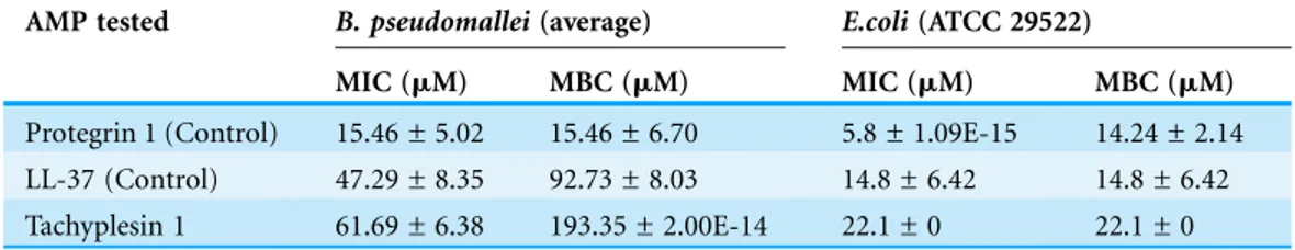
The MIC of TP1 (61.69mM) was threefold lower than its MBC (193.35 mM). Relative to the MIC of TP1, MIC levels of LL37 (47.29mM) and PG1 (15.46mM) were
approximately 1.3 and four-fold lower, respectively (Table 2). Similarly, the MBC of TP1 demonstrated two-fold and 12-fold higher levels compared to LL37 (92.73mM) and PG1 (15.46mM), respectively. Overall, both the MIC and MBC levels of TP1 were found to be higher than that of LL-37 and PG1. On the other hand, the MIC and MBC values ofB. pseudomalleifor all three AMPs were observed higher than E. coli. In conclusion, TP1 exhibitedB. pseudomalleiinhibition, although not as potent as PG1 and LL-37.
Time-kill kinetic assay ofB. pseudomallei
At 221mM of TP1, the growth inhibition ofB. pseudomalleiK96243 was observed within two hours after exposure compared to that of CAZ (8 h) (Fig. 1). At 110mM, TP1 managed to inhibit the growth ofB. pseudomalleiK96243 following eight hours of exposure compared to the growth of the untreated bacteria at the same time point (approximately 9.0 log CFU/ml), similar to CAZ. At 55mM, there was a slight
inhibition of the growth of B. pseudomalleiK96243 (9.96 log CFU/ml) as compared to the untreated bacteria (10.17 log CFU/ml) at 24 h, albeit not significant. Overall, TP1 was able to inhibitB. pseudomalleiK96243 growth at a concentration above at 55mM and in a shorter duration as compared to CAZ.
Inhibition activity of AMPs againstB. pseudomallei in biofilm state
B. pseudomalleiK96423 in biofilm state was exposed to the 11 different AMPs (1 mg/ml) using MBEC assay (Fig. 2). Of the AMPs tested, only TP1, 1018, and PG1
demonstrated significant inhibition (4.2 log CFU/biofilm, 7.2 log CFU/biofilm, and 1.1 log CFU/biofilm, respectively) compared to the untreated bacteria (7.8 log CFU/biofilm). CAZ and MRP demonstrated complete inhibition. At the same, using 1 mg/ml of TP1, LL-37, and PG1 (which was equivalent to 442, 222, and 464mM, respectively), the inhibition activity of TP1 was 3.5-log lower than PG1 but approximately 2.0-log higher
Table 2 Summary of MIC and MBC values of PG1, LL-37 and TP1.
AMP tested B. pseudomallei(average) E.coli(ATCC 29522)
MIC (mM) MBC (mM) MIC (mM) MBC (mM)
than LL-37 compared to the untreated bacteria (7.8 log CFU/biofilm). There was no significant inhibition between LL-37-treated bacteria and the untreated bacteria. The inhibition activity of TP1 was higher that LL-37, but lower than CAZ, MRP, and PG1.
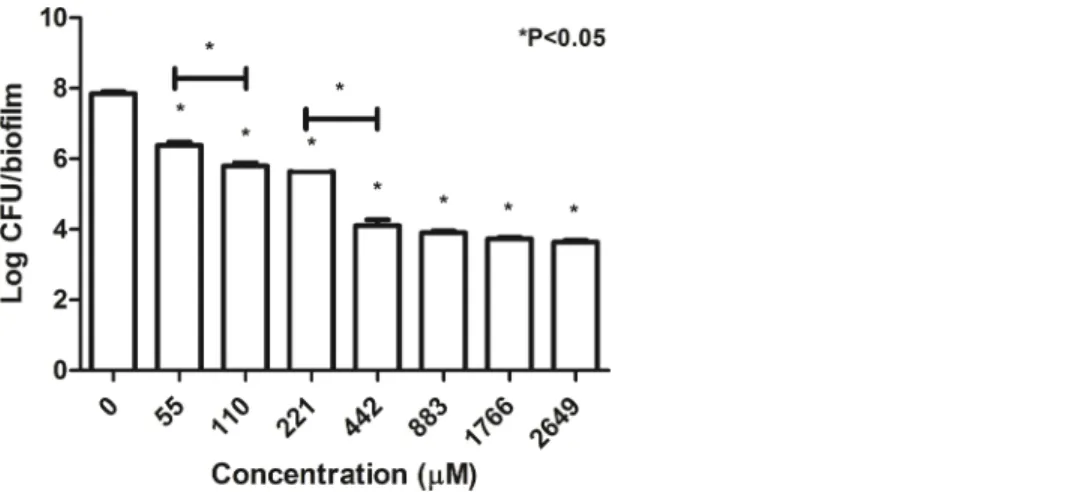
WhenB. pseudomalleiK96243 in biofilm state was exposed to increasing
concentrations of TP1 (from 55 to 2,649mM; two-fold increase), the number of cells gradually decreased from 7.84 log CFU/biofilm to 3.6 log CFU/biofilm (Fig. 3). There was a significant decrease in the number of cells when exposed to all the TP1
Figure 1 Time killing curve of TP1 compared to that of CAZ forB. pseudomalleiK96243.B. pseu-domalleiK96243 was exposed to TP1 (55, 110, and 221mM) and CAZ (54mM). Data was presented as
mean and standard deviation of three independent replicates, analyzed with one-way ANOVA followed by Dunnett’s test to determine the significance relative to the untreated bacteria (P < 0.05).
concentrations as compared to the untreated cells (0mM). In addition, TP1 was observed to suppress the growth of the cells in biofilm state at concentrations above 442mM (∼4.00 log CFU/biofilm;∼2.0-log CFU/biofilm reduction compared to the untreated) although MBEC was not achieved. There were significant growth inhibitions observed when the bacteria were exposed to TP1 at 55 and 100mM (1.0-log reduction) as well as 221 and 442mM (1.3-log reduction). In general, TP1 was able to reduce the number ofB. pseudomalleiin biofilm state from concentrations above 55mM.
Scanning electron microscopy (SEM)
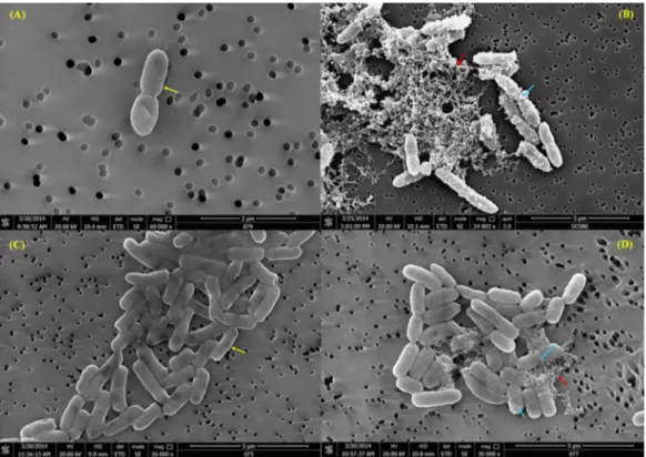
The untreatedB. pseudomalleicells, in basal RPMI medium, displayed a smooth and intact surface (Fig. 4A). When the cells were exposed to TP1, some of the cells looked
corrugated with dimples on the surface, and the appearance of blisters and bubbles were also observed on the membrane (Fig. 4B). Moreover, some of the cells were also observed to lose their original structure, leaving cell debris intermixed with the cell membranes that were blebbing. Similar observation was also seen withE. coli(Figs. 4Cand4D).
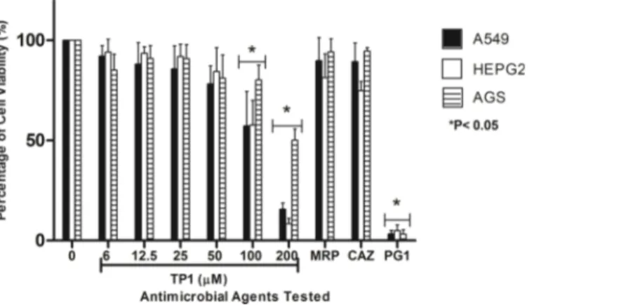
Cytotoxicity of AMPs on mammalian cell lines
As the concentration of TP1 increased (from 2.7 to 110.4mM), the percentage of viable cells for all three cell lines used were found to be decreased (from 92.0 to 15%, respectively for A549, 94.1 to 8.3%, respectively for HEPG2, and 85.1 to 50.1%, respectively for AGS). Besides that, at 48.6 and 110.4 mM of TP1, there was a significant reduction of cell viability in all three cell lines compared to that of lower concentrations of TP1 (from 2.7 to 22.3mM). Among the three cell lines tested, at 48.6 and 110.4mM, AGS showed a higher percentage of viability (80 and 50%, respectively) compared to A549 (57 and 15%, respectively) and HEPG2 (57 and 8%, respectively). On the other hand, at lower concentrations of TP1 from 2.7 to 22.3mM, HEPG2 showed higher percentage of viability (90.5 ± 7.6%) compared to that of AGS (90.0 ± 8.4%) and
Figure 3 Number of survivingB. pseudomalleiK96243 in a biofilm state 24-h post-exposure to TP 1. B. pseudomalleiK96243 was exposed to different concentrations of TP1 (55–2649mM, with twofold
A549 (85.8 ± 9.6%). When the same amount of peptides used (200 mg/ml which is equivalent to 110.4mM of TP1 and 115mM of PG1), TP1 showed lesser cytotoxicity (15.0% for A549, 8.3% for HEPG2, and 50.1% for AGS) compared to that of PG1 (3.3% for A549, 5.0% for HEPG2, and 3.0% for AGS) (Fig. 5). Overall, HEPG2 cells
demonstrated higher percentage of cell viability compared to the AGS at the lower concentrations of TP1 (2.7–11.0 mM), albeit at a not significant level. However, AGS demonstrated significantly higher percentage of cell viability (P < 0.05) compared to HEPG2 at the higher concentrations of TP1 (48.6–110.4mM). In general, TP1 has lower cytotoxicity at concentrations between 2.7 to 22.3mM where HEPG2 demonstrated more tolerance to TP1.
At this stage, TP1 demonstrated inhibition activities on the testedB. pseudomallei strains comparable to LL-37 and PG1. SEM also revealed that TP1 was able to induce membrane blebbing, disrupting the membrane integrity and eventually leading to cell death. To date, the exact target onB. pseudomalleifor TP1 binding has yet to be reported but there were existing reports of TP1 binding to theLPS(Kushibiki et al., 2014) and membrane (Hong et al., 2015) ofE. colias well as the minor groove of the DNA (Yonezawa et al., 1992). Therefore, in silico molecular docking was carried out to predict the binding targets of TP1 onB. pseudomallei.
Figure 4 (A) SEM observation ofB. pseudomalleiK96243 pre-exposure; (B) post-exposure of 62mM
TP1; (C)E. coli ATCC29522 pre-exposure and (D) post-exposure of 22mM of TP1. The bacteria
Possible TP1 interactions with protein targets from in silico molecular docking study
Interaction of TP1 to the LPS of E. coli
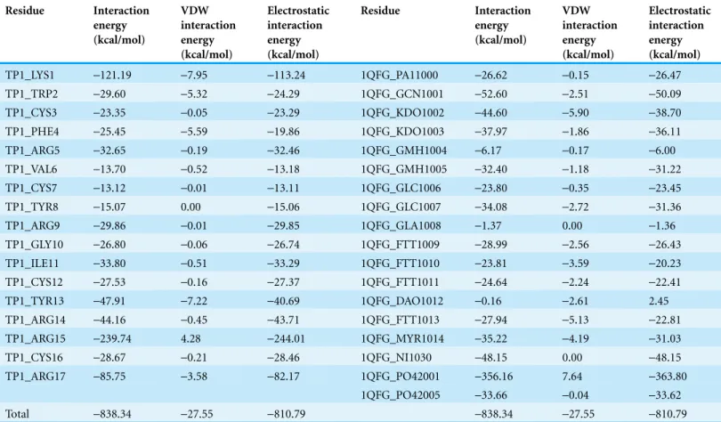
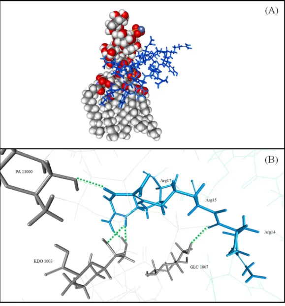
TP1 was observed to interact with the LPS ofE. coliwith a binding affinity from ADV of -5.4 kcal/mol and an IE of-838.34 kcal/mol where the interaction was mostly
contributed by electrostatic-810.79 kcal/mol compared to VDW (-27.55 kcal/mol;
Table 3). The IE of TP1 was mostly contributed by residues LYS1 and ARG 15
(-121.19 and-239.74 kcal/mol, respectively) while the IE of LPS was contributed mostly by the PO4 residue (-356.16 kcal/mol). Moreover, nine hydrogen bonds were formed between TP1 and the LPS of E. coliat TP1:LYS1:HT1–LPS: FTT1010:O3, TP1:ARG15: HH11–LPS: KDO1002:O1A, TP1:ARG15: HH12–LPS: PO42001:O1, TP1:ARG15: HH12–LPS: PO42001:O2, TP1:ARG15: HH22–LPS: GCN1001:O4, TP1:ARG15: HH22–LPS: PO42001:O1, TP1:ARG17: HE–LPS: KDO1003:O7, TP1:ARG17: HE–LPS: KDO1003:O8, and TP1:ARG17: HH22–LPS: KDO1003:O8, where the hydrogen bonds were formed mostly at the terminal ends of TP1 (Fig. 6). However, there were no hydrophobic interactions observed between the TP1 and LPS. As such, the TP1-LPS docked complex was stabilized the intermolecular hydrogen bonds formed.
Interaction of TP1 to Streptococcus pneumoniae protein structures
Based on the molecular docking using ADV, TP1 interacted with all threeS. pneumoniae proteins with a binding affinity of-8.1 kcal/mol for autolysin,-7.7 kcal/mol for pneumolysin, and-7.3 kcal/mol for PspA (Table 4). This indicates that TP1 has a higher binding affinity to autolysin, followed by pneumolysin, and PspA. In addition, the number of hydrogen bonds formed between TP1 and the S. pneumoniaeproteins demonstrated a similar trend to the binding affinity where the autolysin (with the highest binding affinity) formed 13 bonds with TP1, followed by pneumolysin (nine bonds) and PspA (eight bonds). However, based on the IE, TP1 demonstrated stronger interaction
Figure 5 Percentage of viability of A549, HEPG2 and AGS cell lines post-exposure to TP1, PG1, MRP and CAZ.The cell lines were exposed to TP1 (0, 2.7, 5.5, 11.0, 22.3, 48.6, and 110.4mM), PG1 (115mM),
MRP (26mM), and CAZ (54mM). These experiments were conducted in three independent replicates
with PspA (-573.48 kcal/mol), followed by autolysin (-444.14 kcal/mol), and pneumolysin (-135.59 kcal/mol). Moreover, there were additional intermolecular interactions between TP1-penumolysin and TP1-PspA docked complexes but none was detected with TP1-autolysin docked complex. TP1-pneumolysin docked complex was observed to contain two pi-cation interactions between ARG9 of TP1 to TRP433 of pneumolysin. On the other hand, TP1-PspA docked complex demonstrated three types of additional interactions; one pi-pi interaction (TRP2 to TYR 541 of PspA), four pi-cation interactions (LYS1 to TRP 525 of PspA; three bonds from LYS1 to TRP518 molecules), and a pi-sigma interaction between LYS1 to TRP 518 of PspA. Based on the data obtained, among the threeS. pneumoniaeproteins, TP1 was predicted to bind to autolysin at close proximity but was predicted to form the strongest interaction and the most stable docked complex with PspA.
Interaction of TP1 on homology modelled B. pseudomallei protein structure
Homology model of PspAB. pseudomallei(YDP model) was evaluated to ensure acceptable model quality (Supplemental Information 5). TP1 was observed to interact with the YDP model with a binding affinity of-7.6 kcal/mol and an IE of -822.80 kcal/mol where the interaction was mostly contributed by electrostatic
Table 3 Contribution of the interactions energy in kcal/mol of theE. coliLPS binding residues with TP1. Residue Interaction energy (kcal/mol) VDW interaction energy (kcal/mol) Electrostatic interaction energy (kcal/mol) Residue Interaction energy (kcal/mol) VDW interaction energy (kcal/mol) Electrostatic interaction energy (kcal/mol)
TP1_LYS1 -121.19 -7.95 -113.24 1QFG_PA11000 -26.62 -0.15 -26.47
TP1_TRP2 -29.60 -5.32 -24.29 1QFG_GCN1001 -52.60 -2.51 -50.09
TP1_CYS3 -23.35 -0.05 -23.29 1QFG_KDO1002 -44.60 -5.90 -38.70
TP1_PHE4 -25.45 -5.59 -19.86 1QFG_KDO1003 -37.97 -1.86 -36.11
TP1_ARG5 -32.65 -0.19 -32.46 1QFG_GMH1004 -6.17 -0.17 -6.00
TP1_VAL6 -13.70 -0.52 -13.18 1QFG_GMH1005 -32.40 -1.18 -31.22
TP1_CYS7 -13.12 -0.01 -13.11 1QFG_GLC1006 -23.80 -0.35 -23.45
TP1_TYR8 -15.07 0.00 -15.06 1QFG_GLC1007 -34.08 -2.72 -31.36
TP1_ARG9 -29.86 -0.01 -29.85 1QFG_GLA1008 -1.37 0.00 -1.36
TP1_GLY10 -26.80 -0.06 -26.74 1QFG_FTT1009 -28.99 -2.56 -26.43
TP1_ILE11 -33.80 -0.51 -33.29 1QFG_FTT1010 -23.81 -3.59 -20.23
TP1_CYS12 -27.53 -0.16 -27.37 1QFG_FTT1011 -24.64 -2.24 -22.41
TP1_TYR13 -47.91 -7.22 -40.69 1QFG_DAO1012 -0.16 -2.61 2.45
TP1_ARG14 -44.16 -0.45 -43.71 1QFG_FTT1013 -27.94 -5.13 -22.81
TP1_ARG15 -239.74 4.28 -244.01 1QFG_MYR1014 -35.22 -4.19 -31.03
TP1_CYS16 -28.67 -0.21 -28.46 1QFG_NI1030 -48.15 0.00 -48.15
TP1_ARG17 -85.75 -3.58 -82.17 1QFG_PO42001 -356.16 7.64 -363.80
1QFG_PO42005 -33.66 -0.04 -33.62
Total -838.34 -27.55 -810.79 -838.34 -27.55 -810.79
Note:
IE-770.63 kcal/mol compared to VDW IE (-52.17 kcal/mol;Table 5). The binding affinity and the IE indicated that TP1 will bind to the YDP model in close proximity with a strong intermolecular interaction. The IE of TP1 was mostly contributed by residues LYS1, ARG 14, and ARG 15 (-158.78,-160.54, and-124.82 kcal/mol, respectively) while the IE of YDP was contributed mostly by ASP 1220 (-209.81 kcal/mol) and ASP 1169 (-190.00 kcal/mol). Moreover, nine hydrogen bonds were observed between TP1 and YDP model at TP1:LYS1: HZ2 to YDP: THR1113: O, TP1:CYS12:HN to YDP: SER1185: OG, TP1:ARG14: HE to YDP: ASP1169:OD1, TP1:ARG14: HE to YDP: ASP1169:OD2, TP1:ARG14: HH22 to YDP: ASP1169:OD1, TP1:ARG17: HH22 to YDP:
Table 4 Summary of TP1 interaction withS. pneumoniaeprotein structures. Docked complex Binding affinity (kcal/mol) Interaction energy (kcal/mol) VDW interaction energy (kcal/mol) Electrostatic interaction energy (kcal/mol) No. of hydrogen bonds Hydrogen bond details Additional intermolecular interactions TP1-autolysin (B)
-8.1 (-7.6) -444.17 -51.03 -393.14 13 TP1:LYS1:HT1–B:ASP246: O None
TP1:LYS1:HT2–B: ASP246:OD2
TP1:LYS1:HT2–B:ASP246: O TP1:LYS1:HT3–B: ASP246:OD2 TP1:LYS1: HZ1–B: VAL279: O TP1:LYS1: HZ2–B: MET278: O TP1:LYS1: HZ3–B: SER280: OG TP1:TRP2: HE1–B: SER280: OG TP1:ARG15: HH11–B: SER280: O
TP1:ARG15: HH12–B: ASN281:OD1 TP1:ARG15: HH22–B: ASN281:OD1 B: LYS274:HZ2–TP1:ARG5: O B: LYS274:HZ3–TP1:ARG5: O
TP1-pneumolysin (P) -7.7 (-6.2) -135.59 -37.62 -97.97 9 TP1:ARG14: HH22–P: GLU434:OE2 Pi-Cation:
TP1:ARG15: HE–P: ASN400:OD1 P: TRP433–TP1:ARG9:NE
TP1:ARG15: HH21–P: ASN400:OD1 P: TRP433–TP1:ARG9:NE TP1:ARG15: HH21–P: ASN400: O
TP1:ARG17: HH12–P: ASP380:OD2 TP1:ARG17: HH21–P: THR378:OG1 TP1:ARG17: HH22–P: THR378:OG1 P: LYS424:HZ1–TP1:ARG17: O P: GLU434:HN–TP1:GLY10: O
TP1-pspA -7.3 (-6.4) -573.48 -46.10 -527.38 8 TP1:LYS1: HZ2–pspA: ASN571: O Pi-Pi:
TP1:TRP2: HE1–pspA: TYR546: OH PspA: TYR541–TP1:TRP2 TP1:GLY10:HN–pspA: LYS580: O
TP1:TYR13: HH–pspA: ASP573: OD2 Pi-Cation:
pspA: TRP525–TP1:LYS1:NZ pspA: TRP518–TP1:LYS1: N pspA: TRP518–TP1:LYS1: N pspA: TRP518–TP1:LYS1:NZ TP1:ARG15: HH21–pspA: ASN542: O
TP1:ARG15: HH21–pspA: ASN542: O
TP1:ARG17: HH22–pspA: ASN542: O Pi-Sigma:
ASP1120:OD2, YDP: SER1155: HG to TP1:CYS16: O, YDP: ARG1157:HH12–TP1:CYS12: O, and YDP: TYR1166: HH to TP1:CYS12: O (Fig. 7). However, there were no
hydrophobic interactions observed between the TP1 and YDP. In general, the TP1-YDP model was stabilized by the intermolecular hydrogen bonds formed.
Along with the common binding protein targets for peptide and inhibitors, the PDB database search was performed and the availableB. pseudomalleiPDB structures (Supplemental Information 6) were subjected to molecular docking to predict the possible interactions with TP1 (Supplemental Information 7). Of the 26B. pseudomallei proteins retrieved PDB (including stress proteins, secretion needle proteins, and
penicillin binding pump) binding affinities from -5.7 to-11.2 kcal/mol were observed. Besides that, seven proteins with the interaction of amino acids in target receptor from peptide less than-100 kcal/mol (with CHARMm force field) were then further
analysed to identify additional hydrogen bonds and intermolecular pi-interactions (Supplemental Information 8). TP1 demonstrated the most negative interaction energy with the cell inhibiting factor (PDB ID: 3GQM;-230.75 kcal/mol) with a binding
Table 5 Contribution of the interactions energy in kcal/mol of the homology modelledB. pseudomalleiprotein (YDP) binding residues with TP1.
Residue Interaction energy (kcal/mol)
VDW interaction energy (kcal/mol)
Electrostatic interaction energy (kcal/mol)
Residue Interaction
energy (kcal/mol)
VDW interaction energy (kcal/mol)
Electrostatic interaction energy (kcal/ mol)
TP1_LYS1 -158.78 -6.00 -152.78 YDP MODEL_THR1113 -26.00 -1.90 -24.10
TP1_TRP2 -39.15 -4.41 -34.74 YDP MODEL_GLY1114 -9.83 -0.89 -8.94
TP1_CYS3 -22.10 -0.41 -21.69 YDP MODEL_ASN1115 -5.55 -2.25 -3.31
TP1_PHE4 -15.84 -0.22 -15.62 YDP MODEL_ARG1119 115.99 -7.04 123.03
TP1_ARG5 -43.61 -0.24 -43.37 YDP MODEL_ASP1120 -209.81 -0.04 -209.77
TP1_VAL6 -1.99 -0.12 -1.87 YDP MODEL_MET1121 -22.28 -0.85 -21.43
TP1_CYS7 -16.13 -0.66 -15.47 YDP MODEL_ASN1152 -13.83 -0.39 -13.44
TP1_TYR8 -3.87 -0.64 -3.23 YDP MODEL_LEU1153 -15.54 -1.94 -13.59
TP1_ARG9 -37.61 -1.29 -36.32 YDP MODEL_SER1155 -18.76 -2.36 -16.39
TP1_GLY10 -19.93 -1.69 -18.24 YDP MODEL_ALA1156 -9.01 -1.78 -7.23
TP1_ILE11 -6.20 -2.93 -3.27 YDP MODEL_ARG1157 3.56 -6.45 10.01
TP1_CYS12 -19.07 -5.30 -13.77 YDP MODEL_TYR1166 -13.50 -2.83 -10.67
TP1_TYR13 -56.36 -4.94 -51.42 YDP MODEL_GLY1167 -12.56 -0.52 -12.04
TP1_ARG14 -160.54 -11.89 -148.65 YDP MODEL_TYR1168 -13.56 -3.68 -9.88
TP1_ARG15 -67.65 -1.55 -66.10 YDP MODEL_ASP1169 -190.00 -5.01 -184.99
TP1_CYS16 -29.17 -3.82 -25.35 YDP MODEL_ASN1171 -14.29 -0.88 -13.41
TP1_ARG17 -124.82 -6.07 -118.74 YDP MODEL_LEU1177 2.96 -0.34 3.30
YDP MODEL_ASP1179 -81.10 -2.25 -78.86
YDP MODEL_PRO1180 -20.33 -3.95 -16.39
YDP MODEL_SER1185 -21.11 -4.17 -16.94
YDP MODEL_ARG1223 -12.49 -0.41 -12.09
affinity of-8.5 kcal/mol, six hydrogen bonds, and two cation-pi interactions. Based on the total interaction energy of all the 26 of the TP1-B. pseudomalleidocked complex structures, the key amino acid residues of TP1 that displayed strong interaction energy withB. pseudomalleistructures were from the terminal amino acids of peptides; LYS1, ARG5, CYS7, TYR8, ARG9, ILE11, ARG14, CYS16, and ARG17 (Supplemental Information 7).
DISCUSSION
Treatment of melioidosis involves long courses of antibiotics which often lead to resistance (Dance, 2014). Therefore, researchers are aiming for alternative antimicrobial agents such as AMPs in order to contain the situation. AMPs have made a big impact in antimicrobial research, to diminish the inefficacy of antimicrobial therapy in immunocompromised hosts and also ongoing emergence of resistance to conventional antibiotics worldwide (Giuliani, Pirri & Nicoletto, 2007). Among the AMPs which
were reported to demonstrate potential to inhibit B. pseudomalleiinclude LL-37
(Kanthawong et al., 2012), PG1 (Sim et al., 2011), bovine lactoferrin (Puknun et al., 2013), phospholipase A2 (Samy et al., 2015), and SMAP-29 (Blower, Barksdale & van Hoek, 2015). However, there are more potential AMPs that are yet to be tested against B. pseudomallei. In that instance, TP 1, a 17-residue AMP arranged in two anti-parallel
b-sheets connected by two disulfide bonds, isolated from the haemocyte membrane of Japanese horse shoe crab,Tachypleus tridentatus(Nakamura et al., 1988) was shown to exert broad-spectrum antimicrobial activity against a wide range of Gram-negative (i.e. E. coli, andS. typhimurium) and Gram-positive bacteria (i.e.Staphylococcus aureus) (Nakamura et al., 1988;Ohta et al., 1992). To date, the inhibition activity of TP1 on B. pseudomalleiis yet to be reported. As such, our studies focus to understand the mode of action of AMPs, specifically the TP1 and to identify potential interaction targets on B. pseudomallei.
In the preliminary screening, we observed that the activity of the AMPs was not affected by the antimicrobial susceptibility of the B. pseudomalleiisolates. A similar observation was reported by Mataraci & Dosler (2012)where their tested strain, methicillin-resistantS. aureus(MRSA) ATCC 43300 were susceptible to AMPs indolicidin and cecropin (1–7)–melittin A (2–9) amide (CAMA).
Advancing from the preliminary study, the MIC and MBC values of TP1 were determined to be higher than LL-37 and PG1. These high inhibition values of TP1 towards B. pseudomalleimay be due to its non-specific binding where more TP1 molecules were required to bind to theB. pseudomalleiisolates in order to exhibit the inhibition activity. TP1 may be prone to aggregation at inhibitory concentrations where some experimental evidence may assist in reducing the inhibition values of the AMP. The higher B. pseudomalleiinhibition values may result from the bacteria lowering the membrane surface net charge or altering the hydrophobicity (Peschel, 2002;Poole, 2002). In addition, the inhibition values of LL-37 and PG1 in our study were higher that the findings ofKanthawong et al. (2012)andSim et al. (2011). This observation may be due to the strain dependant variation among studies whereby Kanthawong et al. (2012)used B. pseudomalleiisolate 1026b whileSim et al. (2011)usedB. pseudomalleiisolates from Singapore. AlthoughB. pseudomalleiK96243 was exposed to LL-37 and PG1, there is always a possibility that the strains used for this study has adapted to the laboratory environment after many passages, thus displaying properties not observed with the same strain used in other laboratories. However, the antimicrobial activity trend in our study was similar and the values obtained from each replicate were the same. In our study,E. coliATCC 25922 was used as a control microorganism since it was reported susceptible to LL-37 (Baranska-Rybak et al., 2006), PG1 (Aumelas et al., 1996), and TP1 (Hong et al., 2015).
bactericidal effect at a shorter duration compared to conventional antibiotics, which gives the microorganism lesser chances to develop resistance (Hancock, 2001; Rodrı´guez-Rojas, Makarova & Rolff, 2014).
During the biofilm state of the growth, the maximum inhibition ofB. pseudomalleicells in biofilm state (442mM) was higher that the MBC of the planktonic cells (221mM). Similarly,Anutrakunchai et al. (2015) have also reported that the CAZ susceptibilities of B. pseudomalleibiofilm were much higher than those of planktonic cells. Moreover, the protective extracellular polymeric substance encasingB. pseudomalleiin biofilm state prevents uptake by phagocytic cells (Taweechaisupapong et al., 2005) and also causes the bacteria to be refractory to antimicrobial action (Di Luca, Maccari & Nifosı`, 2014). Due to the non-specific binding of AMPs to the components of the EPS, there was insufficient AMPs molecules to exhibit inhibition effect on the bacterial cells. In a study byChan, Burrows & Deber (2004),Pseudomonas aeruginosa in biofilm form secrete alginate which inducesa-helical conformation on modified magainin 2 and cecropin P1, thus diminishing its antimicrobial activity. Based on our data, there is always a
possibility ofB. pseudomalleiAMP resistance in biofilm state but TP1 has shown reproducible results across replicates, comparable to that of PG1 and possibly even more consistent than LL-37. Moreover, both TP1 and PG1 were observed to inhibit B. pseudomalleiin planktonic and biofilm state. However, LL-37 only exerted its antimicrobial activity on planktonicB. pseudomalleicells. Our observation
contradicted with the study conducted byKanthawong et al. (2012), where they reported anti-B. pseudomalleibiofilm activities with 100mM of LL-37 and biofilm inhibition activities with 936mM of CAZ. This could be due to the strains variation, where strains resistant to CAZ but susceptible to LL-37 were used, as compared to the B. pseudomalleiK96243 used in our study (susceptible to CAZ and tolerated LL-37) in biofilm state. In addition, CAZ and MRP appeared most active againstB. pseudomallei K96243 in biofilm state as the isolate was already susceptible to both the antibiotics from the antibiotic susceptibility profile (Supplemental Information 2). Where serial dilution and plating on NA was involved, the CFU/ml or CFU/biofilm can only be detected if there were any bacteria colonies growing on the agar surface. The graph in
Fig. 3 plateaus at approximately 4 log CFU/biofilm was an example where the limits of detection may apply. Therefore, in these studies, although countable colonies are present but below the countable range (standard counting range are between 25–250 colonies), they were still counted and reported.
components which may lead to the non-specific bindings of the AMPs and thus diminishing the AMPs activity.
Our SEM analysis revealed that TP1 acts on the bacterial membrane, suggesting that due to the loss of membrane integrity, the bacteria fail to contain their cytoplasmic components and thus, causing them to lyse. The observations withE. coliwere similar to the findings of Hartmann et al. (2010)withb-hairpin AMPs. To the best of our
knowledge, this is the first report on the effect of TP1 onB. pseudomallei. Furthermore, also usingE. coliATCC 25922,Hong et al. (2015)also observed that TP1 damaged the entire cell, including the structure of the cell wall and membrane. Pore formation and partial disruption of the cell wall and cytoplasmic membrane was also observed, resulting in the outflow of cell contents and the complete collapse of some cells.
The AMPs in this study were synthesized as acetate salts which do not pose any known effects as the presence of trifluoroacetic acid (commonly used in peptide synthesis) is cytotoxic and undesirable in preclinical and clinical studies (LifeTein, 2015). At the moment, TP1 is not suitable for therapy due to its in vitro cytotoxicity unless certain modification was done (i.e. to reduce non-specific binding). As limited data is available to modify the AMP; we believe that our findings will contribute to the existing
literature so that this AMP can be modified to specifically target either bacteria cells or cancer cells. Although LL-37 was reported to possess anti-B. pseudomalleiproperties (Kanthawong et al., 2012), it has been reported to be cytotoxic to both A549 (Aarbiou et al., 2006), and AGS (Wu et al., 2010) and was not included in our study. On the other hand, PG1, was reported cytotoxic against Hep G2 (Niu et al., 2015) but the effect was yet to be reported on A549 and AGS. One of the disadvantages of AMPs is they are cytotoxic will require certain modification to nullify the effect.
In the in silico study, we have successfully docked TP1 on the LPS ofE. coliwith similar parameters as stated inKushibiki et al. (2014)using ADV instead of AutoDock 4.2 where ADV was programmed with significant improvement the average accuracy of the binding mode predictions compared to AutoDock 4.2 (Trott & Olson, 2010). Our findings were similar toKushibiki et al. (2014)where we observe that ARG17 residues of TP1 were involved in the binding to LPS while the PO4 groups of the LPS were involved in the binding to TP1 (hence the hydrogen bonds between both molecules). Moreover, we also observed that the lack of non-covalent interactions (pi-interactions), maybe due to the lack of benzene and other aromatic residues in the LPS molecule to contribute to the interaction. The slight differences in the binding residues of TP1 to LPS maybe due to the differences in the algorithm of both docking software (Trott & Olson, 2010). However, most of binding residues in the LPS ofE. coliin the docking study by
Kushibiki et al. (2014)were reproducible. On the side note, we are aware of the variation of the O-antigen (connected to the LPS in the outer membrane) among Gram-negative bacteria. However, the basic structure of the LPS comprised of repeated oligosaccharide units, where each unit was made up of common sugars such as hexoses and
the verification of the homology modelledB. pseudomalleiprotein based on PspA, and the membrane blebbing observed in the SEM analysis, we hypothesize that the YDP may contribute to a surface protein onB. pseudomalleiwhere it interacted with TP1 molecules.
Taken together, our docking results show negative binding energies indicated
favourable bindings of TP1 with all three receptors, LPS ofE. coliand the homologically modelled YDP. Although the binding affinity of TP1 to the LPS was not as strong as that of autolysin, pneumolysin, and PspA, the IE of TP1-LPS complex was the most negative, indicating a stronger interaction as compared to PspA autolysin, and pneumolysin. Moreover, the binding affinity and IE of TP1 to the autolysin, pneumolysin, and PspA may indicate that the binding sites for the respective molecules were the common binding sites for S. pneumoniaeinhibitors.
The binding affinity and the IE of YDP model was higher than that of the template molecule PspA indicating that TP1 has a stronger interaction with the YDP model. Moreover, the IE of YDP model was observed to be similar than that of LPS ofE. coli, which hinted that TP1 has similar interaction strength for both of the molecules. However, there were no pi-interactions in the TP1-YDP complex as compared to the TP1-PspA complex. This may indicate that TP1-PspA complex was more stable compared to TP1-YDP complex but TP1 has a stronger interaction to YDP model compared to PspA complex.
Based on our docking study withB. pseudomalleiPDB structures, TP1 has the highest probability of binding toB. pseudomalleicycle inhibiting factor (PDB ID: 3GQM) when both molecules are in close proximity since the total interaction energy was the most negative of all the docked proteins. The binding of TP1 to the cycle inhibiting factor of B. pseudomalleiis expected cause the down regulation of this protein during the interaction. Similar down regulation of the universal stress protein was reported by Aanandhi et al. (2014)on a study of the interaction natural polyphenols with
Mycobacterium tuberculosis. Moreover, the binding of AMPs to proteins in general are based on a few key residues on the AMPs themselves. These are often termed as “hot spot” residues (Clackson & Wells, 1995;London, Movshovitz-Attias & Schueler-Furman, 2010). In our study, we have identified key residues in TP1 which react with all the
B. pseudomalleiproteins, summarized and calculated based on their critical contribution to the total binding energy. As such, we propose that any modification done on TP1 specifically forB. pseudomalleishould avoid tempering with those key residues.
CONCLUSIONS
well as autolysin, pneumolysin, and PspA ofS. pneumoniaefrom in silico molecular docking study were also identified. Further modifications of TP1 can be done to enhance its specificity toB. pseudomalleiand to reduce its cytotoxicity. With the current
technology, we also propose further experiments such as coarse-grain molecular dynamics to simulate the interaction between TP1 andB. pseudomalleiouter membrane (Hall, Chetwynd & Sansom, 2011). Besides that, TP1 can also be tagged with a fluorescent compound to visualize TP1 movement in live B. pseudomalleicells (Gee et al., 2013). Proteomic analysis and embedding of TP1 on resins to carry out affinity chromatography on B. pseudomalleiwhole bacteria, coupled with liquid chromatography-mass
spectrometry will enable in-depth understanding on the effect of TP1 onB. pseudomallei protein structures (Casey, Coley & Foley, 2008;Ortiz-Martin et al., 2015).
ACKNOWLEDGEMENTS
We would also like to thank Dr. Ding Jeak Ling and Dr. Roger W. Beuerman (National University of Singapore), and Dr. Bob Hancock (University of British Columbia) for providing the AMPs for this study.
ADDITIONAL INFORMATION AND DECLARATIONS
Funding
This work was supported by the Ministry of Higher Education (MOHE) Malaysia under the High Impact Research (HIR)–MOHE project UM.C/625/1/HIR/MoE/CHAN/02 (H-50001-A000013), the Ministry of Science, Innovation and Technology (MOSTI), the Malaysia under the Science Fund (55-02-03-1002), and the University of Malaya Research Grant (UMRG RP027B-15AFR). The funders had no role in study design, data collection and analysis, decision to publish, or preparation of the manuscript.
Grant Disclosures
The following grant information was disclosed by the authors:
Ministry of Higher Education (MOHE) Malaysia under the High Impact Research (HIR)–MOHE project UM.C/625/1/HIR/MoE/CHAN/02: H-50001-A000013.
Ministry of Science, Innovation and Technology (MOSTI), Malaysia under the Science Fund: 55-02-03-1002.
University of Malaya Research Grant: UMRG RP027B-15AFR.
Competing Interests
The authors declare there are no competing interests.
Author Contributions
Lyn-Fay Lee conceived and designed the experiments, performed the experiments, analyzed the data, wrote the paper, prepared figures and/or tables.
Vanitha Mariappan reviewed drafts of the paper.
Vannajan Sanghiran Lee contributed reagents/materials/analysis tools, reviewed drafts of the paper, funding for in silico work.
Jamuna Vadivelu conceived and designed the experiments, contributed
reagents/materials/analysis tools, reviewed drafts of the paper, funding for in vitro work.
Data Deposition
The following information was supplied regarding data availability: The raw data has been supplied asSupplemental Dataset Files.
Supplemental Information
Supplemental information for this article can be found online athttp://dx.doi.org/ 10.7717/peerj.2468#supplemental-information.
REFERENCES
Aanandhi MV, Bhattacherjee D, George PSG, Ray A. 2014.Natural polyphenols down-regulate universal stress protein in Mycobacterium tuberculosis: an in-silico approach.Journal of Advanced Pharmaceutical Technology & Research5(4):171–178DOI 10.4103/2231-4040.143036.
Aarbiou J, Tjabringa GS, Verhoosel RM, Ninaber DK, White SR, Peltenburg LTC, Rabe KF, Hiemstra PS. 2006.Mechanisms of cell death induced by the neutrophil antimicrobial peptides a-defensins and LL-37.Inflammation Research55(3):119–127DOI 10.1007/s00011-005-0062-9. Alloing G, Trombe MC, Claverys JP. 1990.The ami locus of the gram-positive bacterium
Streptococcus pneumoniae is similar to binding protein-dependent transport operons of gram-negative bacteria.Molecular Microbiology4(4):633–644
DOI 10.1111/j.1365-2958.1990.tb00632.x.
Al-Maleki AR, Mariappan V, Vellasamy KM, Shankar EM, Tay ST, Vadivelu J. 2014.
Enhanced intracellular survival and epithelial cell adherence abilities of Burkholderia pseudomallei morphotypes are dependent on differential expression of virulence-associated proteins during mid-logarithmic growth phase.Journal of Proteomics106:205–220
DOI 10.1016/j.jprot.2014.04.005.
Al-Sohaibani S, Murugan K. 2012.Anti-biofilm activity ofSalvadora persicaon cariogenic isolates ofStreptococcus mutans:in vitroand molecular docking studies.Biofouling28(1):29–38
DOI 10.1080/08927014.2011.647308.
Alves MJ, Ferreira ICFR, Froufe HJC, Abreu RMV, Martins A, Pintado M. 2013.Antimicrobial activity of phenolic compounds identified in wild mushrooms, SAR analysis and docking studies.Journal of Applied Microbiology115(2):346–357DOI 10.1111/jam.12196.
Anutrakunchai C, Sermswan RW, Wongratanacheewin S, Puknun A, Taweechaisupapong S. 2015.Drug susceptibility and biofilm formation of Burkholderia pseudomallei in
nutrient-limited condition.Tropical Biomedicine32(2):300–309.
Aumelas A, Mangoni M, Roumestand C, Chiche L, Despaux E, Grassy G, Calas B, Chavanieu A. 1996.Synthesis and solution structure of the antimicrobial peptide protegrin-1.European Journal of Biochemistry237(3):575–583DOI 10.1111/j.1432-1033.1996.0575p.x.
Baranska-Rybak W, Sonesson A, Nowicki R, Schmidtchen A. 2006. Glycosaminoglycans inhibit the antibacterial activity of LL-37 in biological fluids.Journal of Antimicrobial Chemotherapy57(2):260–265DOI 10.1093/jac/dki460.
Batoni G, Maisetta G, Lisa Brancatisano F, Esin S, Campa M. 2011.Use of antimicrobial peptides against microbial biofilms: advantages and limits.Current Medicinal Chemistry18(2):256–279
Beveridge TJ. 1999.Structures of gram-negative cell walls and their derived membrane vesicles.
Journal of Bacteriology181(16):4725–4733.
Blower RJ, Barksdale SM, van Hoek ML. 2015.Snake cathelicidin NA-CATH and smaller helical antimicrobial peptides are effective against Burkholderia thailandensis.PLoS Neglected Tropical Diseases9(7):e3862DOI 10.1371/journal.pntd.0003862.
Casey JL, Coley AM, Foley M. 2008.Phage display of peptides in ligand selection for use in affinity chromatography.Methods in Molecular Biology421:111–124.
Chan C, Burrows LL, Deber CM. 2004.Helix induction in antimicrobial peptides by alginate in biofilms.Journal of Biological Chemistry279(37):38749–38754DOI 10.1074/jbc.M406044200.
Cheng AC, Currie BJ. 2005.Melioidosis: epidemiology, pathophysiology, and management.
Clinical Microbiology Reviews18(2):383–416DOI 10.1128/CMR.18.2.383-416.2005.
Clackson T, Wells JA. 1995.A hot spot of binding energy in a hormone-receptor interface.Science 267(5196):383–386DOI 10.1126/science.7529940.
Cockerill FR, Wikler MA, Alder J, Dudley MN, Eliopoulos GR, Ferraro MJ, Hardy DJ, Hecht DW, Hindler JA, Patel JB, Powell M, Swenson JM, Thomson RB, Traczewski MM, Turnidge J, Weinstein MP, Zimmer BL. 2012.Methods for dilution antimicrobial susceptibility tests for Bacteria that grow Aerobically; Approved standard. Pennsylvania: Clinical and
Laboratory Standards Institute.
Colovos C, Yeates TO. 1993.Verification of protein structures: patterns of nonbonded atomic interactions.Protein Science2(9):1511–1519DOI 10.1002/pro.5560020916.
Cross JB, Thompson DC, Rai BK, Baber JC, Fan KY, Hu Y, Humblet C. 2009.Comparison of several molecular docking programs: pose prediction and virtual screening accuracy.
Journal of Chemical Information and Modeling49(6):1455–1474DOI 10.1021/ci900056c.
Dance D. 2014.Treatment and prophylaxis of melioidosis.International Journal of Antimicrobial Agents43(4):310–318 DOI 10.1016/j.ijantimicag.2014.01.005.
Di Luca M, Maccari G, Nifosı` R. 2014.Treatment of microbial biofilms in the post-antibiotic era: prophylactic and therapeutic use of antimicrobial peptides and their design by bioinformatics tools.Pathogens and Disease70(3):257–270DOI 10.1111/2049-632X.12151.
Fahrner RL, Dieckmann T, Harwig SSL, Lehrer RI, Eisenberg D, Feigon J. 1996.Solution structure of protegrin-1, a broad-spectrum antimicrobial peptide from porcine leukocytes.
Chemistry & Biology3(7):543–550DOI 10.1016/S1074-5521(96)90145-3.
Fernandez-Recio J, Walas F, Federici L, Venkatesh Pratap J, Bavro VN, Miguel RN, Mizuguchi K, Luisi B. 2004.A model of a transmembrane drug-efflux pump from Gram-negative bacteria.FEBS Letters578(1–2):5–9DOI 10.1016/j.febslet.2004.10.097.
Galdiero S, Falanga A, Cantisani M, Tarallo R, Elena Della Pepa M, D’Oriano V, Galdiero M. 2012.Microbe-host interactions: structure and role of Gram-negative bacterial porins.Current Protein and Peptide Science13(8):843–854.
Gee ML, Burton M, Grevis-James A, Hossain MA, McArthur S, Palombo EA, Wade JD, Clayton AHA. 2013.Imaging the action of antimicrobial peptides on living bacterial cells.
Scientific Reports3:1557DOI 10.1038/srep01557.
Giuliani A, Pirri G, Nicoletto SF. 2007.Antimicrobial peptides: an overview of a promising class of therapeutics.Central European Journal of Biology2(1):1–33
DOI 10.2478/s11535-007-0010-5.
Gupta S, Kapoor P, Chaudhary K, Gautam A, Kumar R, Open Source Drug Discovery Consortium, Raghava GPS. 2013.In silico approach for predicting toxicity of peptides and proteins.PLoS ONE8(9):e73957DOI 10.1371/journal.pone.0073957.
Hall BA, Chetwynd AP, Sansom MSP. 2011.Exploring peptide-membrane interactions with coarse-grained MD simulations.Biophysical Journal100(8):1940–1948
DOI 10.1016/j.bpj.2011.02.041.
Hancock REW. 1999.Hancock laboratory methods.Available athttp://cmdr.ubc.ca/bobh/methods/ MODIFIEDMIC.html.
Hancock REW. 2001.Cationic peptides: effectors in innate immunity and novel
antimicrobials.Lancet Infectious Diseases1(3):156–164DOI 10.1016/S1473-3099(01)00092-5.
Hansen A, Schafer I, Knappe D, Seibel P, Hoffmann R. 2012.Intracellular toxicity
of proline-rich antimicrobial peptides shuttled into mammalian cells by the cell-penetrating peptide penetratin.Antimicrobial Agents and Chemotherapy56(10):5194–5201
DOI 10.1128/aac.00585-12.
Hartmann M, Berditsch M, Hawecker J, Ardakani MF, Gerthsen D, Ulrich AS. 2010.Damage of the bacterial cell envelope by antimicrobial peptides gramicidin S and PGLa as revealed by transmission and scanning electron microscopy.Antimicrobial Agents and Chemotherapy 54(8):3132–3142DOI 10.1128/AAC.00124-10.
Holden MTG, Titball RW, Peacock SJ, Cerdeno-Tarraga AM, Atkins T, Crossman LC, Pitt T, Churcher C, Mungall K, Bentley SD, Sebaihia M, Thomson NR, Bason N, Beacham IR, Brooks K, Brown KA, Brown NF, Challis GL, Cherevach I, Chillingworth T, Cronin A, Crossett B, Davis P, DeShazer D, Feltwell T, Fraser A, Hance Z, Hauser H, Holroyd S, Jagels K, Keith KE, Maddison M, Moule S, Price C, Quail MA, Rabbinowitsch E, Rutherford K, Sanders M, Simmonds M, Songsivilai S, Stevens K, Tumapa S,
Vesaratchavest M, Whitehead S, Yeats C, Barrell BG, Oyston PCF, Parkhill J. 2004.Genomic plasticity of the causative agent of melioidosis, Burkholderia pseudomallei.Proceedings of the National Academy of Sciences of the United States of America101(39):14240–14245
DOI 10.1073/pnas.0403302101.
Hong J, Guan W, Jin G, Zhao H, Jiang X, Dai J. 2015.Mechanism of tachyplesin I injury to bacterial membranes and intracellular enzymes, determined by laser confocal scanning microscopy and flow cytometry.Microbiological Research170:69–77
DOI 10.1016/j.micres.2014.08.012.
Imura Y, Nishida M, Ogawa Y, Takakura Y, Matsuzaki K. 2007.Action mechanism of tachyplesin I and effects of PEGylation.Biochimica et Biophysica Acta (BBA)–Biomembranes 1768(5):1160–1169DOI 10.1016/j.bbamem.2007.01.005.
Kanthawong S, Bolscher JGM, Veerman ECI, van Marle J, de Soet HJJ, Nazmi K,
Wongratanacheewin S, Taweechaisupapong S. 2012.Antimicrobial and antibiofilm activity of LL-37 and its truncated variants against Burkholderia pseudomallei.International Journal of Antimicrobial Agents39(1):39–44DOI 10.1016/j.ijantimicag.2011.09.010.
Kapetanovic IM. 2008.Computer-aided drug discovery and development (CADDD): in silico-chemico-biological approach.Chemico-Biological Interactions171(2):165–176
DOI 10.1016/j.cbi.2006.12.006.
Kitchen DB, Decornez H, Furr JR, Bajorath J. 2004.Docking and scoring in virtual screening for drug discovery: methods and applications.Nature Reviews Drug Discovery3(11):935–949
DOI 10.1038/nrd1549.
peptide derived from horseshoe crab, and lipopolysaccharide.Biochimica et Biophysica Acta (BBA)–Proteins and Proteomics1844(3):527–534DOI 10.1016/j.bbapap.2013.12.017.
Laskowski RA, MacArthur MW, Moss DS, Thornton JM. 1993.PROCHECK: a program to check the stereochemical quality of protein structures.Journal of Applied Crystallography 26(2):283–291DOI 10.1107/S0021889892009944.
Le C-F, Yusof MYM, Hassan MAA, Lee VS, Isa DM, Sekaran SD. 2015.In vivo efficacy and molecular docking of designed peptide that exhibits potent antipneumococcal activity and synergises in combination with penicillin.Scientific Reports5:11886DOI 10.1038/srep11886.
Lengauer T, Rarey M. 1996.Computational methods for biomolecular docking.Current Opinion in Structural Biology6(3):402–406DOI 10.1016/S0959-440X(96)80061-3.
LifeTein. 2015.TFA removal service: switch to acetate or HCl salt form of peptide.Available at
http://www.lifetein.com/TFA_Removal_Peptide_Synthesis_Services.html.
London N, Movshovitz-Attias D, Schueler-Furman O. 2010.The structural basis of
peptide-protein binding strategies.Structure18(2):188–199DOI 10.1016/j.str.2009.11.012.
Lu¨thy R, Bowie JU, Eisenberg D. 1992.Assessment of protein models with three-dimensional profiles.Nature356(6364):83–85DOI 10.1038/356083a0.
Mataraci E, Dosler S. 2012.In vitro activities of antibiotics and antimicrobial cationic peptides alone and in combination against methicillin-resistant Staphylococcus aureus biofilms.
Antimicrobial Agents and Chemotherapy56(12):6366–6371DOI 10.1128/AAC.01180-12.
Miyata T, Tokunaga F, Yoneya T, Yoshikawa K, Iwanaga S, Niwa M, Takao T, Shimonishi Y. 1989. Antimicrobial peptides, isolated from horseshoe crab hemocytes, tachyplesin II, and polyphemusins I and II: chemical structures and biological activity. The Journal of Biochemistry 106(4):663–668.
Momany FA, Rone R. 1992.Validation of the general purpose QUANTAÒ
3.2/CHARMmÒ force field.Journal of Computational Chemistry13(7):888–900DOI 10.1002/jcc.540130714.
Nakamura T, Furunaka H, Miyata T, Tokunaga F, Muta T, Iwanaga S, Niwa M, Takao T, Shimonishi Y. 1988.Tachyplesin, a class of antimicrobial peptide from the hemocytes of the horseshoe crab (Tachypleus tridentatus). Isolation and chemical structure.Journal of Biological Chemistry263(32):16709–16713.
Niu M, Chai S, You X, Wang W, Qin C, Gong Q, Zhang T, Wan P. 2015.Expression of porcine protegrin-1 in Pichia pastoris and its anticancer activity in vitro.Experimental and Therapeutic Medicine9(3):1075–1079DOI 10.3892/etm.2015.2202.
Ohta M, Ito H, Masuda K, Tanaka S, Arakawa Y, Wacharotayankun R, Kato N. 1992.
Mechanisms of antibacterial action of tachyplesins and polyphemusins, a group of antimicrobial peptides isolated from horseshoe crab hemocytes.Antimicrobial Agents and Chemotherapy36(7):1460–1465.
Ortiz-Martin L, Benavente F, Medina-Casanellas S, Gime´nez E, Sanz-Nebot V. 2015.Study of immobilized metal affinity chromatography sorbents for the analysis of peptides by on-line solid-phase extraction capillary electrophoresis-mass spectrometry.Electrophoresis 36(6):962–970DOI 10.1002/elps.201400374.
Peschel A. 2002.How do bacteria resist human antimicrobial peptides?Trends in Microbiology 10(4):179–186DOI 10.1016/S0966-842X(02)02333-8.
Poole K. 2002.Mechanisms of bacterial biocide and antibiotic resistance.Journal of Applied Microbiology92(s1):55S–64SDOI 10.1046/j.1365-2672.92.5s1.8.x.