Our Dermatol Online. 2012; 3(3): 228-230 Date of submission: 30.04.2012 / acceptance: 20.05.2012 Conlicts of interest: None Letter to the Editor
Sir,
Disseminated supericial Porokeratosis is a heterogeneous group of disorders characterized by a distinct clinical inding of keratotic ridge with central groove that corresponds to cornoid lamella in histology. Anetoderma is characterised by localised loss of elastic tisssue resulting in herniation of subcutaneous tissue. We describe a rare association of disseminated supericial porokeratosis and anetoderma, which was developed after an episode of acute pancreatitis.
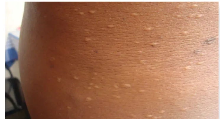
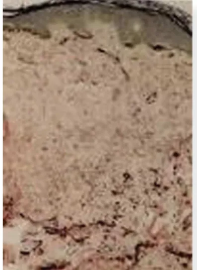
A 56yr old male patient, farmer by occupation, was presented with multiple asymptomatic hyperpigmented annular lesions over the trunk since 8 months and multiple discrete yellowish sac like protrusions over trunk since 6 months. He gives h/o acute pancreatitis 10 months back for which he was treated conservatively. On examination there were multiple discrete annular hyperpigmented plaques of varying sizes with hyperkeratotic grooved borders over anterior and posterior aspects of the trunk and both the upper limbs and neck (Fig. 1). Multiple discrete yellowish sac like protrusions of sizes varying from 5mm to 10mm were seen over both the lanks and posterior aspect of the trunk. Button hole sign was positive (Fig. 2).Routine blood investigations and biochemical investigations were within normal limits. HIV and HBS Ag was negative. Histopathology of keratotic plaques showed foci of epidermal invagination illed with keratin and parakeratotic coronoid lamella ,dermis showed a mild to moderate iniltrate of lymphocytes and evidence of pigment incontinence which was consistent with porokeratosis (Fig. 3). Histopathology of sac like protrusions showed decreased elastic ibres in the papillary and reticular dermis. Staining with verhoeff–van Gieson was consistent with anetoderma (Fig. 4). With the above clinical and histopathological indings the diagnosis of disseminated
supericial porokeratosis and anetoderma was conirmed. For larger lesions were treated with topical 5-lurouracil was given and smaller lesions with topical Tretinoin 0.05% cream. Signiicant improvement was seen within 3 months.
DISSEMINATED SUPERFICIAL POROKERATOSIS AND
ANETODERMA DEVELOPING AFTER ACUTE
PANCREATITIS
ROZWÓJ ROZSIANEJ POWIERZCHOWNEJ POROKERATOZY I
ANETODERMII PO OSTRYM ZAPALENIU TRZUSTKI
Pratyusha Kolanuvada, Chankramath Sujatha,
Hariharasubramony Ambika
Department of Dermatology, MVJ Medical College and Research Hospital, Hoskote, Bangalore, India
Corresponding author: Dr Kolanuvada Pratyusha [email protected]
DOI: 10.7241/ourd.20123.53
Figure 1. Multiple discrete annular hyperpigmented plaques with hyperkeratotic grooved borders
Figure 2. Multiple discrete yellowish sac like protrusions
were seen over both the lanks and posterior aspect of the
trunk
Porokeratosis is a heterogeneous group of disorders that are inherited in a autosomal dominant fashion. Different types of Porokeratosis are Porokeratosis of Mibelli, Disseminated supericial Porokeratosis (DSP), Disseminated supericial porokeratosis of immunosuppression, Disseminated supericial actinic porokeratosis (DSAP), linear porokeratosis, porokeratosis Palmaris plantaris et Disseminata and Punctate Porokeratosis. DSP has its onset in 3rd and 4th decade of life. There is a female predominance with 3:1. Early lesions of disseminated supericial porokeratosis (DSP) are small keratotic papules with central dell. They may be erythematous or pigmented. They enlarge to form supericial ring like lesions with slight central atrophy surrounded by discrete ridge topped by furrow. It mainly involves extremities in a bilateral symmetric fashion. Lesions are distributed symmetrically over extremities and trunk with predilection for the extensor surface. Involvement of face is rare. Lesions spare the axillary vaults, inguinal folds, perigenital region, palms, soles and mucous membranes. Dermoscopy reveals a „white track” structure with brown pigmentation on the inside of the track that can be seen at the edge of an individual lesion, corresponding to the cornoid lamella [1]. Centrally, a white area with red dots, globules, and lines is present that corresponds to capillary vessels; it is more easily observed through the atrophic epithelium. Clonal hyperproliferation of atypical keratinocytes leads to the formation of the cornoid lamella, which expands peripherally and forms the raised boundary between abnormal and normal keratinocytes. Local or systemic changes in immune function may allow the development of atypical clones of keratinocytes [2]. The risk factors for the development of porokeratosis include genetic inheritance, ultraviolet radiation, and immunosuppression. Immunosuppression associated with porokeratosis may be secondary to a disease process such as HIV infection or lymphoma or an iatrogenic suppression such as with immune-modulating drugs used to prevent organ transplant rejection or to treat autoimmune diseases. An autosomal dominant mode of inheritance has been established for familial cases of all forms of porokeratosis. O. Ferreira has reported an association of disseminated supericial porokeratosis with acute pancreatitis and suggested that it may be because of immunosuppression [3].
Anetoderma is a rare skin disease with loss of dermal elastic tissue resulting in clinically localised areas of laccid or herniated sac like skin. It is classiied into primary and secondary. Primary anetoderma appears on previously normal skin with unknown pathogenesis. It is divided in to Schweninger-Buzzi type which has no preceding erythema, and Jadassohn-Pellizari type which is preceded by macular erythema or papular urticaria. Secondary anetoderma occurs at sites of skin diseases such as acne, varicella, xanthoma, discoid lupus erythematosus, granuloma annulare, syphilis [4]. The association of lupus erythematosus, and other autoimmune disorders, and primary anetoderma is often cited in the literature. Whether these indings are coincidental or related is unknown. The exact cause of anetoderma is unknown. Possible explanations for loss of elastic tissue include defective elastin synthesis, uncontrolled production of elastolytic enzymes, loss of elastolytic enzyme inhibitors, elastophagocytosis, or degeneration of elastic ibers secondary to local ischemia induced by microthromboses in dermal vessels [5]. Diagnosis relays on histopathology indings which shows decrease in dermal thickness and normal collagen ibres and decrease in elastin ibres in upper dermis. Speciic elastin staining can demonstrate a marked reduction in elastin content compared with adjacent normal dermis. No treatment has been found to be beneicial once atrophy is advanced. Colchicine has a preventive role.
A rare association of disseminated supericial porokeratosis and anetoderma developing after an episode of acute pancreatitis is being reported for irst time to the best our knowledge.
Acknowledgements
Dr. Padmini Jeyachandran, Prof and Head Department of Pathology, MVJ Medical College and Research Hospital. Dr.Vasantha Kumar S, Principal, MVJ Medical College and Research Hospital.
Figure 3. Foci of epidermal invagination illed with keratin
and parakeratotic coronoid lamella, dermis showed a mild
to moderate iniltrate of lymphocytes (H&E, 40X)
Figure 4. Verhoeff -van Gieson staining showing decreased elastic ibres in the papillary and reticular dermis
Copyright by Pratyusha Kolanuvada, et al. This is an open access article distributed under the terms of the Creative Commons Attribution License,
which permits unrestricted use, distribution, and reproduction in any medium, provided the original author and source are credited.
REFERENCES
1. Delino M, Argenziano G, Nino M: Dermoscopy for the diagnosis of porokeratosis. J Eur Acad Dermatol Venereol. 2004; 18: 194-195.
2. Ito M, Fujiwara H, Maruyama T, Oguro K, Ishihara O, Sato Y: Morphogenesis of the cornoid lamella: histochemical, immunohistochemical, and ultrastructural study of porokeratosis. J Cutan Pathol. 1991; 18: 247-256.
3. Ferreira O, Durate AF: Development of disseminated supericial porokeratosis in a patient with complicated acute pancreatitis. Dermatol Online J. 2011; 17: 5-7.
4. Venencie PY, Winkelmann RK, Moore BA: Anetoderma: clinical indings, associations and long-term follow-up evaluations. Arch Dermatol 1984; 120: 1032–1039.
5. Weinstein S, Piette W: Cutaneous manifestations of antiphospholipid antibody syndrome. Hematol Oncol Clin North Am. 2008; 22: 67-77.