rev bras hematol hemoter. 2016;38(1):86–87
w w w . r b h h . o r g
Revista
Brasileira
de
Hematologia
e
Hemoterapia
Brazilian
Journal
of
Hematology
and
Hemotherapy
Images
in
Clinical
Hematology
Systemic
fungal
infection
by
Histoplasma
capsulatum
:
intracellular
fungus
in
peripheral
leukocytes
Valéria
Salgado,
Mayara
Caldas
Ramos,
Ricardo
Ambrósio
Fock
∗HospitalUniversitário,UniversidadedeSãoPaulo(USP),SãoPaulo,Brazil
a
r
t
i
c
l
e
i
n
f
o
Articlehistory:
Received18September2015 Accepted15December2015 Availableonline3February2016
Amiddle-agedwomanwasadmittedtotheTeaching Hospi-taloftheUniversidadedeSãoPaulo,Brazilwithpneumonia symptoms. Shehad fever,cough and weakness associated withepigastric pain. Anabdominal computed tomography scanshowedextensiveadenopathywithanenlargedspleen.
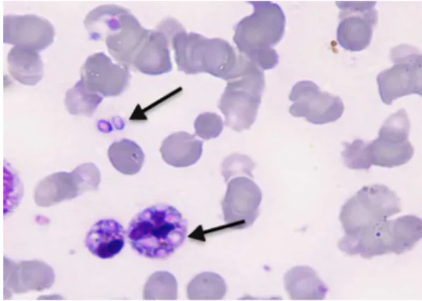
Figure1–Arrowsshowphagocytescontainingoneormore intracellularHistoplasmacapsulatum(magnification:1000×;
May-Grünwaldstain).
∗ Correspondingauthorat:DivisãodeLaboratóriodeAnálisesClínicas,HospitalUniversitário,UniversidadedeSãoPaulo(USP),Av.Lineu
Prestes,2565,05508-900SãoPaulo,SP,Brazil. E-mailaddress:[email protected](R.A.Fock).
SerologyforHIVwaspositiveandyeastformswereobserved intheperipheralbloodsmear(Figures1and2).
Diagnosis of histoplasmosis is made by the detection of Histoplasma capsulatum in sputum, blood or liquor but it is common to observe negative resultsdue to technical
Figure2–Arrowsshowintracellularandextracellular
Histoplasmacapsulatum(magnification:1000×;
May-Grünwaldstain).
http://dx.doi.org/10.1016/j.bjhh.2015.12.002
revbrashematolhemoter.2016;38(1):86–87
87
constraints wherein a reduced number of fungal forms are found in samples.1 Imaging, diagnostic scanning tests
and biopsy of target organs are importantto differentiate fromtuberculosis, sarcoidosisormetastatic carcinomaand lymphomaandusuallythecultureofbiologicalsamplesand serologicalassaysareperformedtoconfirmthediagnosis.1,2
However,abloodsmearisimportanttoestablishthediagnosis ofdisseminatedhistoplasmosisand,afterinvitro contamina-tionisexcluded,thepresenceofbothfreeandintracellular H.capsulatumshouldbereported.
Conflicts
of
interest
Theauthorsdeclarenoconflictsofinterest.
r
e
f
e
r
e
n
c
e
s
1.deOliveiraRB,AtobeJH,deSouzaSA,CastroLimaSantosDW. Epidemiologyofinvasivefungalinfectionsinpatientswith acquiredimmunodeficiencysyndromeatareferencehospital forinfectiousdiseasesinBrazil.Mycopathologia.
2014;178(1–2):71–8.
2.Carreto-BinaghiLE,DamascenoLS,PitanguiNdeS, Fusco-AlmeidaAM,Mendes-GianniniMJ,Zancopé-Oliveira RM,etal.CouldHistoplasmacapsulatumberelatedto